Anatomy & Physiology: Muscles—Fibularis (Peroneus) Brevis.
Structure.
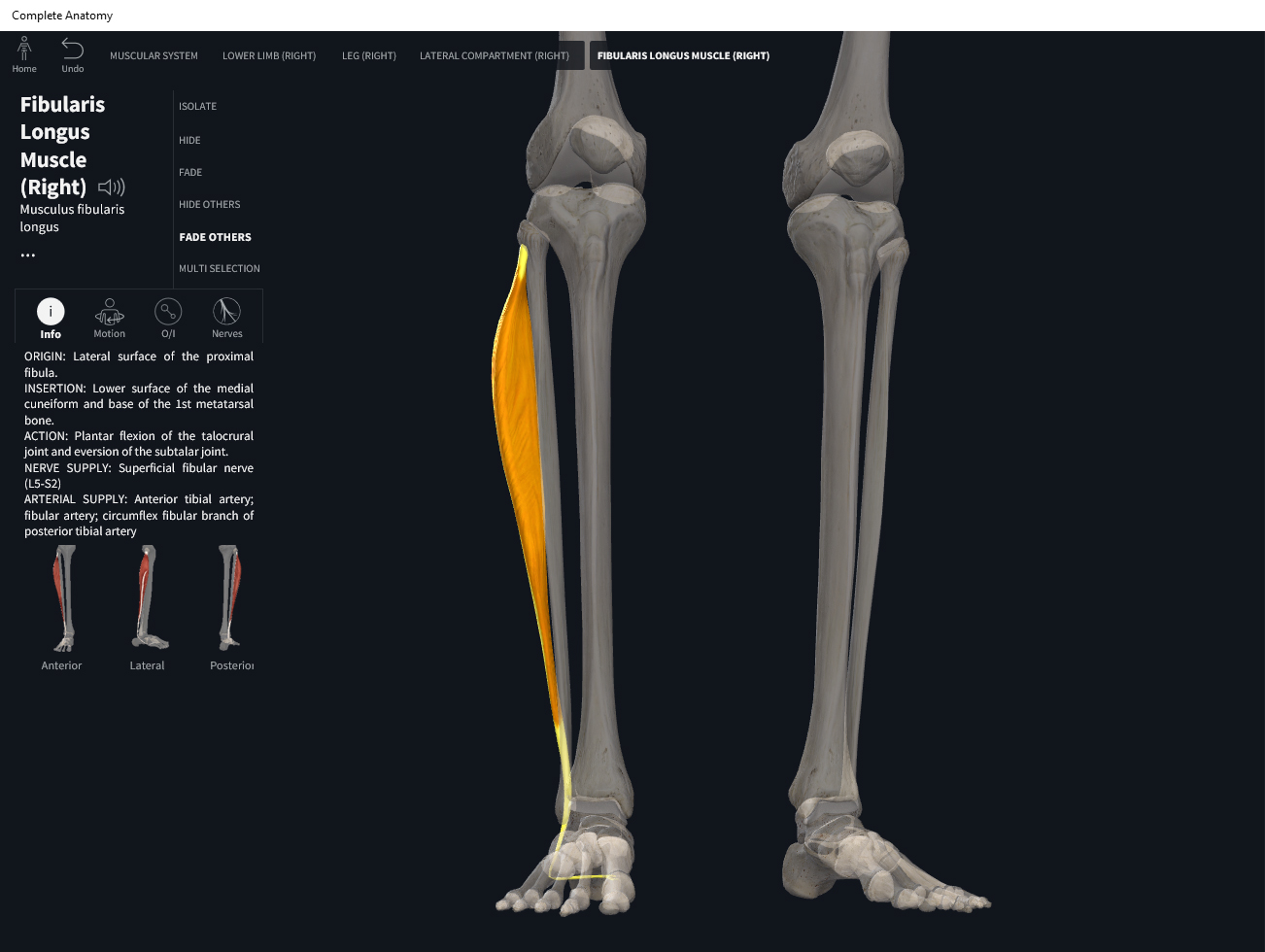
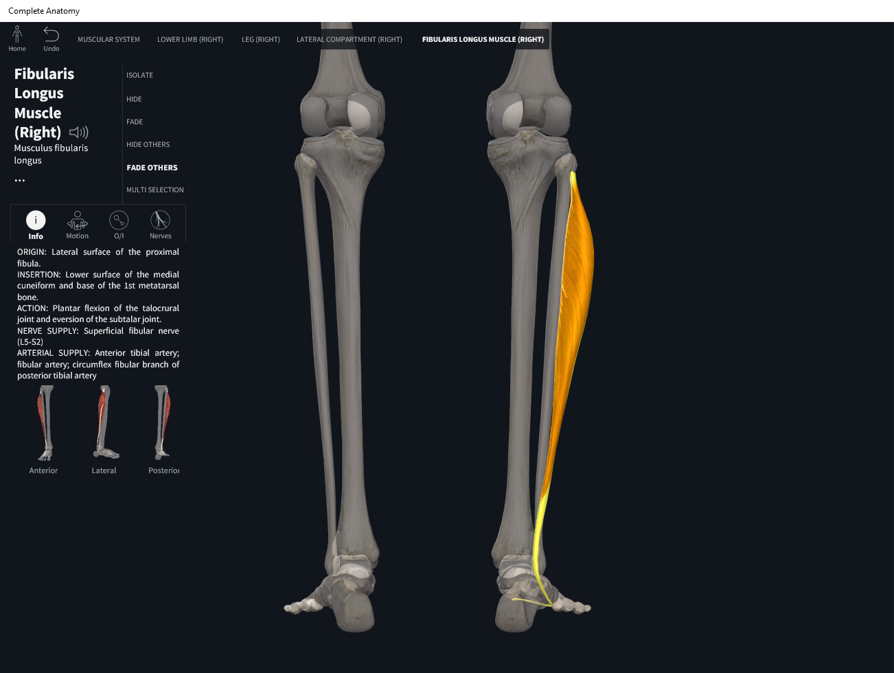
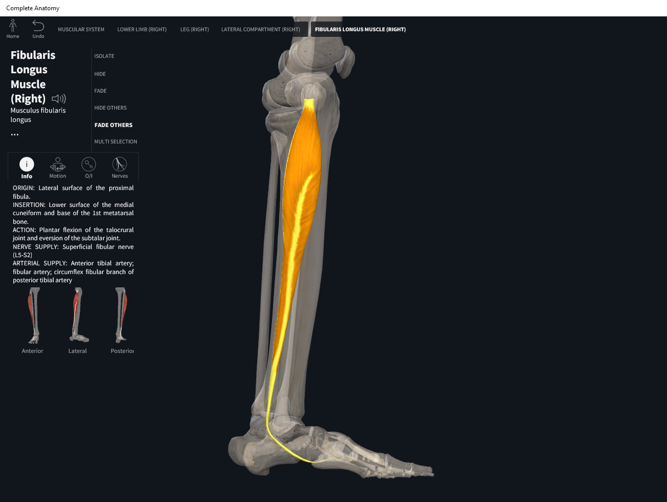
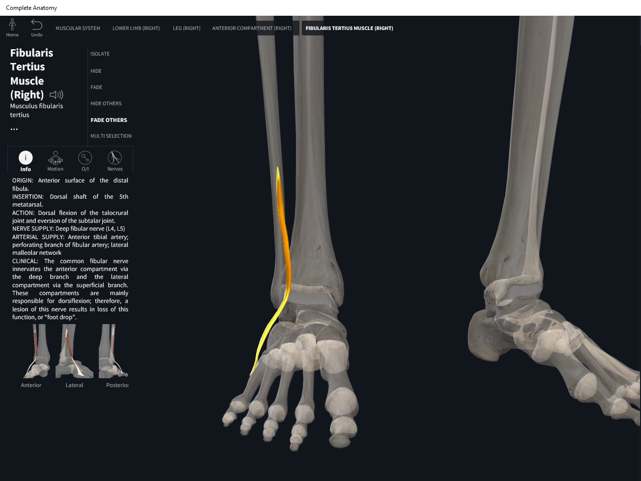
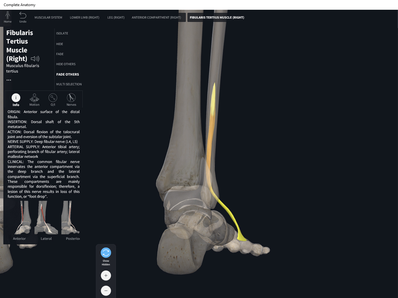
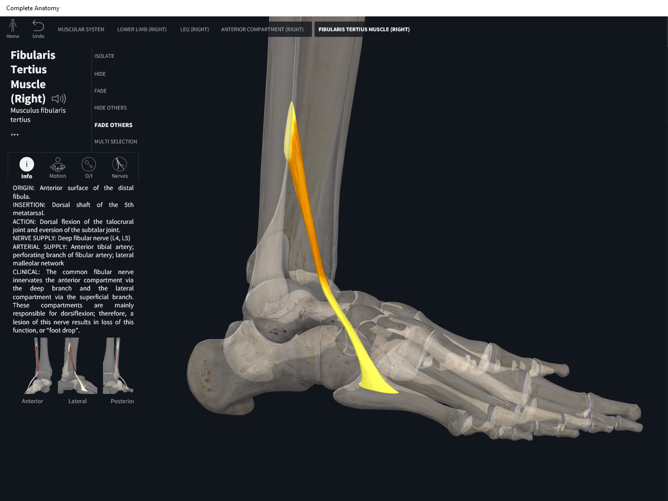
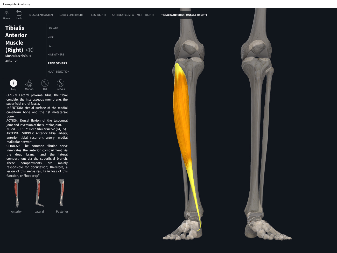
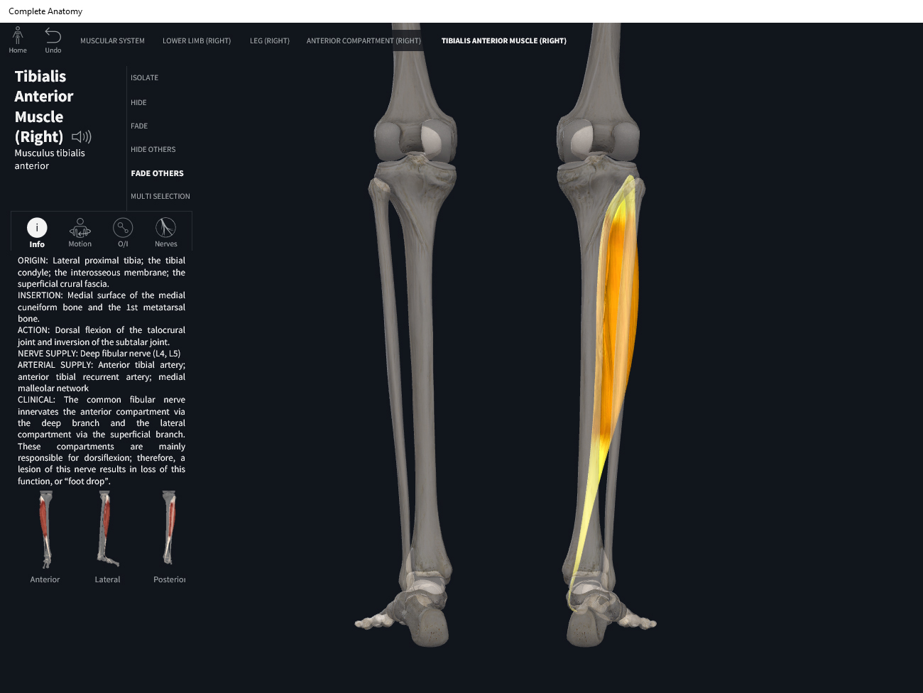
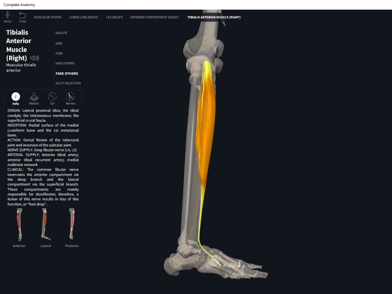
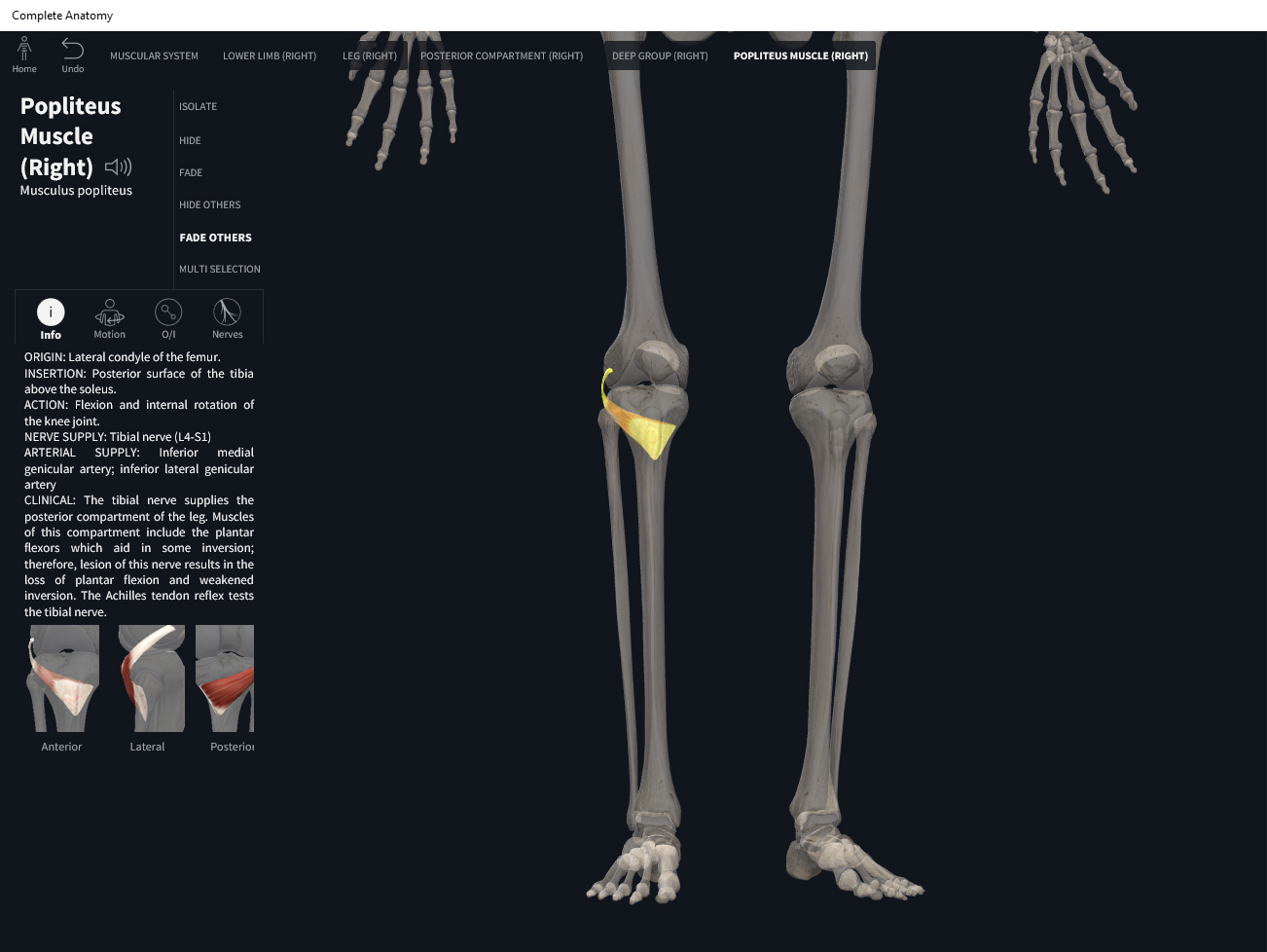
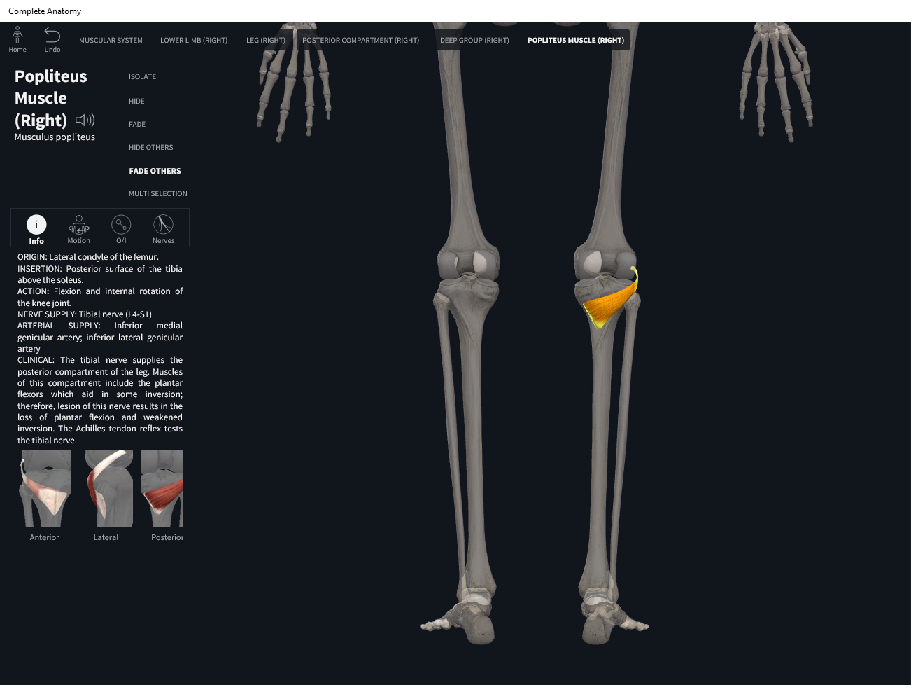
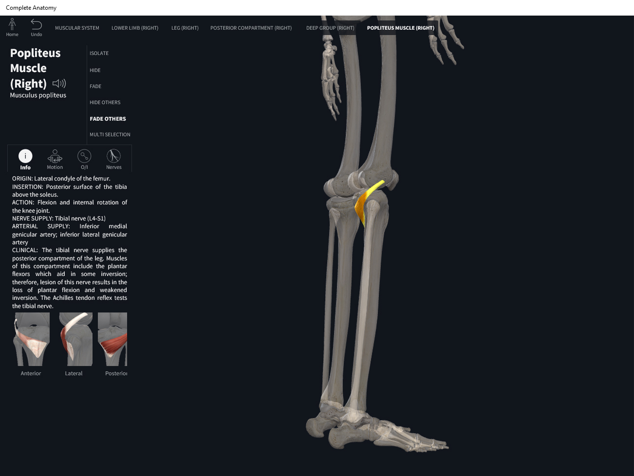
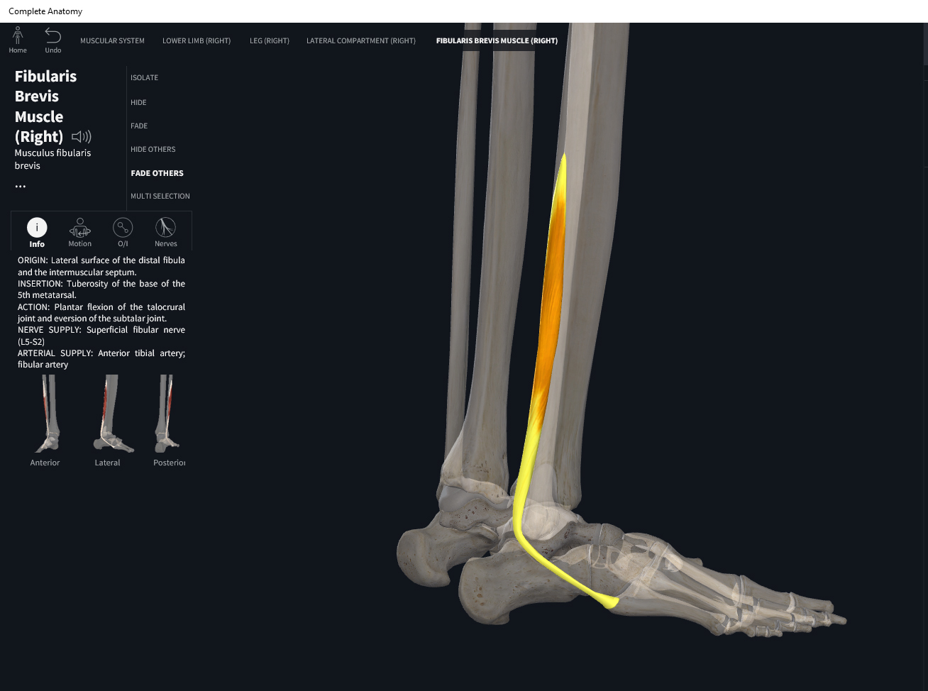
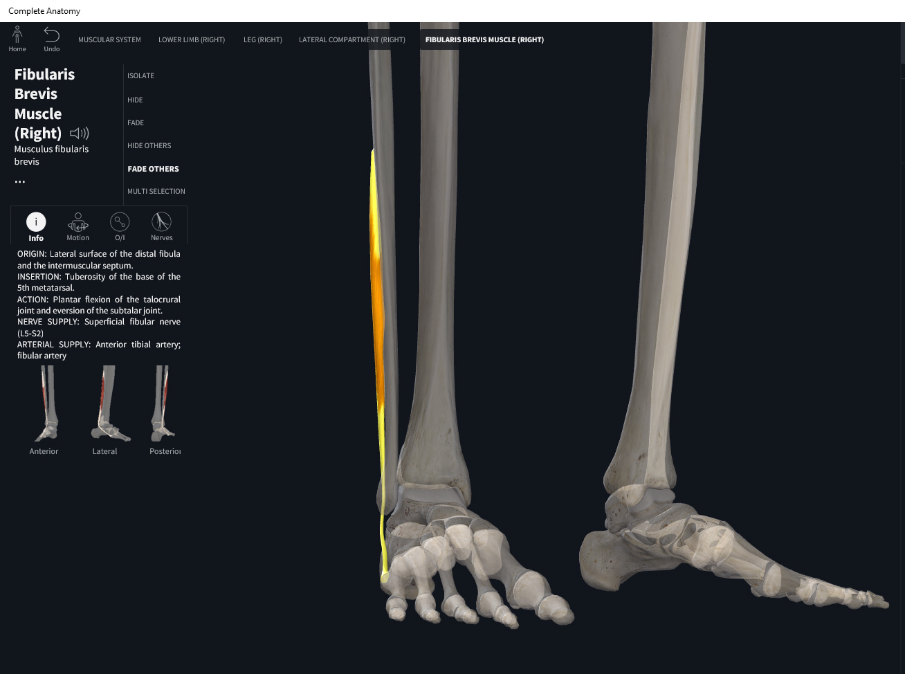
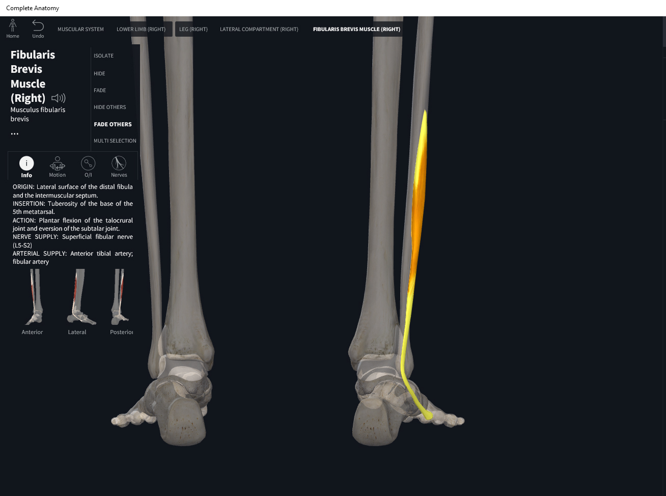
- Origin: body of fibula.
- Insertion: base of metatarsal V.
Function.
- Concentric action: plantar flexion; eversion at intertarsal joints.
- Reverse mover action: evert/pronates talus at subtalar joint.
- Eccentric action: controls/restrains/decelerates inversion/supination at subtalar joint; ankle dorsiflexion.
- Isometric action: stabilization of ankle and subtalar joints.
- Innervation: superficial fibular (peroneal) nerve.
- Arterial supply: fibular artery.
Clinical Significance.
More.
- https://www.anatomynext.com/peroneus-brevis/
- https://www.youtube.com/watch?v=WpDcqdCQIk0
- https://www.youtube.com/watch?v=_wqkhm5I6bw
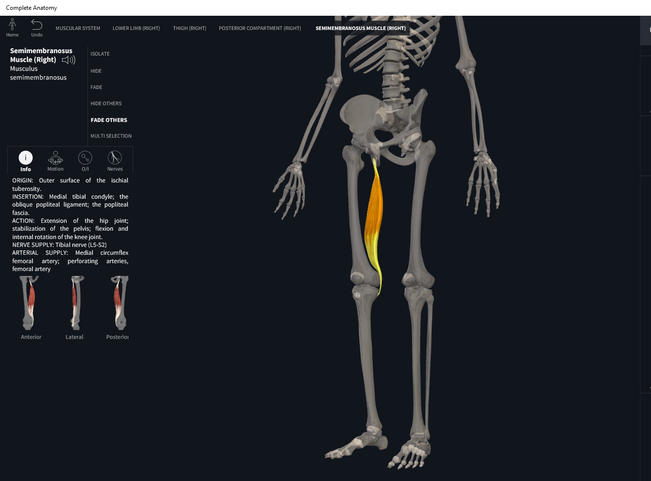
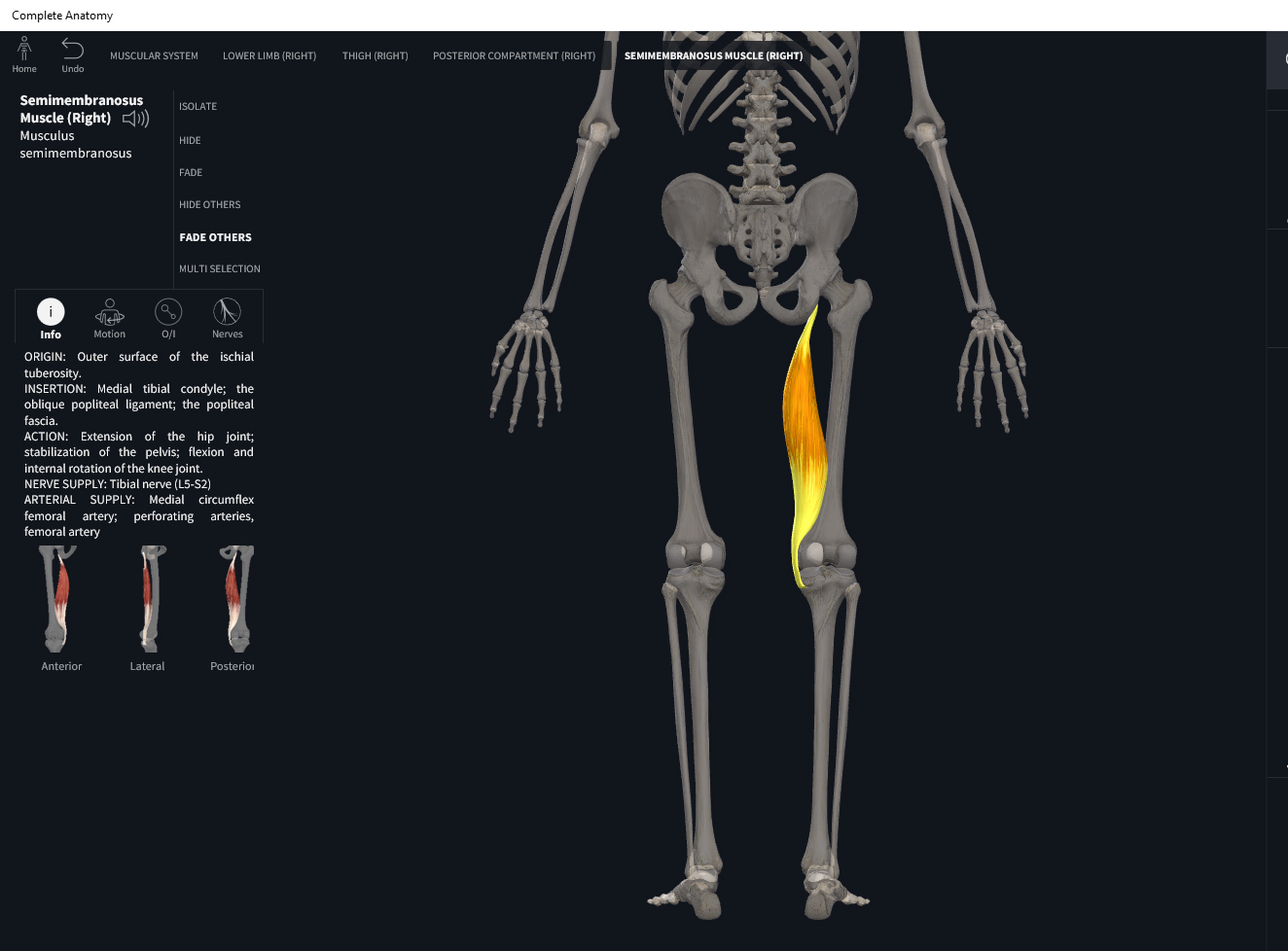
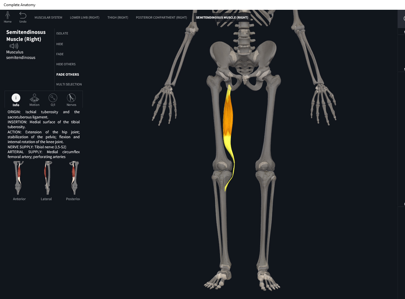
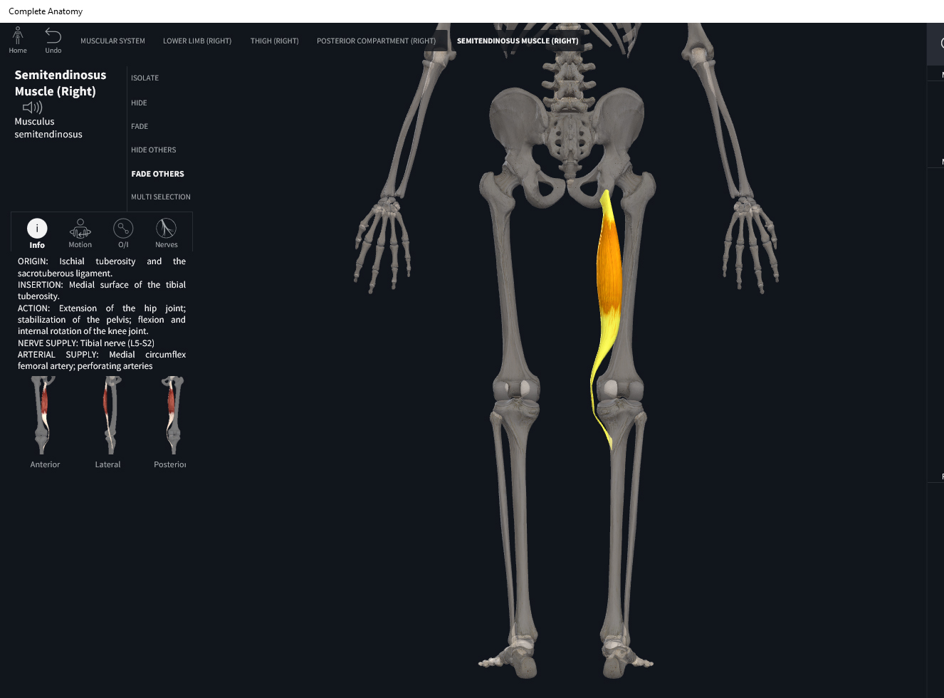
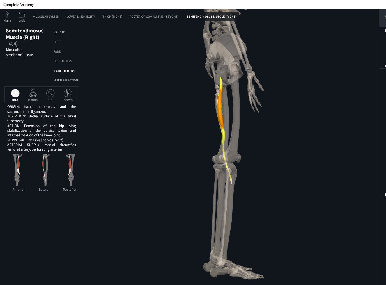
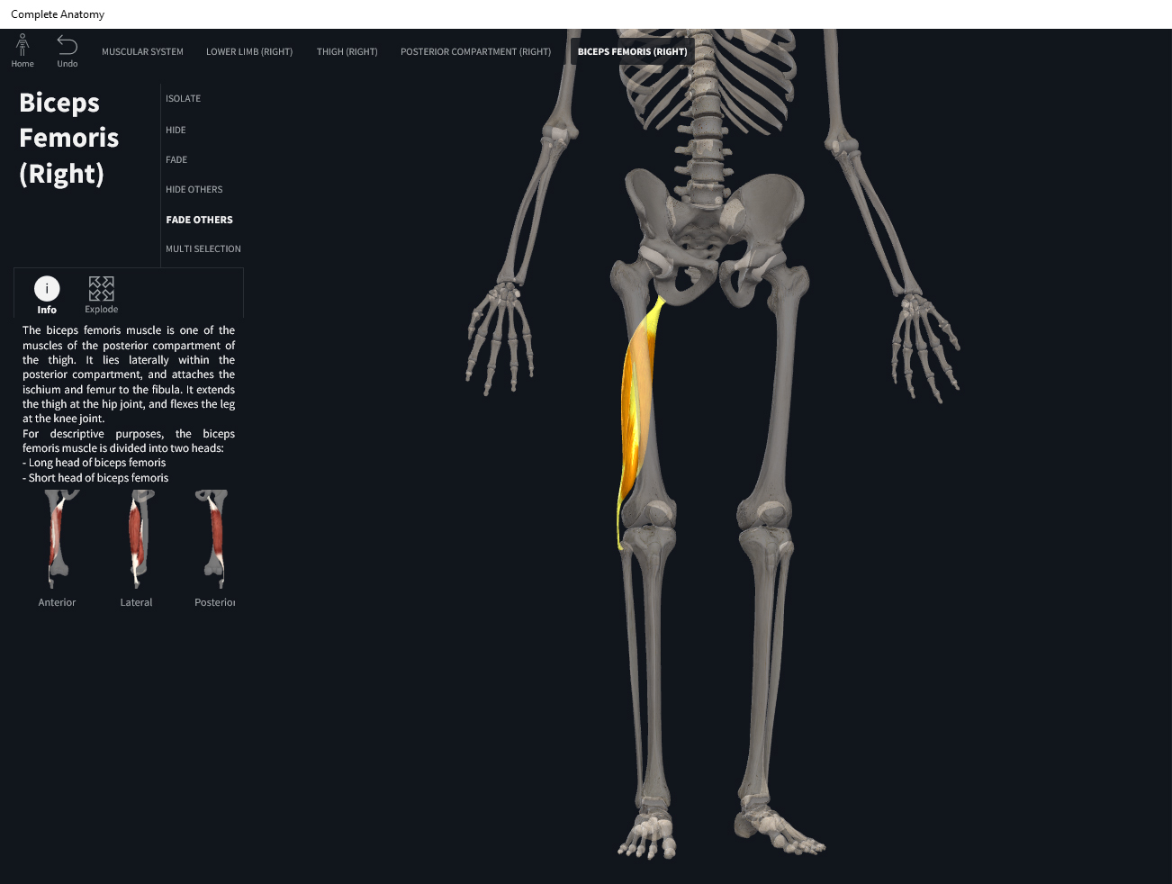
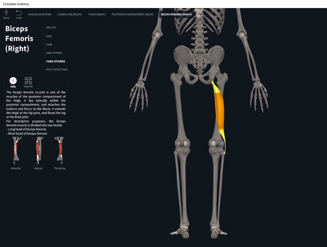
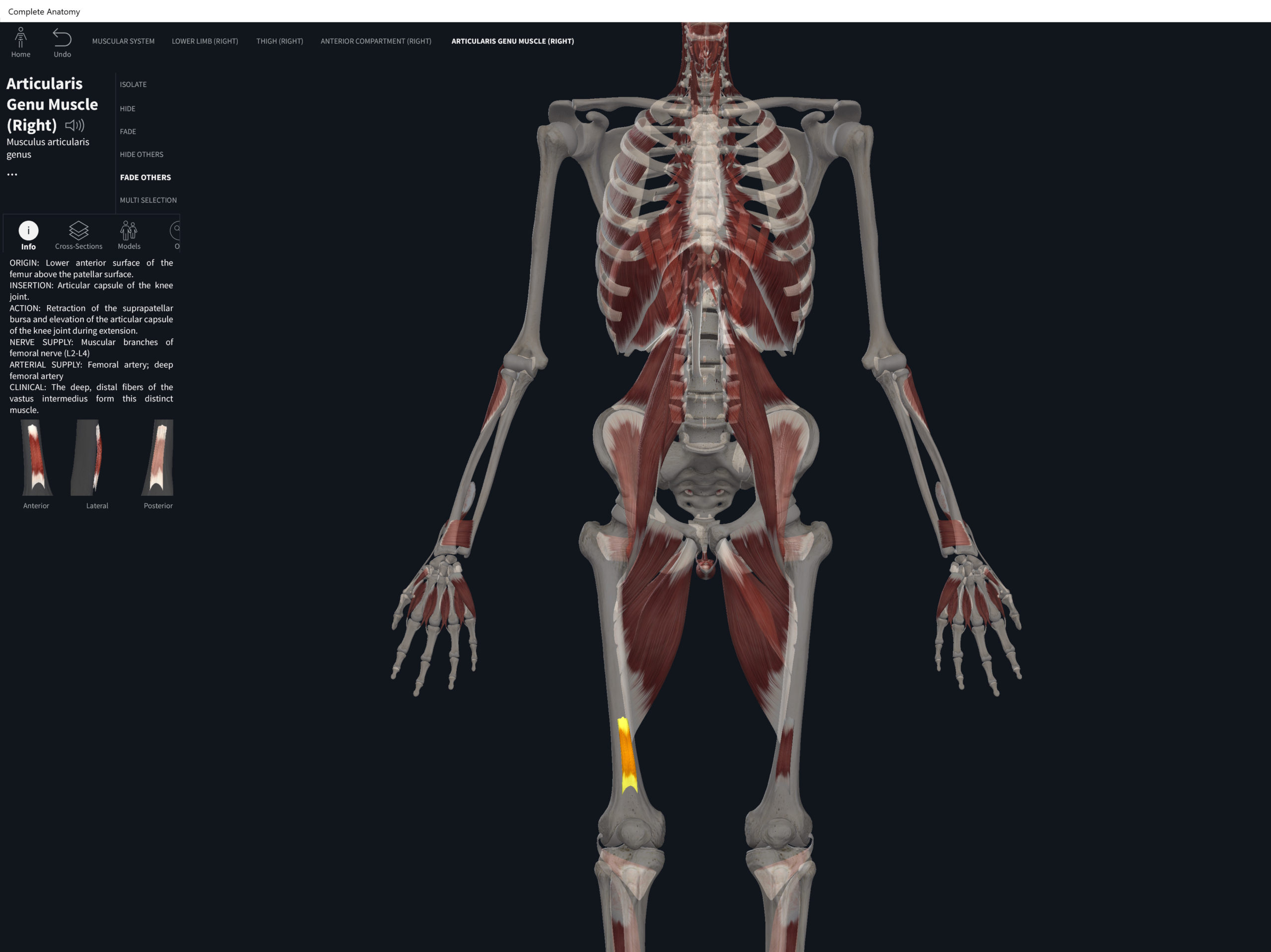
Fibularis brevis. Used with permission by 3D4Medical.
References
Biel, A. (2015). Trail guide to the body: A hands-on guide to locating muscles, bones and more.
Clark, M., Lucett, S., Sutton, B. G., & National Academy of Sports Medicine. (2014). NASM essentials of corrective exercise training. Burlington, MA: Jones & Bartlett Learning.
Jenkins, G., & Tortora, G. J. (2012). Anatomy and Physiology: From Science to Life, 3rd Edition International Stu. John Wiley & Sons.
Muscolino, J. E. (2017). The muscular system manual: The skeletal muscles of the human body.