Posted on

Muscles: Levator Palpebrae Superioris.

Download these notes.

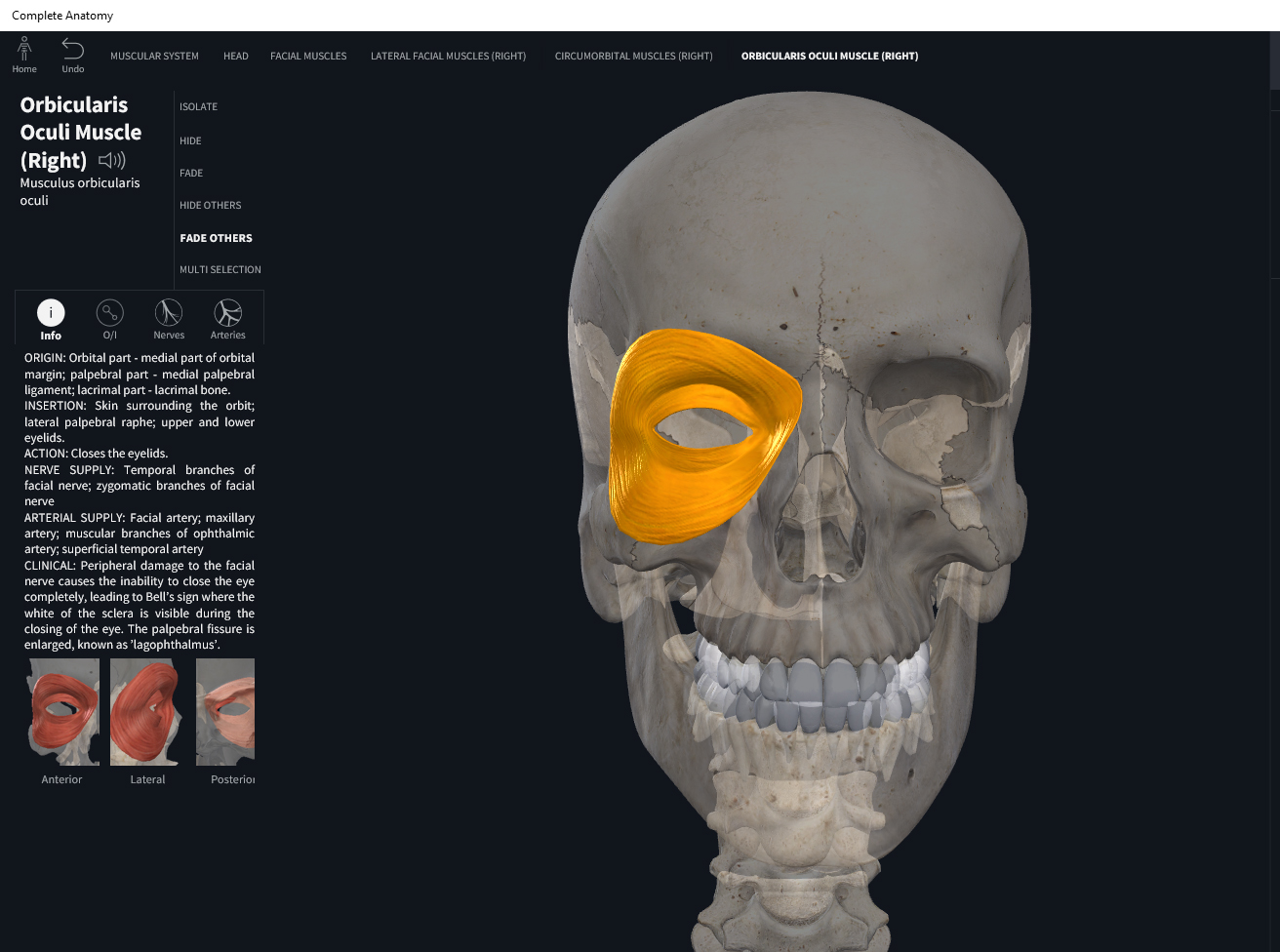
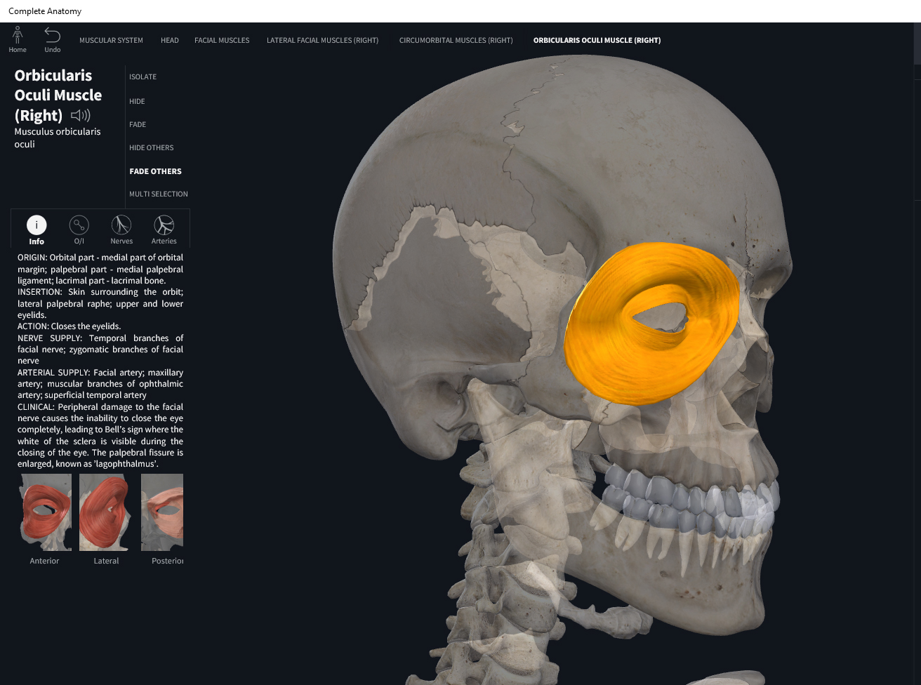
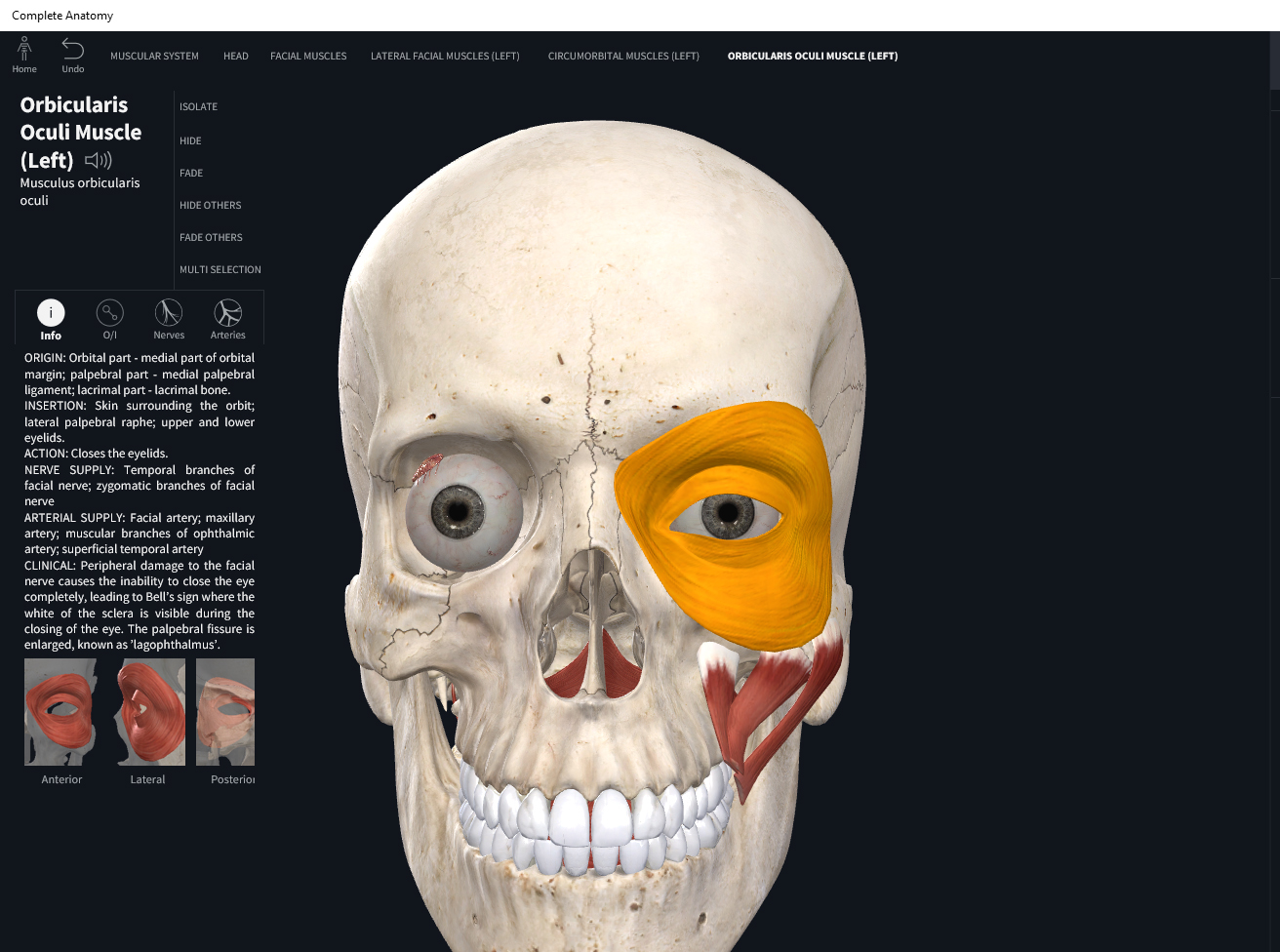
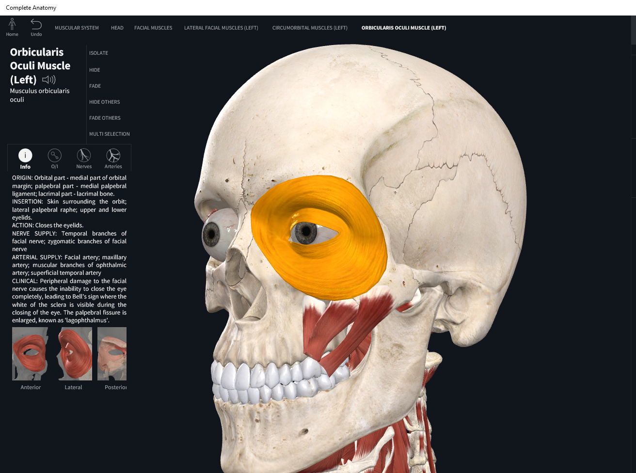
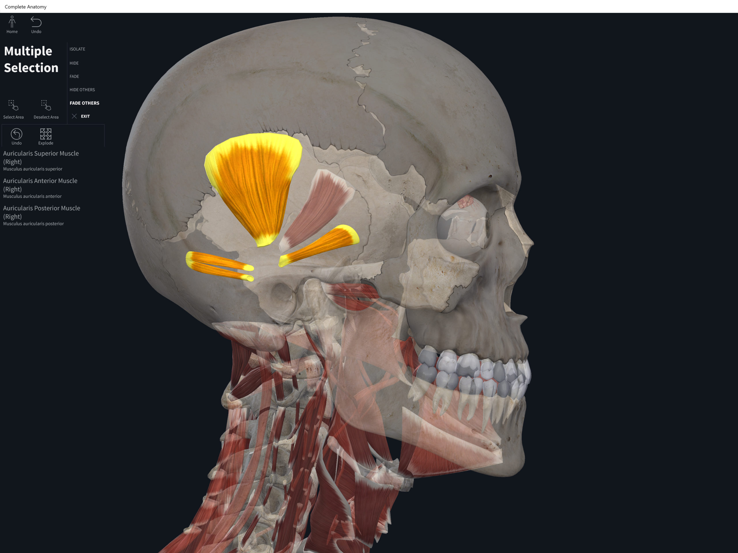
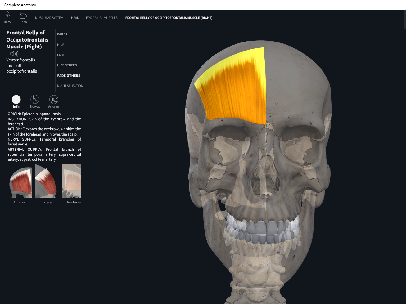
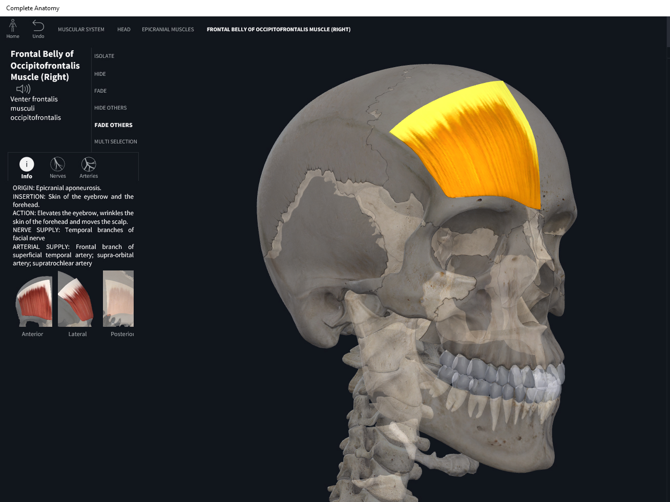
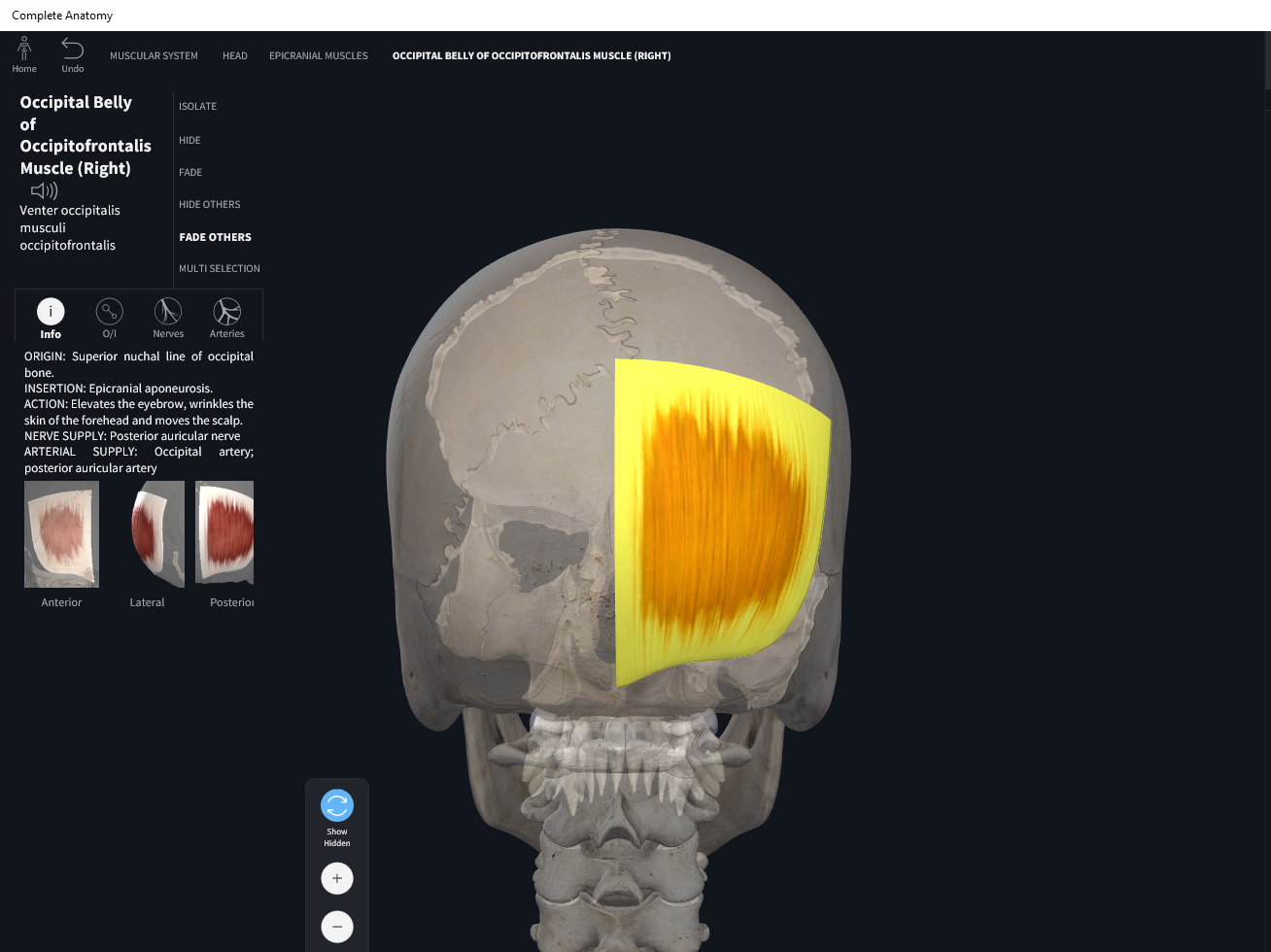
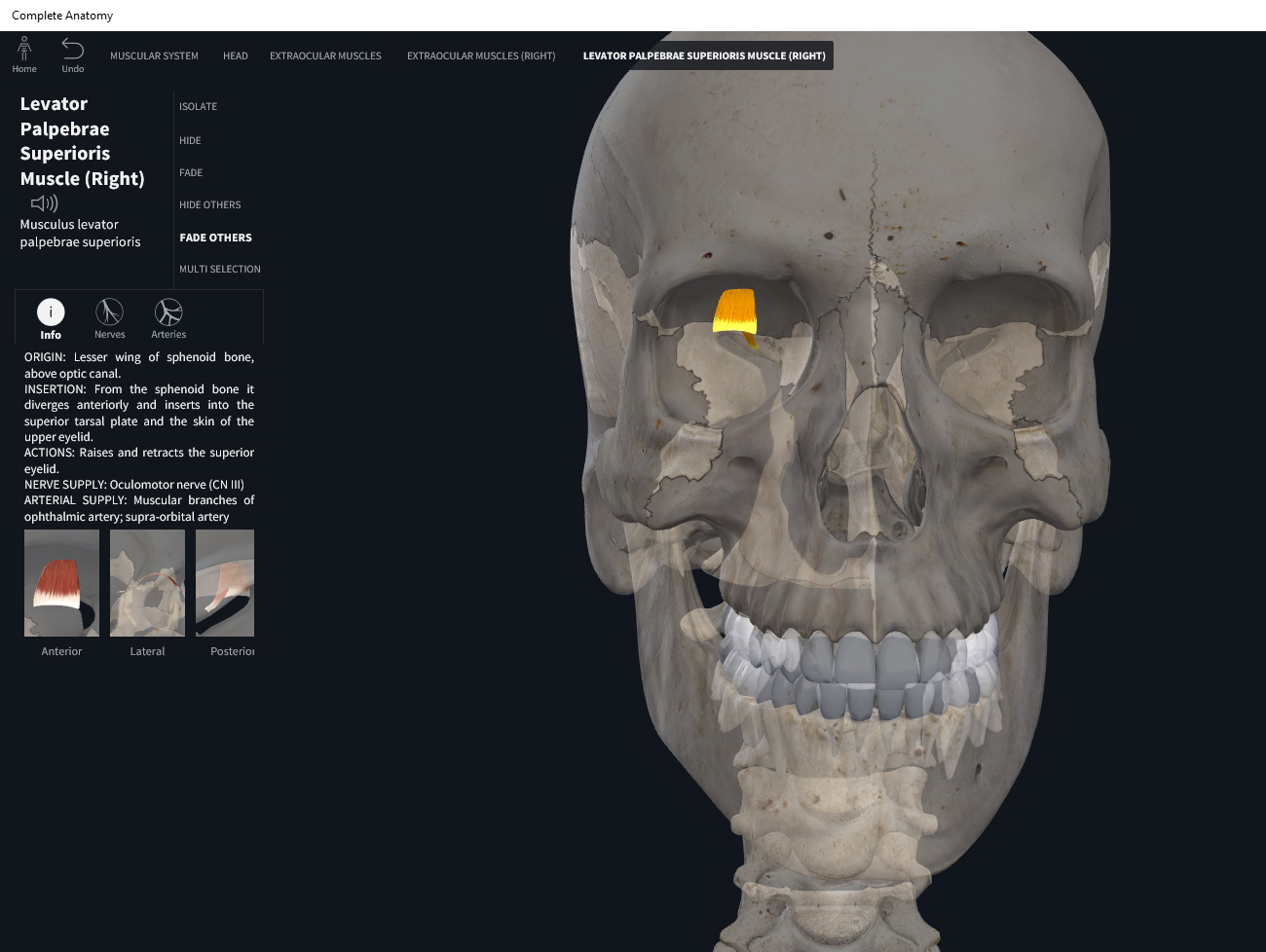
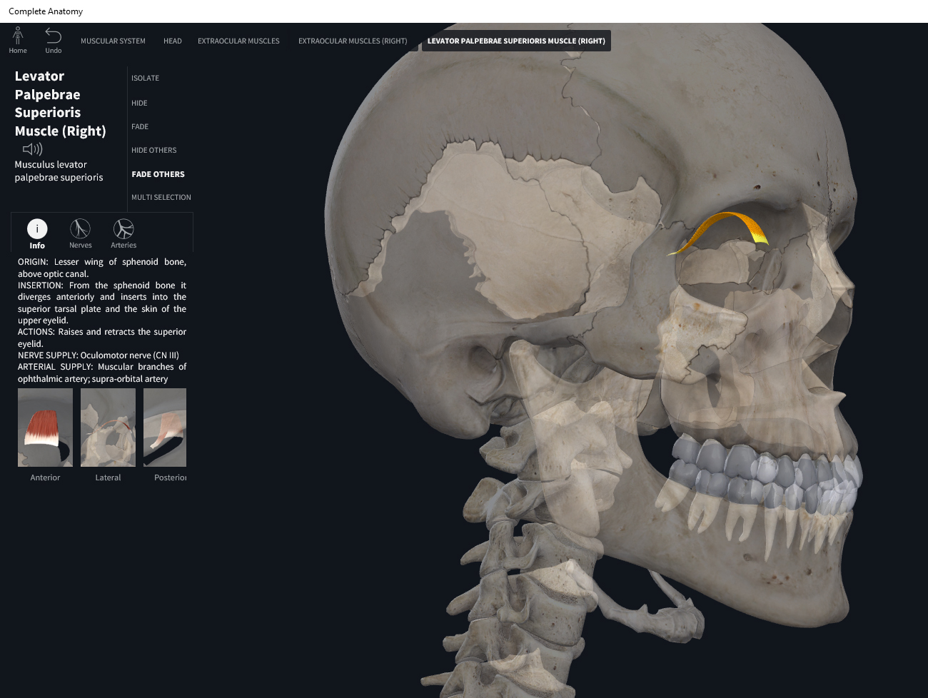
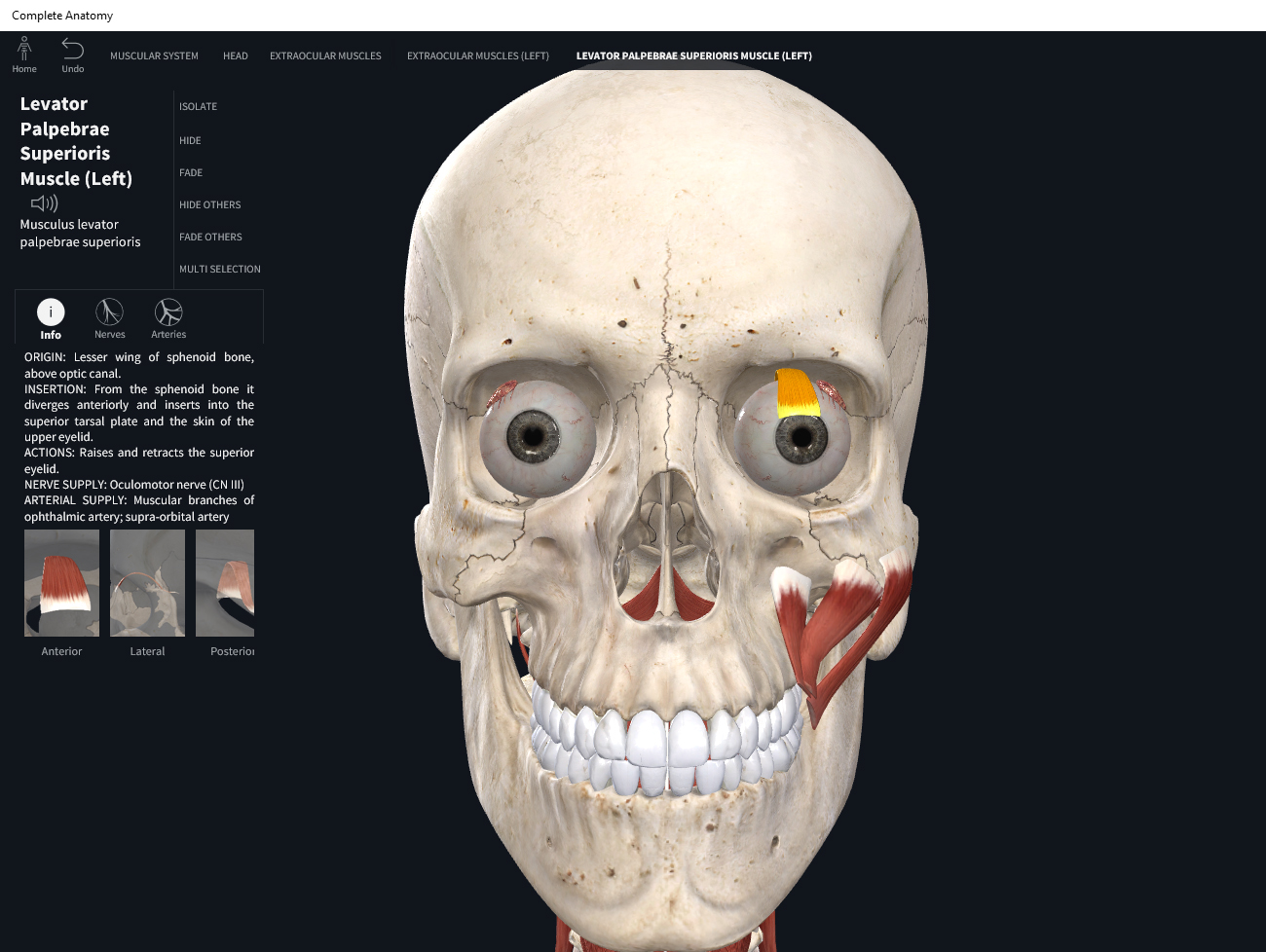
Anatomy & Physiology: Muscles—Levator Palpebrae Superioris.

Structure.

  • Origin: root of orbit (smaller wing of sphenoid).
  • Insertion: skin of upper eyelid.

Function.

  • Concentric action: elevates upper eyelid, opens eye.
  • Reverse mover action:
  • Eccentric action:
  • Isometric action:
  • Innervation: oculomotor III nerve.
  • Arterial supply: ophthalmic artery.

Clinical Significance.

More.

References

Biel, A. (2015). Trail guide to the body: A hands-on guide to locating muscles, bones and more.

Cedars-Sinai. (2018). Vertebrae of the spine. Retrieved from https://www.cedars-sinai.org/health-library/diseases-and-conditions/v/vertebrae-of-the-spine.html

Clark, M., Lucett, S., Sutton, B. G., & National Academy of Sports Medicine. (2014). NASM essentials of corrective exercise training. Burlington, MA: Jones & Bartlett Learning.

Jenkins, G., & Tortora, G. J. (2012). Anatomy and Physiology: From Science to Life, 3rd Edition International Stu. John Wiley & Sons.

Muscolino, J. E. (2017). The muscular system manual: The skeletal muscles of the human body.

Posted on

Muscles: Orbicularis Oculi.

Download these notes.

Anatomy & Physiology: Muscles—Orbicularis Oculi.

Structure.

  • Origin: medial wall of orbit.
  • Insertion: circular, around orbit.
  • Orbital, palpebral, and lacrimal parts.

Function.

  • Concentric action: closes/squints eye. Lesser: depress upper eyelid (palpebral); elevate lower eyelid (palpebral); retract lower eyelid (lacrimal); tear transport and drainage (lacrimal)
  • Eccentric action:
  • Isometric action:
  • Innervation: facial VII nerve.
  • Arterial supply: facial artery and superficial temporal artery.

Clinical Significance.

More.

References

Biel, A. (2015). Trail guide to the body: A hands-on guide to locating muscles, bones and more.

Cedars-Sinai. (2018). Vertebrae of the spine. Retrieved from https://www.cedars-sinai.org/health-library/diseases-and-conditions/v/vertebrae-of-the-spine.html

Clark, M., Lucett, S., Sutton, B. G., & National Academy of Sports Medicine. (2014). NASM essentials of corrective exercise training. Burlington, MA: Jones & Bartlett Learning.

Jenkins, G., & Tortora, G. J. (2012). Anatomy and Physiology: From Science to Life, 3rd Edition International Stu. John Wiley & Sons.

Muscolino, J. E. (2017). The muscular system manual: The skeletal muscles of the human body.

Posted on

Muscles: Auricularis Group.

Download these notes.

Anatomy & Physiology: Muscles—Auricularis Group.

Structure.

  • Auricularis Anterior
    • Origin: temporal fascia/galea aponeurotica (lateral margin).
    • Insertion: major helix/spine of helix (ear).
  • Auricularis Superior
    • Origin: temporal fascia/galea aponeurotica (lateral margin).
    • Insertion: superior (to the ear) aspect of cranial surface.
  • Auricularis Posterior
    • Origin: the mastoid area of the temporal bone.
    • Insertion: ponticulus (ridge) of eminentia conchae.

Function.

  • Concentric action: brings/draws the ear anteriorly (auricularis anterior); elevates ear (auricularis superior); brings/draws ear posteriorly (auricularis posterior).
  • Reverse mover action:
  • Eccentric action:
  • Isometric action:
  • Innervation: facial VII nerve.
  • Arterial supply: superficial temporal and posterior auricular arteries.

Clinical Significance.

References

Biel, A. (2015). Trail guide to the body: A hands-on guide to locating muscles, bones and more.

Cedars-Sinai. (2018). Vertebrae of the spine. Retrieved from https://www.cedars-sinai.org/health-library/diseases-and-conditions/v/vertebrae-of-the-spine.html

Clark, M., Lucett, S., Sutton, B. G., & National Academy of Sports Medicine. (2014). NASM essentials of corrective exercise training. Burlington, MA: Jones & Bartlett Learning.

Jenkins, G., & Tortora, G. J. (2012). Anatomy and Physiology: From Science to Life, 3rd Edition International Stu. John Wiley & Sons.

Muscolino, J. E. (2017). The muscular system manual: The skeletal muscles of the human body.

Posted on

Muscles: Temporoparietalis.

Download these notes.

Anatomy & Physiology: Muscles— Temporoparietalis.

Structure.

  • Origin: aponeurosis above auricularis.
  • Insertion: galeal aponeurosis.

Function.

  • Concentric action: elevates ear; tightens scalp.
  • Reverse mover action:
  • Eccentric action:
  • Isometric action:
  • Innervation: facial VII nerve.
  • Arterial supply: superficial temporal and posterior auricular arteries.

Clinical Significance.

References

Biel, A. (2015). Trail guide to the body: A hands-on guide to locating muscles, bones and more.

Cedars-Sinai. (2018). Vertebrae of the spine. Retrieved from https://www.cedars-sinai.org/health-library/diseases-and-conditions/v/vertebrae-of-the-spine.html

Clark, M., Lucett, S., Sutton, B. G., & National Academy of Sports Medicine. (2014). NASM essentials of corrective exercise training. Burlington, MA: Jones & Bartlett Learning.

Jenkins, G., & Tortora, G. J. (2012). Anatomy and Physiology: From Science to Life, 3rd Edition International Stu. John Wiley & Sons.

Loyola University Medical Education Network. (n.d.). Temporoparietalis. Retrieved from http://www.meddean.luc.edu/lumen/meded/grossanatomy/dissector/mml/tepa.htm

Muscolino, J. E. (2017). The muscular system manual: The skeletal muscles of the human body.

Posted on

Muscles: Occipitofrontalis.

Download these notes.

Anatomy & Physiology: Muscles—Occipitofrontalis.

Structure.

  • Origin: frontal belly—epicranial aponeurosis; occipital belly—occipital bone and mastoid process of temporal bone.
  • Insertion: frontal belly—skin superior to supraorbital margin; occipital belly—epicranial aponeurosis.

Function.

  • Concentric action: frontal belly—raises eyebrows, draw scalp anteriorly, wrinkles skin of forehead horizontally (surprised look); occipital belly—draws scalp posteriorly.
  • Reverse mover action:
  • Eccentric action:
  • Isometric action:
  • Innervation: facial VII nerve.
  • Arterial supply: occipitalis–occipital and posterior auricular arteries; frontalis-supraorbital and supratrochlear branches of ophthalmic artery.

Clinical Significance.

More.

References

Biel, A. (2015). Trail guide to the body: A hands-on guide to locating muscles, bones and more.

Cedars-Sinai. (2018). Vertebrae of the spine. Retrieved from https://www.cedars-sinai.org/health-library/diseases-and-conditions/v/vertebrae-of-the-spine.html

Clark, M., Lucett, S., Sutton, B. G., & National Academy of Sports Medicine. (2014). NASM essentials of corrective exercise training. Burlington, MA: Jones & Bartlett Learning.

Jenkins, G., & Tortora, G. J. (2012). Anatomy and Physiology: From Science to Life, 3rd Edition International Stu. John Wiley & Sons.

Muscolino, J. E. (2017). The muscular system manual: The skeletal muscles of the human body.

Posted on

Muscles: Omohyoid.

Download these notes.

Anatomy & Physiology: Muscles—Omohyoid.

Structure.

  • Origin: superior border of scapula and superior transverse ligament.
  • Insertion: body of hyoid.

Function.

  • Concentric action: depress hyoid.
  • Reverse mover action: elevate scapula.
  • Eccentric action: controls/restrains/slows hyoid elevation, scapular depression.
  • Isometric action: stabilize hyoid, scapula, and clavicle.
  • Innervation: branches of spinal nerves C1-C3.
  • Arterial supply: superior thyroid artery and transverse cervical artery.

Clinical Significance.

More.

References

Biel, A. (2015). Trail guide to the body: A hands-on guide to locating muscles, bones and more.

Cedars-Sinai. (2018). Vertebrae of the spine. Retrieved from https://www.cedars-sinai.org/health-library/diseases-and-conditions/v/vertebrae-of-the-spine.html

Clark, M., Lucett, S., Sutton, B. G., & National Academy of Sports Medicine. (2014). NASM essentials of corrective exercise training. Burlington, MA: Jones & Bartlett Learning.

Jenkins, G., & Tortora, G. J. (2012). Anatomy and Physiology: From Science to Life, 3rd Edition International Stu. John Wiley & Sons.

Muscolino, J. E. (2017). The muscular system manual: The skeletal muscles of the human body.

Posted on

Muscles: Thyrohyoid.

Download these notes.

Anatomy & Physiology: Muscles—Thyrohyoid.

Structure.

  • Origin: thyroid cartilage of larynx.
  • Insertion: greater horn of hyoid.

Function.

  • Concentric action: elevates thyroid cartilage of larynx; depress hyoid.
  • Reverse mover action: elevate thyroid cartilage.
  • Eccentric action: controls/restrains/slows elevation of hyoid, depression of thyroid cartilage.
  • Isometric action: stabilize hyoid bone and thyroid cartilage.
  • Innervation: branches of spinal nerves C1-C3; descending hypoglossal XII nerve.
  • Arterial supply: superior thyroid artery.

Clinical Significance.

More.

References

Biel, A. (2015). Trail guide to the body: A hands-on guide to locating muscles, bones and more.

Cedars-Sinai. (2018). Vertebrae of the spine. Retrieved from https://www.cedars-sinai.org/health-library/diseases-and-conditions/v/vertebrae-of-the-spine.html

Clark, M., Lucett, S., Sutton, B. G., & National Academy of Sports Medicine. (2014). NASM essentials of corrective exercise training. Burlington, MA: Jones & Bartlett Learning.

Jenkins, G., & Tortora, G. J. (2012). Anatomy and Physiology: From Science to Life, 3rd Edition International Stu. John Wiley & Sons.

Muscolino, J. E. (2017). The muscular system manual: The skeletal muscles of the human body.

Posted on

Muscles: Sternohyoid.

Download these notes.

Anatomy & Physiology: Muscles—Sternohyoid.

Structure.

  • Origin: medial end of clavicle, manubrium.
  • Insertion: body of hyoid.

Function.

  • Concentric action: depress hyoid.
  • Reverse mover action: elevate sternum.
  • Eccentric action: controls/restrains/slows elevation of hyoid, depression of sternum.
  • Isometric action: stabilize hyoid.
  • Innervation: branches of spinal nerves C1-C3.
  • Arterial supply: superior thyroid artery.

Clinical Significance.

More.

References

Biel, A. (2015). Trail guide to the body: A hands-on guide to locating muscles, bones and more.

Cedars-Sinai. (2018). Vertebrae of the spine. Retrieved from https://www.cedars-sinai.org/health-library/diseases-and-conditions/v/vertebrae-of-the-spine.html

Clark, M., Lucett, S., Sutton, B. G., & National Academy of Sports Medicine. (2014). NASM essentials of corrective exercise training. Burlington, MA: Jones & Bartlett Learning.

Jenkins, G., & Tortora, G. J. (2012). Anatomy and Physiology: From Science to Life, 3rd Edition International Stu. John Wiley & Sons.

Muscolino, J. E. (2017). The muscular system manual: The skeletal muscles of the human body.

Posted on

Muscles: Stylohyoid.

Download these notes.

Anatomy & Physiology: Muscles—Stylohyoid.

Structure.

  • Origin: styloid process of temporal bone.
  • Insertion: body of hyoid.

Function.

  • Concentric action: elevates and posteriorly draws hyoid.
  • Reverse mover action: extends head.
  • Eccentric action: controls/restrains/slows depression of hyoid.
  • Isometric action: stabilizes hyoid.
  • Innervation: facial VII nerve.
  • Arterial supply: occipital, posterior auricular, and facial arteries.

Clinical Significance.

More.

References

Biel, A. (2015). Trail guide to the body: A hands-on guide to locating muscles, bones and more.

Cedars-Sinai. (2018). Vertebrae of the spine. Retrieved from https://www.cedars-sinai.org/health-library/diseases-and-conditions/v/vertebrae-of-the-spine.html

Clark, M., Lucett, S., Sutton, B. G., & National Academy of Sports Medicine. (2014). NASM essentials of corrective exercise training. Burlington, MA: Jones & Bartlett Learning.

Jenkins, G., & Tortora, G. J. (2012). Anatomy and Physiology: From Science to Life, 3rd Edition International Stu. John Wiley & Sons.

Muscolino, J. E. (2017). The muscular system manual: The skeletal muscles of the human body.

Posted on

Muscles: Geniohyoid.

Download these notes.

Anatomy & Physiology: Muscles—Geniohyoid.

Structure.

  • Origin: inner mandibular surface.
  • Insertion: body of hyoid.

Function.

  • Concentric action: elevates and hyoid; protracts hyoid and tongue; depress mandible.
  • Reverse mover action: elevate hyoid.
  • Eccentric action: controls/restrains/slows elevation of mandible, depression of hyoid.
  • Isometric action: stabilize hyoid.
  • Innervation: first cervical spinal nerve.
  • Arterial supply: lingual artery.

Clinical Significance.

More.

References

Biel, A. (2015). Trail guide to the body: A hands-on guide to locating muscles, bones and more.

Cedars-Sinai. (2018). Vertebrae of the spine. Retrieved from https://www.cedars-sinai.org/health-library/diseases-and-conditions/v/vertebrae-of-the-spine.html

Clark, M., Lucett, S., Sutton, B. G., & National Academy of Sports Medicine. (2014). NASM essentials of corrective exercise training. Burlington, MA: Jones & Bartlett Learning.

Jenkins, G., & Tortora, G. J. (2012). Anatomy and Physiology: From Science to Life, 3rd Edition International Stu. John Wiley & Sons.

Muscolino, J. E. (2017). The muscular system manual: The skeletal muscles of the human body.