Posted on

Muscles: Diaphragm.

Download these notes.

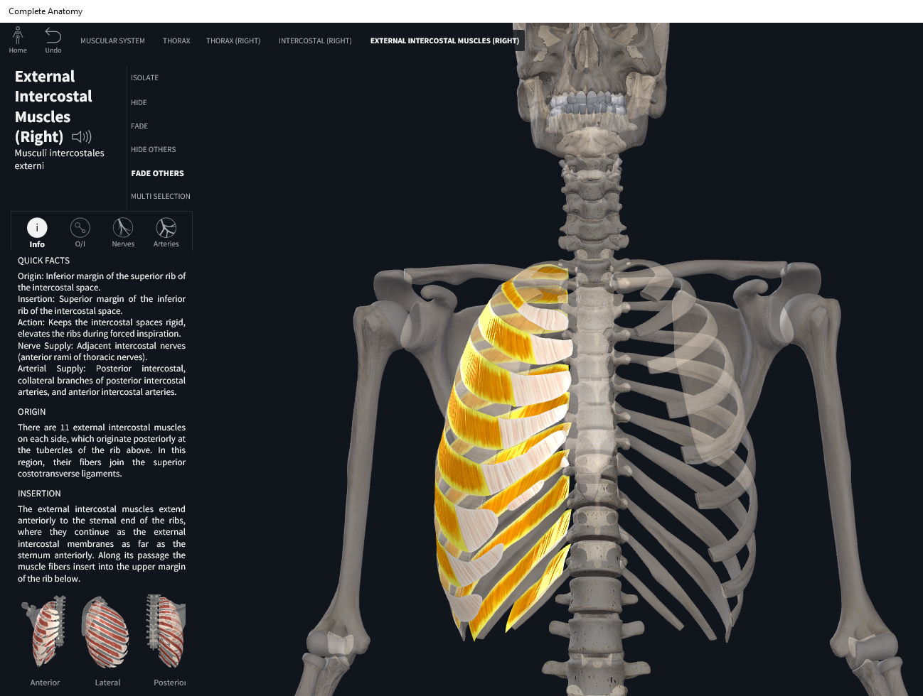
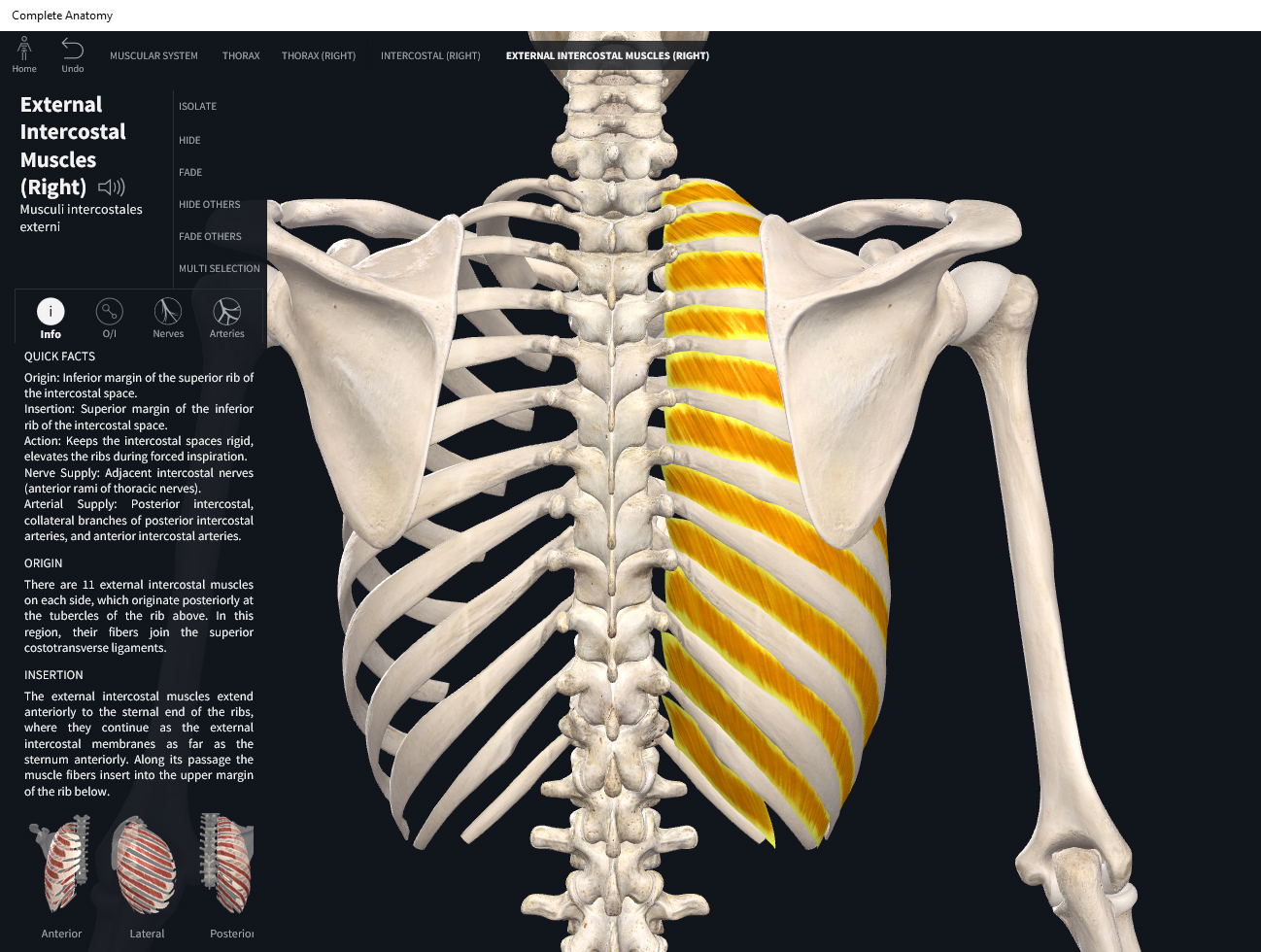
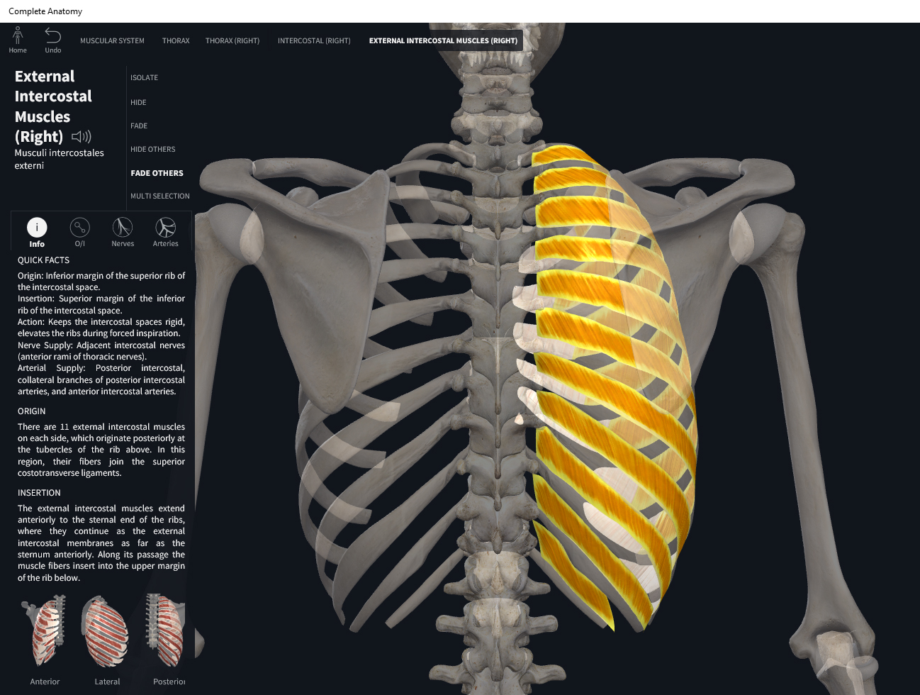
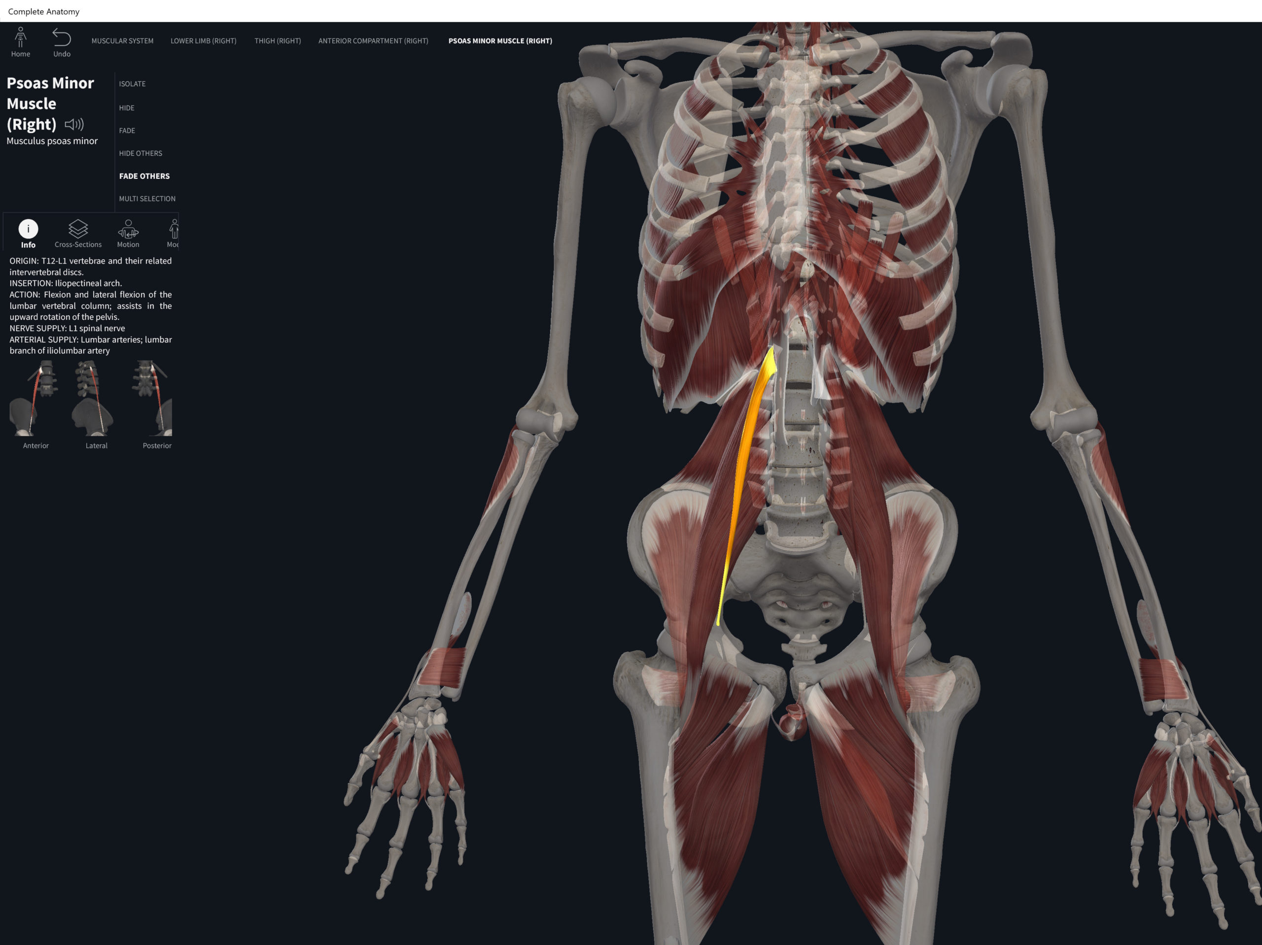
Anatomy & Physiology: Muscles—Diaphragm.

Structure.

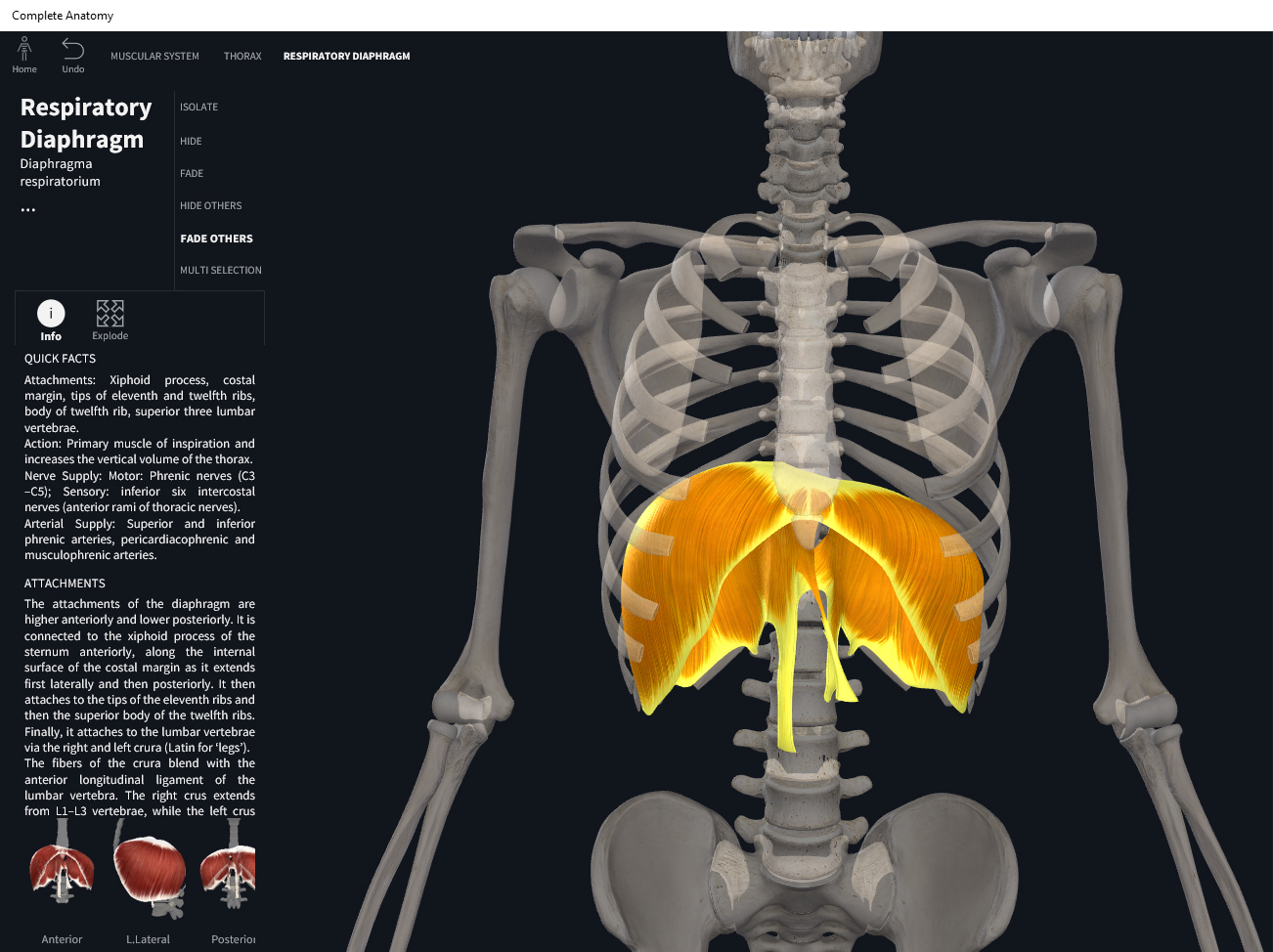
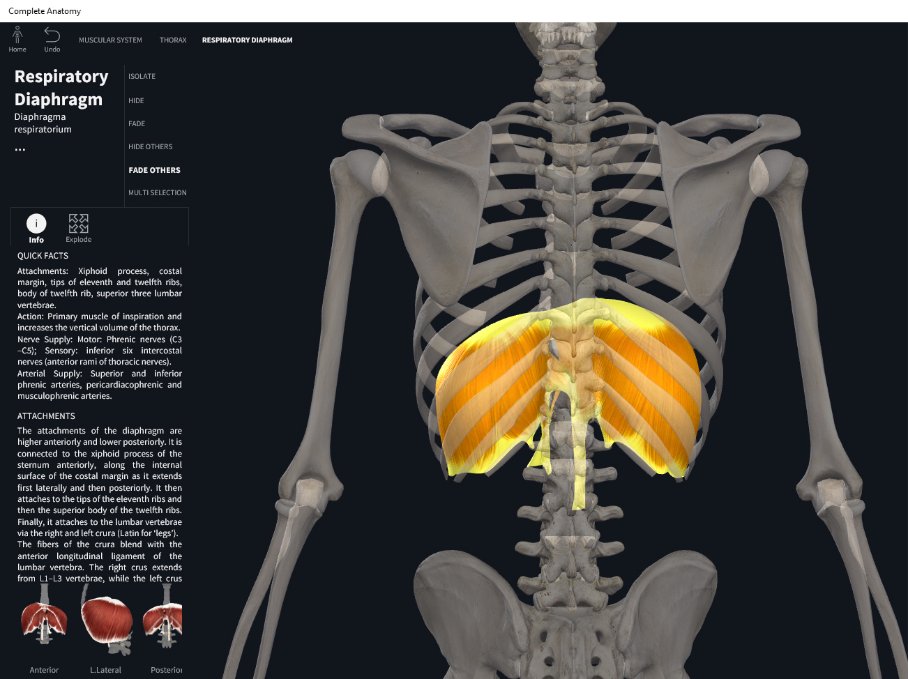
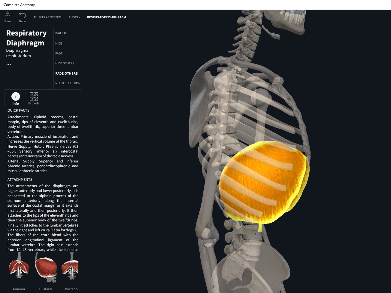
  • Origin: costal region– inner surfaces of cartilages and adjacent bony regions of ribs 6-12; sternal region–posterior side of xiphoid; crural (lumbar region)–2 aponeurotic arches covering external surfaces of the quadratus lumborum and psoas major; right & left crus–from bodies of L1-L3 and their intervertebral discs.
  • Insertion: central tendon.

Function.

  • Concentric action: pull central tendon inferiorly, increase thoracic cavity volume vertically (resulting in inhalation).
  • Reverse mover action: increase thoracic cavity volume.
  • Eccentric action: controls/restrains/slows thoracic cavity compression.
  • Isometric action: stabilization of trunk.
  • Innervation: phrenic nerve, C3-C5.
  • Arterial supply: branches of aorta and internal thoracic artery; superior and inferior phrenic arteries, musculophrenic and pericardiacophrenic arteries.

Clinical Significance.

More.

References

Biel, A. (2015). Trail guide to the body: A hands-on guide to locating muscles, bones and more.

Cedars-Sinai. (2018). Vertebrae of the spine. Retrieved from https://www.cedars-sinai.org/health-library/diseases-and-conditions/v/vertebrae-of-the-spine.html

Clark, M., Lucett, S., Sutton, B. G., & National Academy of Sports Medicine. (2014). NASM essentials of corrective exercise training. Burlington, MA: Jones & Bartlett Learning.

Jenkins, G., & Tortora, G. J. (2012). Anatomy and Physiology: From Science to Life, 3rd Edition International Stu. John Wiley & Sons.

Muscolino, J. E. (2017). The muscular system manual: The skeletal muscles of the human body.

Posted on

Muscles: Transversus Thoracis.

Download these notes.

Anatomy & Physiology: Muscles—Transversus Thoracis.

Structure.

  • Origin: lower 1/3 of inner side of sternum and lower three costosternal junctions.
  • Insertion: costal cartilages 2-6.

Function.

  • Concentric action: depression of ribs 2-6.
  • Reverse mover action: sternum elevation.
  • Eccentric action: controls/restrains/slows elevation of ribs 2-6, depression of sternum.
  • Isometric action: stabilization rib cage and sternum.
  • Innervation: intercostal nerves.
  • Arterial supply: anterior intercostal arteries.

Clinical Significance.

References

Biel, A. (2015). Trail guide to the body: A hands-on guide to locating muscles, bones and more.

Cedars-Sinai. (2018). Vertebrae of the spine. Retrieved from https://www.cedars-sinai.org/health-library/diseases-and-conditions/v/vertebrae-of-the-spine.html

Clark, M., Lucett, S., Sutton, B. G., & National Academy of Sports Medicine. (2014). NASM essentials of corrective exercise training. Burlington, MA: Jones & Bartlett Learning.

Jenkins, G., & Tortora, G. J. (2012). Anatomy and Physiology: From Science to Life, 3rd Edition International Stu. John Wiley & Sons.

Muscolino, J. E. (2017). The muscular system manual: The skeletal muscles of the human body.

Posted on

Muscles: Internal Intercostals.

Download these notes.

Anatomy & Physiology: Muscles—Internal Intercostals.

Structure.

  • Origin: superior border of rib below.
  • Insertion: inferior border of rib above.

Function.

  • Concentric action: draws adjacent ribs together to decrease anteroposterior and lateral dimensions of thoracic cavity (forced exhalation); ipsilateral trunk rotation. Lesser: trunk flexion, lateral flexion, and extension.
  • Reverse mover action: elevate ribs 2-12; trunk contralateral rotation, flexion, lateral flexion, and extension.
  • Eccentric action: control/restrain/slows rib 1-11 elevation and depression; trunk contralateral rotation and ipsilateral rotation; flexion, extension, and contralateral lateral flexion of trunk.
  • Isometric action: stabilization of ribs and spine.
  • Innervation: thoracic nerve T2-T12.
  • Arterial supply: anterior intercostal arteries, posterior intercostal arteries, costocervical trunk and superior thoracic artery.

Clinical Significance.

More.

References

Biel, A. (2015). Trail guide to the body: A hands-on guide to locating muscles, bones and more.

Cedars-Sinai. (2018). Vertebrae of the spine. Retrieved from https://www.cedars-sinai.org/health-library/diseases-and-conditions/v/vertebrae-of-the-spine.html

Clark, M., Lucett, S., Sutton, B. G., & National Academy of Sports Medicine. (2014). NASM essentials of corrective exercise training. Burlington, MA: Jones & Bartlett Learning.

Jenkins, G., & Tortora, G. J. (2012). Anatomy and Physiology: From Science to Life, 3rd Edition International Stu. John Wiley & Sons.

Muscolino, J. E. (2017). The muscular system manual: The skeletal muscles of the human body.

Posted on

Muscles: External Intercostals.

Download these notes.

Anatomy & Physiology: Muscles—External Intercostals.

Structure.

  • Origin: inferior border of rib above.
  • Insertion: superior border of rib below.

Function.

  • Concentric action: elevate ribs and increase anteroposterior and lateral dimensions of the thoracic cavity (inhalation); contralateral rotation of trunk. Lesser: trunk flexion, extension, lateral flexion.
  • Reverse mover action: depress ribs 1-11; trunk rotation, lateral flexion, flexion, ipsilateral rotation.
  • Eccentric action: controls/restrains/slows depression of ribs 2-12; trunk ipsilateral rotation, flexion, extension, and contralateral lateral flexion.
  • Isometric action: stabilization of ribs and trunk.
  • Innervation: thoracic nerve T2-T12.
  • Arterial supply: anterior intercostal arteries, posterior intercostal arteries, costocervical trunk and superior thoracic arteries.

Clinical Significance.

More.

References

Biel, A. (2015). Trail guide to the body: A hands-on guide to locating muscles, bones and more.

Cedars-Sinai. (2018). Vertebrae of the spine. Retrieved from https://www.cedars-sinai.org/health-library/diseases-and-conditions/v/vertebrae-of-the-spine.html

Clark, M., Lucett, S., Sutton, B. G., & National Academy of Sports Medicine. (2014). NASM essentials of corrective exercise training. Burlington, MA: Jones & Bartlett Learning.

Jenkins, G., & Tortora, G. J. (2012). Anatomy and Physiology: From Science to Life, 3rd Edition International Stu. John Wiley & Sons.

Muscolino, J. E. (2017). The muscular system manual: The skeletal muscles of the human body.

Posted on

Muscles: Psoas Minor.

Download these notes.

Anatomy & Physiology: Muscles—Psoas Minor.

Structure.

  • Origin: bodies of T12 and L1 and intervening intervertebral disc.
  • Insertion: fascia covering psoas major and iliacus.

Function.

  • Concentric action: accelerates hip flexion and external/lateral rotation; extends and rotates lumbar spine.
  • Reverse mover action: pelvis posterior tilt, lower trunk flexion, ipsilateral pelvic elevation.
  • Eccentric action: controls/restrains/decelerates hip extension and internal rotation.
  • Isometric action: stabilization of lumbo-pelvic hip complex.
  • Innervation: spinal nerve branches of L2-L3.
  • Arterial supply: lumbar arteries, arteria lumbalis ima of median sacral artery, lumbar part of iliolumbar artery, common iliac artery.

Clinical Significance.

References

Biel, A. (2015). Trail guide to the body: A hands-on guide to locating muscles, bones and more.

Cedars-Sinai. (2018). Vertebrae of the spine. Retrieved from https://www.cedars-sinai.org/health-library/diseases-and-conditions/v/vertebrae-of-the-spine.html

Clark, M., Lucett, S., Sutton, B. G., & National Academy of Sports Medicine. (2014). NASM essentials of corrective exercise training. Burlington, MA: Jones & Bartlett Learning.

Jenkins, G., & Tortora, G. J. (2012). Anatomy and Physiology: From Science to Life, 3rd Edition International Stu. John Wiley & Sons.

Muscolino, J. E. (2017). The muscular system manual: The skeletal muscles of the human body.

Posted on

Muscles: Transverse Abdominis.

Download these notes.

Anatomy & Physiology: Muscles—Transverse Abdominis.

Structure.

  • Origin: iliac crest, inguinal ligament, lumbar fascia, and cartilages of ribs 5-10.
  • Insertion: xiphoid process, linea alba, and pubis.

Function.

  • Concentric action: increase intra-abdominal pressure; support abdominal viscera.
  • Reverse mover action: increase intra-abdominal pressure; support abdominal viscera.
  • Eccentric action: controls/restrains/slows abdominalpelvic cavity expansion.
  • Isometric action: stabilization of lumbo-pelvic hip complex (synergistically with internal oblique, multifidus, and deep erector spinae).
  • Innervation: thoracic spinal nerves T8-T12 iliohypogastric nerve, ilioinguinal nerve.
  • Arterial supply: subcostal and posterior intercostal arteries, deep circumflex iliac artery, inferior epigastric artery.

Clinical Significance.

More.

References

Biel, A. (2015). Trail guide to the body: A hands-on guide to locating muscles, bones and more.

Cedars-Sinai. (2018). Vertebrae of the spine. Retrieved from https://www.cedars-sinai.org/health-library/diseases-and-conditions/v/vertebrae-of-the-spine.html

Clark, M., Lucett, S., Sutton, B. G., & National Academy of Sports Medicine. (2014). NASM essentials of corrective exercise training. Burlington, MA: Jones & Bartlett Learning.

Jenkins, G., & Tortora, G. J. (2012). Anatomy and Physiology: From Science to Life, 3rd Edition International Stu. John Wiley & Sons.

Muscolino, J. E. (2017). The muscular system manual: The skeletal muscles of the human body.

Posted on

Muscles: Internal Oblique.

Download these notes.

Anatomy & Physiology: Muscles—Internal Oblique.

Structure.

  • Origin: anterior 2/3 of iliac crest, inguinal ligament, and thoracolumbar fascia.
  • Insertion: cartilage of ribs 9-12; linea alba, and contralateral rectus sheaths.

Function.

  • Concentric action: spinal bilateral flexion, lateral flexion, and ipsilateral rotation; compress abdomen.
  • Reverse mover action: pelvic posterior tilt, lower trunk flexion; ipsilateral pelvic elevation; lateral trunk flexion; contralateral rotation of pelvis, contralateral rotation of lower trunk; compress abdominopelvic cavity.
  • Eccentric action: control/restrains/slows spinal extension, lateral flexion, and rotation.
  • Isometric action: stabilization of lumbo-pelvic hip complex.
  • Innervation: thoracic spinal nerves T8-T12; iliohypogastric nerve; ilioinguinal nerve.
  • Arterial supply: subcostal and posterior intercostal arteries, deep circumflex iliac artery, inferior epigastric artery.

Clinical Significance.

More.

References

Biel, A. (2015). Trail guide to the body: A hands-on guide to locating muscles, bones and more.

Cedars-Sinai. (2018). Vertebrae of the spine. Retrieved from https://www.cedars-sinai.org/health-library/diseases-and-conditions/v/vertebrae-of-the-spine.html

Clark, M., Lucett, S., Sutton, B. G., & National Academy of Sports Medicine. (2014). NASM essentials of corrective exercise training. Burlington, MA: Jones & Bartlett Learning.

Jenkins, G., & Tortora, G. J. (2012). Anatomy and Physiology: From Science to Life, 3rd Edition International Stu. John Wiley & Sons.

Muscolino, J. E. (2017). The muscular system manual: The skeletal muscles of the human body.

Posted on

Muscles: External Oblique.

Download these notes.

Anatomy & Physiology: Muscles—External Oblique.

Structure.

  • Origin: external surface of ribs 5-12.
  • Insertion: anterior iliac crest of pelvis, linea alba, and contralateral rectus.

Function.

  • Concentric action: spinal flexion (especially lumbar region), lateral flexion, and contralateral rotation; compress abdomen.
  • Reverse mover action: pelvic posterior tilt, lower trunk flexion, ipsilateral elevation of pelvis, lower trunk lateral flexion, ipsilateral rotation of pelvis.
  • Eccentric action: controls/restrains/slows spinal extension, lateral flexion, and rotation.
  • Isometric action: stabilization of lumbo-pelvic hip complex.
  • Innervation: thoracic spinal nerves T7-T12; iliohypogastric L1.
  • Arterial supply: subcostal and posterior intercostal arteries, deep circumflex iliac artery, inferior epigastric artery.

Clinical Significance.

More.

References

Biel, A. (2015). Trail guide to the body: A hands-on guide to locating muscles, bones and more.

Cedars-Sinai. (2018). Vertebrae of the spine. Retrieved from https://www.cedars-sinai.org/health-library/diseases-and-conditions/v/vertebrae-of-the-spine.html

Clark, M., Lucett, S., Sutton, B. G., & National Academy of Sports Medicine. (2014). NASM essentials of corrective exercise training. Burlington, MA: Jones & Bartlett Learning.

Jenkins, G., & Tortora, G. J. (2012). Anatomy and Physiology: From Science to Life, 3rd Edition International Stu. John Wiley & Sons.

Muscolino, J. E. (2017). The muscular system manual: The skeletal muscles of the human body.

Posted on

Muscles: Rectus Abdominis.

Download these notes.

Anatomy & Physiology: Muscles—Rectus Abdominis.

Structure.

  • Origin: pubic crest and pubic symphysis.
  • Insertion: cartilage of ribs 5-7; xiphoid process.

Function.

  • Concentric action: spinal flexion (especially lumbar portion), lateral flexion, and rotation; compress abdomen (defecation, urination, forced exhalation, childbirth).
  • Reverse mover action: pelvic posterior tilt; lower trunk flexion; ipsilateral pelvic elevation; abdominopelvic conpression.
  • Eccentric action: controls/restrains/slows spinal extension, lateral flexion, and rotation.
  • Isometric action: stabilization of lumbo-pelvic hip complex.
  • Innervation: thoracic spinal nerves T7-T12. Intercostal nerves.
  • Arterial supply: superior epigastric artery inferior epigastric artery, terminal branches of subcostal and posterior intercostal arteries.

Clinical Significance.

More.

References

Biel, A. (2015). Trail guide to the body: A hands-on guide to locating muscles, bones and more.

Cedars-Sinai. (2018). Vertebrae of the spine. Retrieved from https://www.cedars-sinai.org/health-library/diseases-and-conditions/v/vertebrae-of-the-spine.html

Clark, M., Lucett, S., Sutton, B. G., & National Academy of Sports Medicine. (2014). NASM essentials of corrective exercise training. Burlington, MA: Jones & Bartlett Learning.

Jenkins, G., & Tortora, G. J. (2012). Anatomy and Physiology: From Science to Life, 3rd Edition International Stu. John Wiley & Sons.

Muscolino, J. E. (2017). The muscular system manual: The skeletal muscles of the human body.

Posted on

Muscles: Quadratus Lumborum.

Download these notes.

Anatomy & Physiology: Muscles—Quadratus Lumborum.

Structure.

  • Origin: iliac crest, iliolumbar ligament.
  • Insertion: 12th rib, transverse processes L2-L5.

Function.

  • Concentric action: spinal lateral flexion; pulls rib 12 inferiorly during forced exhalation; fix ribs to prevent elevation during deep inhalation; help lumbar extension. Anterior pelvic tilt and lower spine extension relative to upper spine.
  • Reverse mover action: trunk lateral flexion; rib 12 depression; trunk extension.
  • Eccentric action: controls/restrains/decelerates contralateral and lateral spinal flexion; rib 12 elevation; ipsilateral depression and posterior tilt of pelvis.
  • Isometric action: stabilization of the lumbo-pelvic-hip complex.
  • Innervation: spinal nerves T12-L3.
  • Arterial supply: branches of subcostal and lumbar arteries; iliolumbar artery.

Clinical Significance.

More.

References

Biel, A. (2015). Trail guide to the body: A hands-on guide to locating muscles, bones and more.

Cedars-Sinai. (2018). Vertebrae of the spine. Retrieved from https://www.cedars-sinai.org/health-library/diseases-and-conditions/v/vertebrae-of-the-spine.html

Clark, M., Lucett, S., Sutton, B. G., & National Academy of Sports Medicine. (2014). NASM essentials of corrective exercise training. Burlington, MA: Jones & Bartlett Learning.

Jenkins, G., & Tortora, G. J. (2012). Anatomy and Physiology: From Science to Life, 3rd Edition International Stu. John Wiley & Sons.

Muscolino, J. E. (2017). The muscular system manual: The skeletal muscles of the human body.