Posted on

Muscles: Pectoralis Major.

Download these notes.

Anatomy & Physiology: Muscles—Pectoralis Major.

Structure.

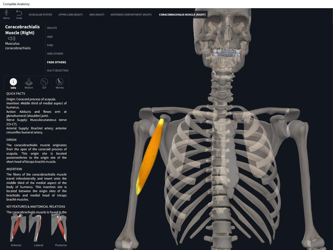
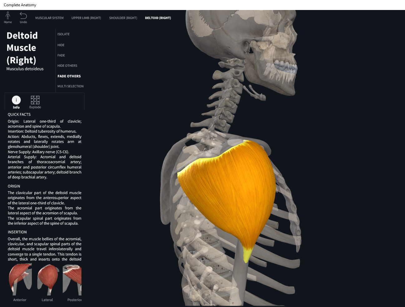
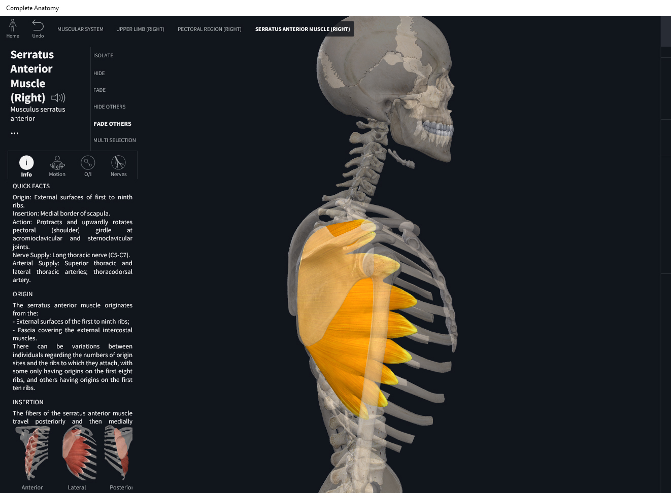
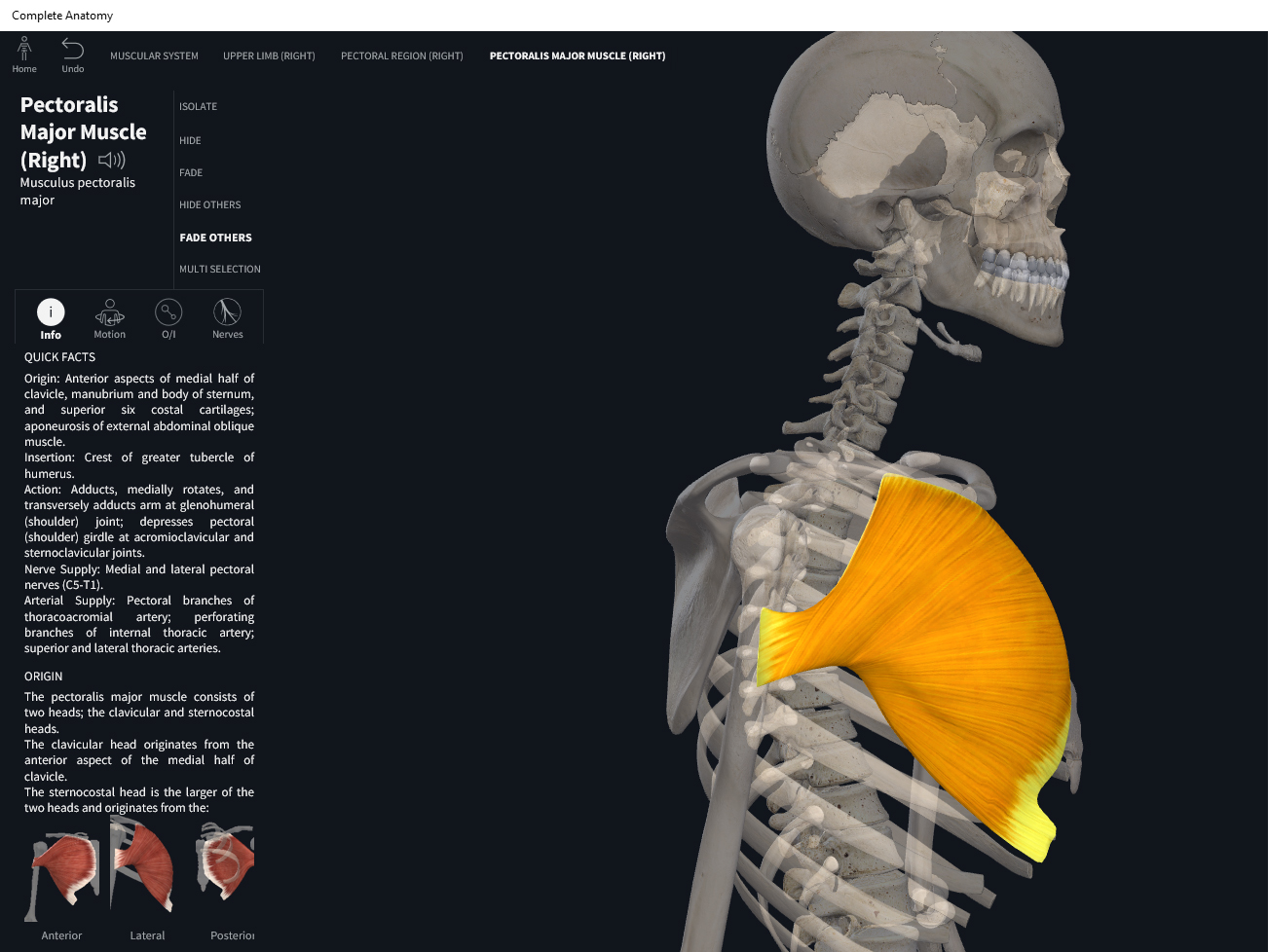
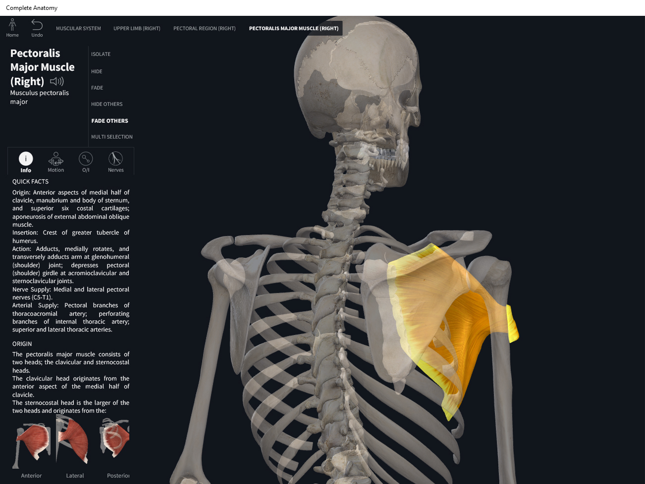
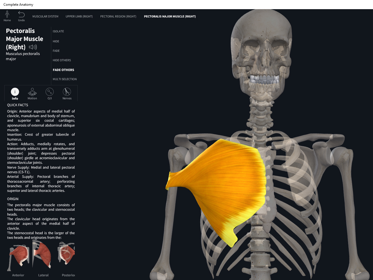
  • Origin: anterior surface of clavicular head; anterior surface of sternum, costal cartilage of ribs 2-6 or sometimes 1-7.
  • Insertion: greater tubercle and lateral lip of the intertubercular sulcus of of humerus.

Function.

  • Concentric action: shoulder flexion (clavicular fibers), horizontal adduction, and internal/medial rotation, protraction.
  • Reverse mover action: lateral deviation of trunk; trunk flexion at spinal joints; anterior translation of the trunk; scapular downward rotation; ipsilateral rotation of trunk; trunk elevation.
  • Eccentric action: shoulder extension, horizontal abduction, and external rotation, retraction. Controls/restrains/slows arm abduction, lateral rotation, extension, horizontal extension, flexion, and adduction. Controls/restrains/slows scapular retraction, protraction, elevation, and upward rotation. Controls/restrains/slows contralateral rotation, contralateral lateral deviation, posterior translation and depression of trunk.
  • Isometric action: stabilization of the shoulder girdle, clavicle, and scapula.
  • Innervation: medial and lateral pectoral nerves.
  • Arterial supply: pectoral branches of thoracoacromial trunk; posterior intercostal arteries (branches of aorta) and lateral thoracic artery (branch of axillary artery).

Clinical Significance.

More.

References

Biel, A. (2015). Trail guide to the body: A hands-on guide to locating muscles, bones and more.

Cedars-Sinai. (2018). Vertebrae of the spine. Retrieved from https://www.cedars-sinai.org/health-library/diseases-and-conditions/v/vertebrae-of-the-spine.html

Clark, M., Lucett, S., Sutton, B. G., & National Academy of Sports Medicine. (2014). NASM essentials of corrective exercise training. Burlington, MA: Jones & Bartlett Learning.

Jenkins, G., & Tortora, G. J. (2012). Anatomy and Physiology: From Science to Life, 3rd Edition International Stu. John Wiley & Sons.

Muscolino, J. E. (2017). The muscular system manual: The skeletal muscles of the human body.

Posted on

Muscles: Coracobrachialis.

Download these notes.

Anatomy & Physiology: Muscles—Coracobrachialis.

Structure.

  • Origin: coracoid process of scapula.
  • Insertion: middle of medial surface of shaft of humerus.

Function.

  • Concentric action: shoulder flexion and adduction. Horizontal arm flexion.
  • Reverse mover action: upward tilt of the scapula at the glenohumeral (GH) and scapulocostal (ScC) joints. Downward scapular rotation. Scapular protraction.
  • Eccentric action: controls/restrains/slows arm extension, abduction, and horizontal extension. Controls/restrains/slows scapular upward rotation, retraction, and downward tilt.
  • Isometric action: stabilization of the shoulder girdle and scapula.
  • Innervation: musculocutaneous nerve, C5, C6, C7.
  • Arterial supply: muscular branches of brachial artery; anterior circumflex humeral artery.

Clinical Significance.

More.

References

Biel, A. (2015). Trail guide to the body: A hands-on guide to locating muscles, bones and more.

Cedars-Sinai. (2018). Vertebrae of the spine. Retrieved from https://www.cedars-sinai.org/health-library/diseases-and-conditions/v/vertebrae-of-the-spine.html

Clark, M., Lucett, S., Sutton, B. G., & National Academy of Sports Medicine. (2014). NASM essentials of corrective exercise training. Burlington, MA: Jones & Bartlett Learning.

Jenkins, G., & Tortora, G. J. (2012). Anatomy and Physiology: From Science to Life, 3rd Edition International Stu. John Wiley & Sons.

Muscolino, J. E. (2017). The muscular system manual: The skeletal muscles of the human body.

Posted on

Muscles: Deltoid.

Download these notes.

Anatomy & Physiology: Muscles—Deltoid.

Structure.

  • Origin: lateral 1/3 (acromial extremity) of clavicle.
  • Insertion: deltoid tuberosity of humerus.

Function.

  • Concentric action: abduction at the glenohumeral (GH) joint. Arm flexion at GH joint (anterior deltoid). Medial rotation of the arm at GH (anterior deltoid). Horizontal flexion at the GH joint (anterior deltoid). Arm extension (posterior deltoid). Lateral rotation (posterior deltoid). Horizonal extension at GH (posterior deltoid).
  • Reverse mover actions: downward rotation of scapula at glenohumeral and scapulocostal joints; ipsilateral rotation of the trunk; contralateral rotation of the trunk.
  • Eccentric action: controls/restrains/slows arm adduction, extension, horizontal extension, flexion, horizontal flexion, lateral rotation, and medial rotation. Upward tilt of scapula such that inferior angle lifts off the thoracic wall.
  • Isometric action: stabilization of the shoulder girdle. Stabilize clavicle.
  • Innervation: axillary nerve.
  • Arterial supply: anterior and posterior circumflex humeral arteries (branches of axillary artery). Pectoral and deltoid branches of thoracoacromial trunk (branch of axillary artery).

Clinical Significance.

More.

References

Biel, A. (2015). Trail guide to the body: A hands-on guide to locating muscles, bones and more.

Cedars-Sinai. (2018). Vertebrae of the spine. Retrieved from https://www.cedars-sinai.org/health-library/diseases-and-conditions/v/vertebrae-of-the-spine.html

Clark, M., Lucett, S., Sutton, B. G., & National Academy of Sports Medicine. (2014). NASM essentials of corrective exercise training. Burlington, MA: Jones & Bartlett Learning.

Jenkins, G., & Tortora, G. J. (2012). Anatomy and Physiology: From Science to Life, 3rd Edition International Stu. John Wiley & Sons.

Muscolino, J. E. (2017). The muscular system manual: The skeletal muscles of the human body.

Posted on

Muscles: Trapezius Upper.

Download these notes.

Anatomy & Physiology: Muscles—Trapezius, Upper.

Structure.

  • Origin: superior nuchal line, ligamentum nuchae; spinous process of C7, T1-T12.
  • Insertion: lateral 1/3 of clavicle; acromion process of scapula.

Function.

  • Concentric action: cervical extension, lateral flexion and rotation; scapular elevation at scapulocostal joint (ScC); scapular retraction at ScC; upward rotation of the scapula at the ScC.
  • Reverse mover actions: head and neck extension at spinal joints; contralateral rotation of the head and neck at spinal joints; lateral flexion of the head and neck at spinal joints.
  • Eccentric action: cervical flexion, lateral flexion, and rotation; scapular depression. Controls/restrains/slows scapular protraction at ScC and ipsilateral rotation of the trunk at the spinal joints.
  • Isometric action: stabilization of the medial border of scapula and cervical spine.
  • Innervation: accessory XI nerve; cervical spinal nerves C3-C5.
  • Arterial supply: transverse cervical artery (branch from thyrocervical trunk), dorsal scapular artery (branch from subclavian artery).

Clinical Significance.


References

Biel, A. (2015). Trail guide to the body: A hands-on guide to locating muscles, bones and more.

Cedars-Sinai. (2018). Vertebrae of the spine. Retrieved from https://www.cedars-sinai.org/health-library/diseases-and-conditions/v/vertebrae-of-the-spine.html

Clark, M., Lucett, S., Sutton, B. G., & National Academy of Sports Medicine. (2014). NASM essentials of corrective exercise training. Burlington, MA: Jones & Bartlett Learning.

Jenkins, G., & Tortora, G. J. (2012). Anatomy and Physiology: From Science to Life, 3rd Edition International Stu. John Wiley & Sons.

Muscolino, J. E. (2017). The muscular system manual: The skeletal muscles of the human body.

Posted on

Muscles: Trapezius Middle.

Download these notes.

Anatomy & Physiology: Muscles—Trapezius, Middle.

Structure.

  • Origin: superior nuchal line, ligamentum nuchae; spinous process of C7, T1-T12.
  • Insertion: acromion process of scapula; superior aspect of scapular spine.

Function.

  • Concentric action: scapular retraction.
  • Reverse mover action: contralateral rotation of the trunk at spinal joints.
  • Eccentric action: scapular protraction and elevation. Controls/restrains/slows scapular protraction at ScC and ipsilateral rotation of trunk at spinal joints.
  • Isometric action: stabilization of the scapula.
  • Innervation: accessory XI nerve; cervical spinal nerves C3-C5.
  • Arterial supply: transverse cervical artery (branch from thyrocervical trunk), dorsal scapular artery (branch from subclavian artery).

Clinical Significance.


References

Biel, A. (2015). Trail guide to the body: A hands-on guide to locating muscles, bones and more.

Cedars-Sinai. (2018). Vertebrae of the spine. Retrieved from https://www.cedars-sinai.org/health-library/diseases-and-conditions/v/vertebrae-of-the-spine.html

Clark, M., Lucett, S., Sutton, B. G., & National Academy of Sports Medicine. (2014). NASM essentials of corrective exercise training. Burlington, MA: Jones & Bartlett Learning.

Jenkins, G., & Tortora, G. J. (2012). Anatomy and Physiology: From Science to Life, 3rd Edition International Stu. John Wiley & Sons.

Muscolino, J. E. (2017). The muscular system manual: The skeletal muscles of the human body.

Posted on

Muscles: Trapezius Lower.

Download these notes.

Anatomy & Physiology: Muscles—Trapezius, Lower.

Structure.

  • Origin: superior nuchal line, ligamentum nuchae; spinous process of C7, T1-T12.
  • Insertion: spine of scapula.

Function.

  • Concentric action: scapular depression at scapulocostal joint (ScC). Retracts scapula at ScC. Upwardly rotates the scapula at the ScC.
  • Reverse mover actions: extends trunk at spinal joints; contralateral rotation of the trunk at spinal joints; lateral flexion of the trunk at the spinal joints.
  • Eccentric action: controls/restrains/slows scapular elevation, protraction, and downward rotation at the ScC. Controls/restrains/slows flexion, ipsilateral rotation, and contralateral flexion of trunk at spinal joints.
  • Isometric action: stabilization of the scapula and clavicle. Stabilizes head, neck, trunk at the spinal joints.
  • Innervation: accessory XI nerve; cervical spinal nerves C3-C5.
  • Arterial supply: transverse cervical artery (branch from thyrocervical trunk), dorsal scapular artery (branch from subclavian artery).

Clinical Significance.


References

Biel, A. (2015). Trail guide to the body: A hands-on guide to locating muscles, bones and more.

Cedars-Sinai. (2018). Vertebrae of the spine. Retrieved from https://www.cedars-sinai.org/health-library/diseases-and-conditions/v/vertebrae-of-the-spine.html

Clark, M., Lucett, S., Sutton, B. G., & National Academy of Sports Medicine. (2014). NASM essentials of corrective exercise training. Burlington, MA: Jones & Bartlett Learning.

Jenkins, G., & Tortora, G. J. (2012). Anatomy and Physiology: From Science to Life, 3rd Edition International Stu. John Wiley & Sons.

Muscolino, J. E. (2017). The muscular system manual: The skeletal muscles of the human body.

Posted on

Muscles: Rhomboid Major.

Download these notes.

Anatomy & Physiology: Muscles—Rhomboid Major.

Structure.

  • Origin: spinous process of T2-T5.
  • Insertion: vertebral border of scapula inferior to spine.

Function.

  • Concentric action: scapular retraction at scapulocostal joint (ScC), elevates scapula at ScC, and downward rotation.
  • Reverse mover action: contralateral rotation of the trunk at spinal joints; extends trunk at spinal joints.
  • Eccentric action: controls/restrains/slows scapular protraction depression, upward rotation, and lateral tilt; controls/restrains/slows flexion and ipsilateral rotation of the trunk.
  • Isometric action: stabilization of the scapula; stabilization of C7-T5 vertebrae.
  • Innervation: dorsal scapular nerve, C4, C5.
  • Arterial supply: dorsal scapular artery (branch of subclavian artery).

Clinical Significance.


References

Biel, A. (2015). Trail guide to the body: A hands-on guide to locating muscles, bones and more.

Cedars-Sinai. (2018). Vertebrae of the spine. Retrieved from https://www.cedars-sinai.org/health-library/diseases-and-conditions/v/vertebrae-of-the-spine.html

Clark, M., Lucett, S., Sutton, B. G., & National Academy of Sports Medicine. (2014). NASM essentials of corrective exercise training. Burlington, MA: Jones & Bartlett Learning.

Jenkins, G., & Tortora, G. J. (2012). Anatomy and Physiology: From Science to Life, 3rd Edition International Stu. John Wiley & Sons.

Muscolino, J. E. (2017). The muscular system manual: The skeletal muscles of the human body.

Posted on

Muscles: Rhomboid Minor.

Download these notes.

Anatomy & Physiology: Muscles—Rhomboid Minor.

Structure.

  • Origin: spinous process of C7-T1.
  • Insertion: vertebral border of scapula superior to spine.

Function.

  • Concentric action: scapular retraction and downward rotation. Medially tilts scapula at the scapulocostal joint (ScC).
  • Reverse mover action: contralateral rotation of trunk at spinal joints.
  • Eccentric action: controls/restrains/slows scapular protraction depression, upward rotation, and lateral tilt; controls/restrains/slows flexion and ipsilateral rotation of the trunk.
  • Isometric action: stabilization of the scapula; stabilization of C7-T5 vertebrae.
  • Innervation: dorsal scapular nerve.
  • Arterial supply: dorsal scapular artery (branch of subclavian artery).

Clinical Significance.


References

Biel, A. (2015). Trail guide to the body: A hands-on guide to locating muscles, bones and more.

Cedars-Sinai. (2018). Vertebrae of the spine. Retrieved from https://www.cedars-sinai.org/health-library/diseases-and-conditions/v/vertebrae-of-the-spine.html

Clark, M., Lucett, S., Sutton, B. G., & National Academy of Sports Medicine. (2014). NASM essentials of corrective exercise training. Burlington, MA: Jones & Bartlett Learning.

Jenkins, G., & Tortora, G. J. (2012). Anatomy and Physiology: From Science to Life, 3rd Edition International Stu. John Wiley & Sons.

Muscolino, J. E. (2017). The muscular system manual: The skeletal muscles of the human body.

Posted on

Muscles: Levator Scapulae.

Download these notes.

Anatomy & Physiology: Muscles—Levator Scapulae.

Structure.

  • Origin: transverse processes of C1-C4.
  • Insertion: superior vertebral border of scapulae.

Function.

  • Concentric action: cervical extension, lateral flexion and ipsilateral rotation when scapulae are anchored; elevation and downward rotation of scapulae. Scapular retraction at scapulocostal joint (ScC).
  • Reverse mover action: neck extension at spinal joints; lateral flexion of the neck at spinal joints; ipsilateral rotation of the neck at spinal joints.
  • Eccentric action: cervical flexion, contralateral rotation, lateral flexion; scapular depression and upward rotation when neck is stabilized.
  • Isometric action: stabilization of the scapulae and cervical spine.
  • Innervation: dorsal scapular nerve; cervical spinal nerves C3-C5.
  • Arterial supply: dorsal scapular artery (branch of the subclavian artery).

Clinical Significance.


References

Biel, A. (2015). Trail guide to the body: A hands-on guide to locating muscles, bones and more.

Cedars-Sinai. (2018). Vertebrae of the spine. Retrieved from https://www.cedars-sinai.org/health-library/diseases-and-conditions/v/vertebrae-of-the-spine.html

Clark, M., Lucett, S., Sutton, B. G., & National Academy of Sports Medicine. (2014). NASM essentials of corrective exercise training. Burlington, MA: Jones & Bartlett Learning.

Jenkins, G., & Tortora, G. J. (2012). Anatomy and Physiology: From Science to Life, 3rd Edition International Stu. John Wiley & Sons.

Muscolino, J. E. (2017). The muscular system manual: The skeletal muscles of the human body.

Posted on

Muscles: Serratus Anterior.

Download these notes.

Anatomy & Physiology: Muscles—Serratus Anterior.

Structure.

  • Origin: ribs 1-8 or ribs 1-9.
  • Insertion: vertebral border and inferior angle of scapula.

Function.

  • Concentric action: scapular protraction/abduct scapula, elevation, depression, medial tilt, downward tilt, and upward rotation; elevates ribs when scapula is stabilized; “boxer’s muscle” (horizontal arm movements, punching, pushing).
  • Reverse mover action: retracts trunk at scapulocostal joint (ScC); depresses trunk at ScC; elevates trunk at ScC.
  • Eccentric action: controls/restrains/slows scapular retraction, downward rotation, depresion, elevation, lateral tilt, and upward tilt. Controls/restrains/slows protraction, elevation, and depression of the trunk.
  • Isometric action: stabilization of the scapula and stabilization of the rib cage.
  • Innervation: long thoracic nerve C5-C7.
  • Arterial supply: dorsal scapular artery (branch of subclavian artery), lateral thoracic artery (branch of axillary artery), superior thoracic artery (branch of axillary artery).

Clinical Significance.

References

Biel, A. (2015). Trail guide to the body: A hands-on guide to locating muscles, bones and more.

Cedars-Sinai. (2018). Vertebrae of the spine. Retrieved from https://www.cedars-sinai.org/health-library/diseases-and-conditions/v/vertebrae-of-the-spine.html

Clark, M., Lucett, S., Sutton, B. G., & National Academy of Sports Medicine. (2014). NASM essentials of corrective exercise training. Burlington, MA: Jones & Bartlett Learning.

Jenkins, G., & Tortora, G. J. (2012). Anatomy and Physiology: From Science to Life, 3rd Edition International Stu. John Wiley & Sons.

Muscolino, J. E. (2017). The muscular system manual: The skeletal muscles of the human body.