Gram Staining (Cappuccino & Welsh, 2017).
Description. Gram staining is a differential staining technique that differentiates bacteria based on their cell wall construction. Gram-positive bacteria have a thick peptidoglycan cell wall (stains purple) and its teichoic acid cross-linkages resist the decolorizing agent (Microbe Online, 2015). Gram-negative bacteria have a thin peptidoglycan cell wall which lies between two membranous layers (outer membrane and cell membrane, or in acid-fast bacteria the lipid layer and cell membrane) (Black & Black, 2015). This test helps to classify bacteria into two major categories: gram-negative and gram-positive.
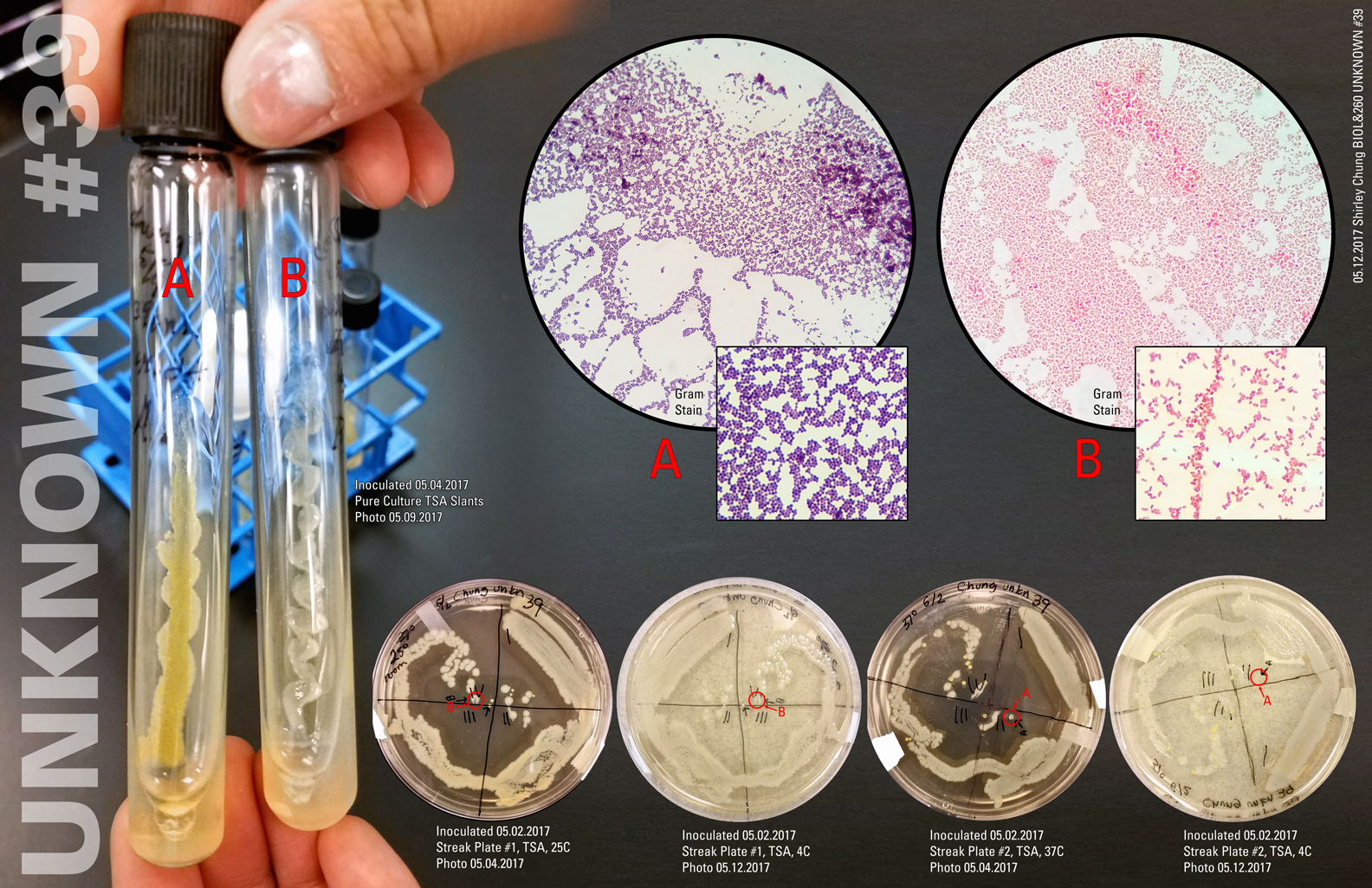
Clinical significance. The Gram stain is the predominant differential stain—especially used in the clinical setting. It quickly assesses organisms that fall into one of two categories—Gram positive (purple) and Gram negative (pink/reddish).
Summary of main steps. It is assumed that the reader knows aseptic technique and how to heat fix.
- Inoculate clean slide. Use aseptic technique throughout. If using a broth culture, place 2-3 loopfuls of culture onto the glass slide. If using a solid medium, first place 2 loopfuls of deionized (DI) water onto the slide. Then place 1 loopful of culture into the DI water puddle on the slide (repeat if necessary). Spread the loopful of culture around on the slide in a circular pattern.
- Allow the slide to air dry. Then heat fix.
- Flood slide with crystal violet (primary stain, Hucker’s, stains all cells purple). Wait 1 minute.
- Flush gently with DI water.
- Flood slide with Gram’s iodine mordant (intensifies primary stain by binding to the primary stain and forming an insoluble complex, crystal-violet-iodine or CV-I). Gram’s iodine is also a killing agent. Wait 1 minute.
- Flush gently with DI water.
- Quickly and carefully (be stingy) cover the slide with 95% ethyl alcohol decolorizer (selectively removes color from cell components/structures). You should see some pale blue run-off. The decolorizer is a lipid solvent and also dehydrates protein.In Gram-negative cells, the decolorizer makes the cell wall more porous by dissolving the lipid components—aiding in the removal of the CV-I complex.In Gram-positive cells, the decolorizer makes the cell wall pores smaller—making the removal of CV-I more difficult (i.e. makes the CV-I “stick” better).
- Flush immediately with DI water.
- Flood slide with safranin (counterstain for contrast those structures that have been decolorized). Wait 45 seconds.
- Flood gently with DI water.
- Blot gently with bibulous paper.
Reference
Black, J. G., & Black, L. J. (2015). Microbiology: Principles and explorations (9th ed.). Hoboken, NJ: John Wiley & Sons.
Cappuccino, J. G., & Welsh, C. (2018). Microbiology: A laboratory manual.
Microbe Online. (2015, February 2). Gram staining: Principle, procedure and results. Retrieved from http://microbeonline.com/gram-staining-principle-procedure-results/