Posted on

BC4139 Intro to Biochem Week 1.

Last edited: 08.08.2019

See Appendix A Amino acid abbreviations; Appendix B Glucogenic, Ketogenic, and Glucoketogenic Amino acids; Appendix C 20 Amino Acids to Know Drawn at Physiological pH 7.4.

Review of Macromolecules and Functional Groups.

  • What is a monomer? A monomer is a base unit of something. A base unit is something that cannot be broken down further without losing is character, traits, physical/chemical properties, behavior, etc. A monomer of a chocolate bar (a polymer) is one square of chocolate. Several squares of chocolate (monomers) make up that one large chocolate bar (polymer).
  • What is a polymer? A polymer is a larger compound built up from base units of monomers.
  • Examples of polymers and monomers.

Condensation (dehydration) synthesis.

Hydrolysis.

Carbohydrates.

A. Structure.

A sugar monomer is a single sugar unit called a monosaccharide. Two monosaccharides make up a disaccharide via glycosidic linkage. Sucrose = glucose + fructose. Maltose = glucose + glucose.

Glycosidic linkages are condensation/dehydration reactions in which H2O is a product (is produced in the reaction). The formula for a typical sugar is CnH2nOn.

Sugars may be aldoses (aldehyde group at top) or ketoses (ketone group at top). On a Fischer projection, L-sugars have the hydroxyl group on the left of the last chiral carbon (carbon farthest away from the aldehyde or ketone group). D-sugars have the hydroxyl group to the right of the last chiral carbon. The human body almost exclusively uses D-sugars.

Sugars can have the same molecular formula but still be different due to stereochemistry. Galactose and glucose can have the same molecular formula. They are different sugars because they are arranged differently. It’s like having 5 red legos and 5 blue legos; you can put them together in many different combinations.

B.Function.

Examples of polysaccharides: starch (plant energy storage), cellulose (fiber-like used for plant cell walls), glycogen (animals’s energy storage in muscles and liver), chitin (exoskeletons and fungal cell walls). Example function of carbohydrates and used for energy storage and structural support.

Proteins.

A. Structure.

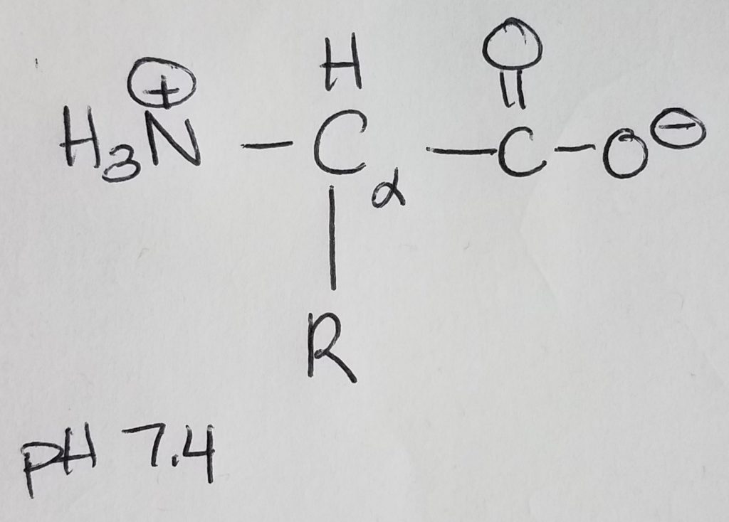
Amino acids are monomers for proteins which are 1+ polypeptide chains (polymer). 2 AA’s are joined via peptide bonding. Polypeptide chains can get longer and longer and you can put multiples of them together to form complex molecules. Once AA’s become part of a chain they are called “residues”. AA’s are made up of: an amine group, a carboxylic acid group, a hydrogen at the alpha-carbon, and a R-sidechain.

At physiological pH of 7.4, the amino group is protonated (+NH3) and the carboxylic acid group is deprotonated (COO-). Because the molecule has a dual charge, it’s called a zwitterion. The charged amino group at the leftmost position of the chain is called the N-terminus; the carboxylate ion at the rightmost position of the chain is called the C-terminus. AA’s are joined together to form a chain via peptide bonding.

Remember that if the pH < pKa, then that group will be protonated (e.g. +NH3, COOH). If pH > pKa, then that group will be deprotonated (e.g. NH2, and COO-). Physiological pH is 7.4; pKa is ~9.0 for amines; pKa is ~4.75 for carboxylic acid. Histidine’s pKa is very close but a little on the basic side at physiological pH.

Amino acids have stereochemistry (they have mirror images).

https://youtu.be/HG4bqgGR3DI

The “L” designation in L-amino acids denotes that the zwitterion is oriented with the protonated amine group at the left and the carboxylate ion on the right. The human body uses L-amino acids with the exception of glycine which is achiral. This is also important because the L stereochemistry is what our body uses in ligand-receptor binding (e.g. enzymes).

The alpha-carbon is the carbon directly bonded to the carboxylic acid group. The alpha amine is the amine group directly bonded to the alpha carbon.
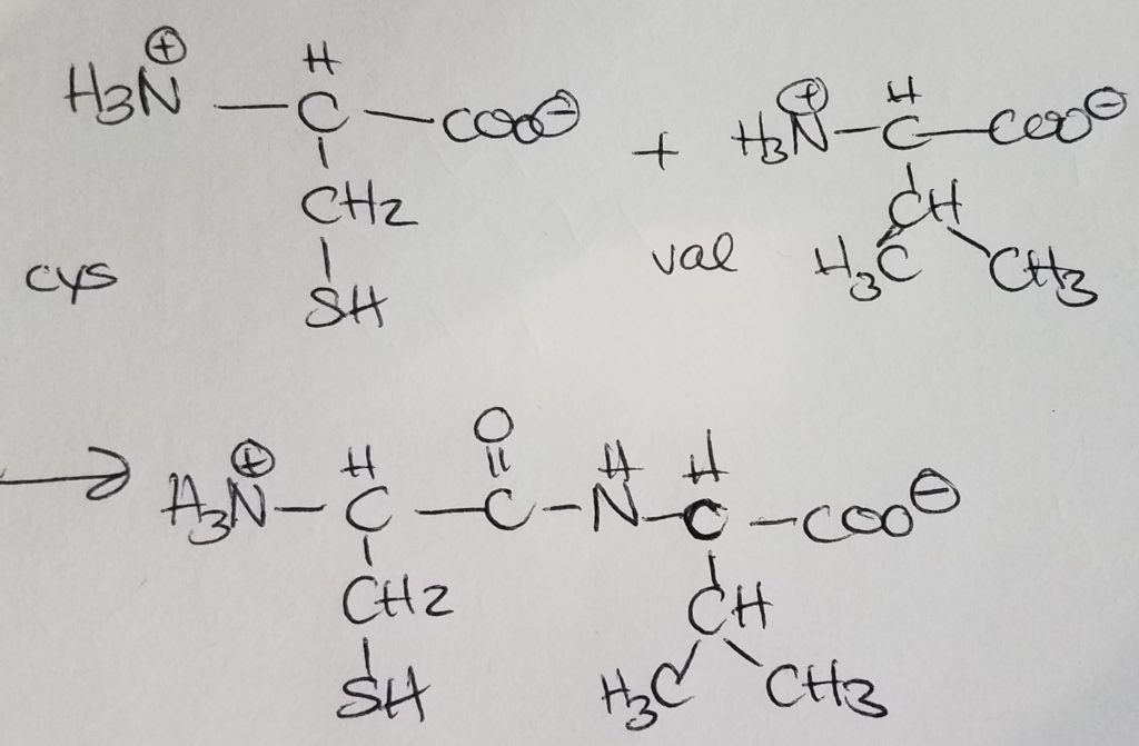
Peptide bonding is when the carboxylate end of one AA covalently bonds with the charged amino end of another AA giving off a water molecule. Peptide bonding is a special form of dehydration synthesis (condensation reaction) between two amino acids. A more general term for peptide bonding is amide bonding. Bonding sequence matters. If amino A is bonded to amino B and then to amino C, the charged amino group of A is retained. In the order A-B-C, the carboxylate ion of amino C is retained. Bonding order matters. A-B-C is not the same as B-C-A.
Proteins have primary, secondary, tertiary, and quaternary structures.

Primary structure is characterized by an unique sequence of amino acids in a polypeptide. Structure and function go hand in hand. Any tiny change in structure could impact the function of the protein.

Secondary structure is characterized by repeated folding of the polypeptide BACKBONE. The keyword is backbone. Hydrogen bonds (H-bonds) stabilize peptide bonds. The alpha helix and B-pleated sheets (parallel or antiparallel). Antiparallel configuration is more stable as steric and angle strain.

http://www.chem.ucla.edu/~harding/notes/strain_01.pdf

https://chem.libretexts.org/Bookshelves/Organic_Chemistry/Supplemental_Modules_(Organic_Chemistry)/Alkanes/Properties_of_Alkanes/Cycloalkanes/Ring_Strain_and_the_Structure_of_Cycloalkanes

Tertiary structure is characterized by lots more globular folding and interactions between R-sidechains/groups (H bonds, ionic bonds, sulfhydral bonds, disulfide bridges, salt bridges). This is where the polar/nonpolar, acidic/basic, etc. properties of amino residues becomes an important consideration.

Quaternary structure is characterized by 1+ separate tertiary structures assembling together to form a more complex molecule. Often the “core” or pockets created may hold inorganic ions and some of the gaps created may be ligand-receptor sites. The act of binding and releasing can cause conformational changes altering the function of the protein.

B. Function.

Amino acids may be classified as essential (ones the human body cannot make and must come from exogenous sources). Nonessential amino acids are ones the human body can make.

Amino acids may be classified based on the polar/nonpolar characteristics of the sidechain: polar, nonpolar; acidic/basic/uncharged or neutral.

Amino acids may be classified based on aromatic or aliphatic (structural comparison).

Amino acids may be classified based on what they can make: glucogenic (make glucose); ketogenic (make ketones); glucoketogenic (make both glucose and ketones).

Proteins can have all sorts of functions: acting as enzymes, structural support, antibodies, cell signalling, transport, and movement.

See Appendix A Amino acid abbreviations; Appendix B Glucogenic, Ketogenic, and Glucoketogenic Amino acids; Appendix C 20 Amino Acids to Know Drawn at Physiological pH 7.4.

Lipids.

A. Structure.

A functional unit of lipids is a triglyceride which is a glycerol (trihydric alcohol) and fatty acids (acyl groups which can be saturated or unsaturated long hydrocarbon with a carboxylic acid head. The carboxylate head bonds with the hydroxyl (of the glycerol) via ester linkages. The fatty acids that get attached to the glycerol do not have to be the same. Triglycerides are amphipathic (the key to this structure is its hydrophilic and hydrophobic duality).

Phospholipid = glycerol + 2 FA chains + phosphate + head-group. The key to this structure is its hydrophilic and hydrophobic duality. This is important to cell membranes, micelles, etc.

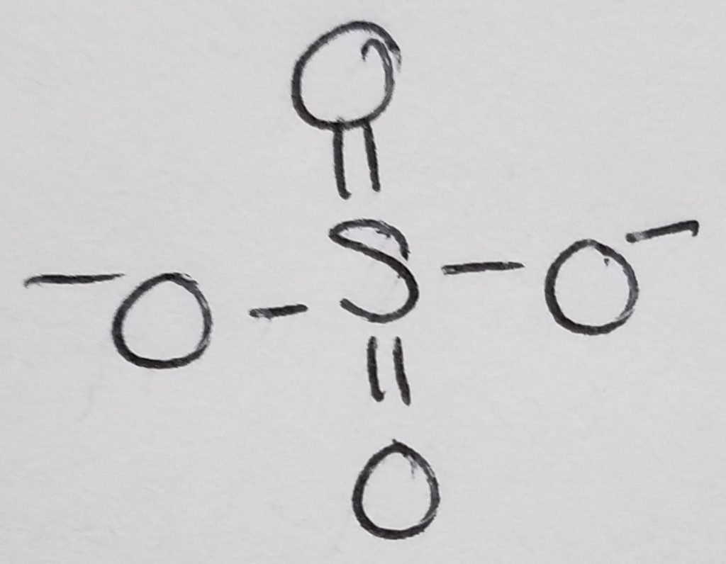
Know phosphate and how it generally hooks up to form a chain.
Likewise, recognize sulfate as well.

Waxes = 2 long hydrocarbon tails connected via ester bond.

Steroids = 4 carbon rings with no fatty acid tails.

http://www.biologie.ens.fr/~mthomas/L3/intro_biologie/2-sucres-lipides-acides-nucleiques.pdf
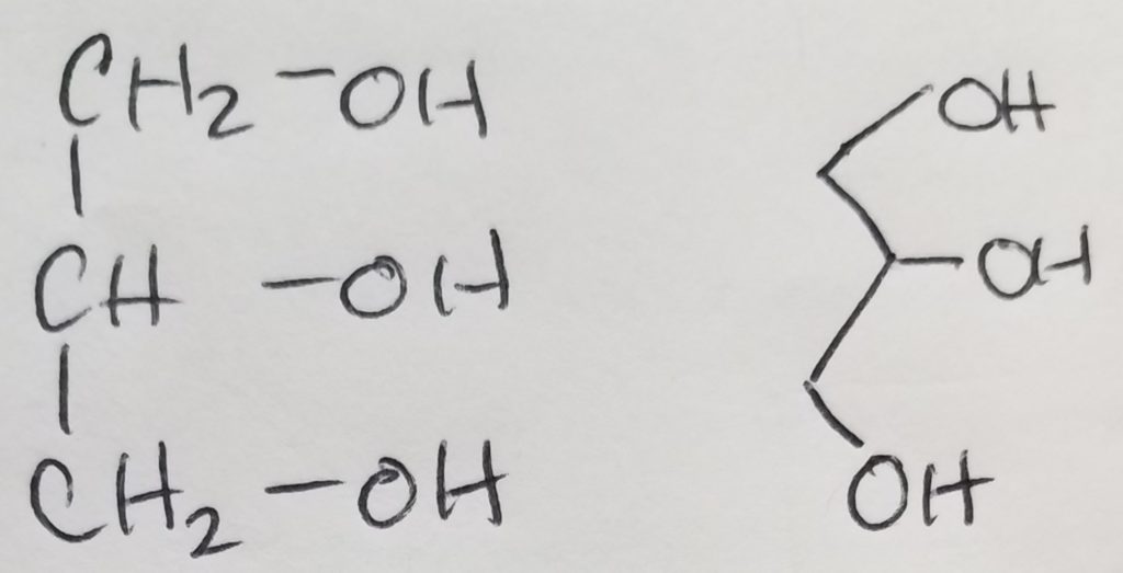
Recognize and know the glycerol structure.

**Circle the ester bond(s) and circle the glycerol component. BE CAREFUL that the ester bonds will overlap with the glycerol oxygen.
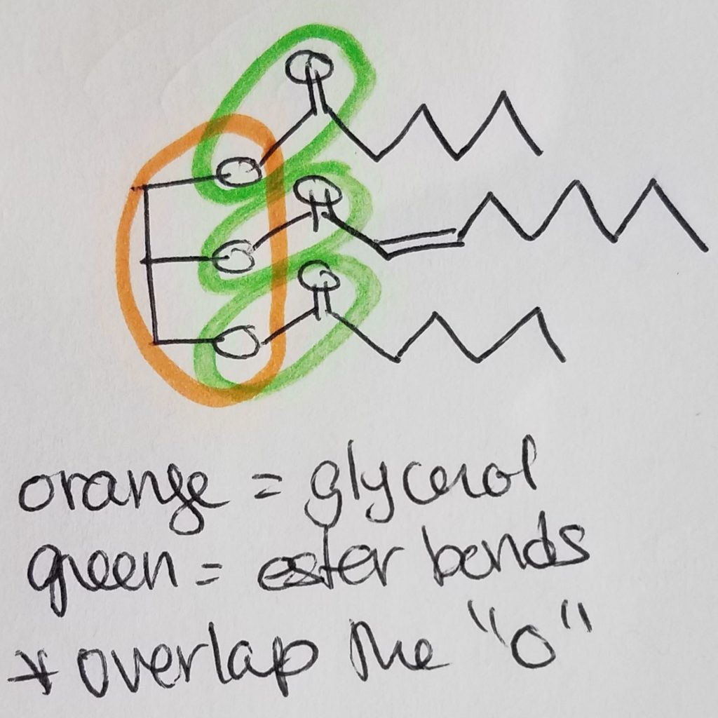
B. Function.

Some functions include (but not limited to): creating a barrier; insulation; protection; energy storage; hormones, carriers (e.g. fat soluble vitamins) and signalling.

Nucleic Acids.

A. Structure

Nucleotides are the monomers for nucleic acids. A nucleotide = pentose sugar (e.g. deoxyribose or ribose) + phosphate (mono, di, or tri) + nitrogenous base. “Deoxyribose” just means that the second carbon in the ring does not have a hydroxyl group (i.e. carbon 2 is bonded to two hydrogens). A nucleoSide = pentose sugar (e.g. deoxyribose or ribose) + nitrogenous base = nucleoTide without the phosphates.
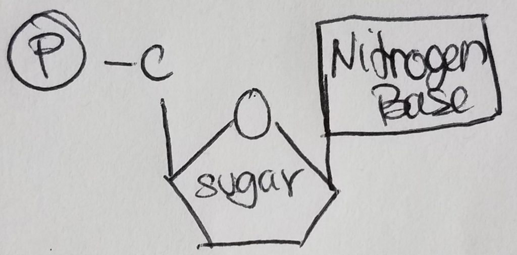
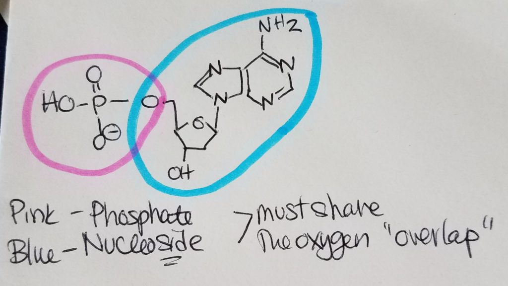
The pentose sugar is bonded to the nitrogenous base via N-glycosidic bond. The phosphates are bonded to the pentose sugar via phosphoester bonding.

https://chem.libretexts.org/Courses/Sacramento_City_College/SCC%3A_Chem_309_-_General%2C_Organic_and_Biochemistry_(Bennett)/Text/13%3A_Functional_Group_Reactions/13.10%3A_Phosphoester_Formation
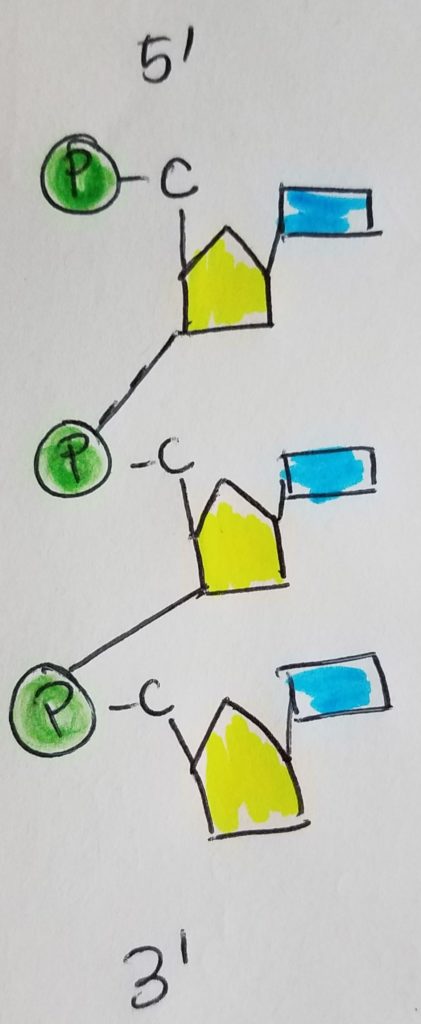
The nitrogen bases used are guanine, adenosine, cytosine, thymine, and uracil (DNA pairs are C-G and A-T, RNA pairs are C-G and A-U). Adenine and guanine are purines (5 ring connected to a 6 ring); cytosine, thymine, and uracil are pyrimidines. Base pairs between C and G are stronger due to an extra H-bond.

How to tell them apart?

Cytosine has an amine group bonded to one of the carbons on the ring. Adenosine has a carbonyl group with the carbonyl carbon as part of the ring. Uracil has two carbonyl groups, each with the carbonyl carbon as part of the ring.

Adenine has an amine group hanging off the 6-ring structure; guanine has a carbonyl group with the carbonyl carbon as part of the ring structure.

The nucleotide monomers are connected via covalent bonding carbon 5 of one sugar to the carbon 3 of the other sugar (phosphate group sandwiched inbetween). We call this the 5′ (five prime) position and 3′ (three prime) position. It is a condensation reaction forming a phosphodiester bond between the sugars and phosphate groups (alternating sugar-phosphate-sugar-phosphate etc.) with water given off as a product.

Differences between DNA and RNA.

1. Sugar. DNA has deoxyribose (missing -OH on 2nd carbon); RNA has ribose.
2. Base pairs. DNA’s base pairs are C-G, A-T. RNA’s base pairs are C-G, A-U.
3. # of strands. DNA has two strands in a double helix configuration (H-bonds help to stabilize). RNA is single-stranded.

B. Function.

Nucleic acids are in genetic material like DNA and RNA.

Central Dogma.

As far as our understanding today, scientists believe that genes determine the sequence of mRNA which in turn determines the sequence of proteins.

https://www.khanacademy.org/science/biology/gene-expression-central-dogma/central-dogma-transcription/a/the-genetic-code-discovery-and-properties

Basic Steps: From Gene to the Primary Structure of Protein.

Step 1. Transcription.

In the nucleus, RNA polymerase unzips DNA and reads DNA in order to make and spit out pre-mRNA (transcribed from the DNA) using nucleoside triphosphates as building materials. Emphasis on triphosphates because the process requires lots of energy which is available in those phosphate bonds (e.g. each time phosphate gets cleaved).

Step 2. Editing the pre-mRNA.

In the nucleus, the pre-mRNA needs to be edited. A methylated structure called a “cap” is placed on pre-mRNA’s 5′ end. A poly-adenosine monophosphate tail is added at the 3′ end. Introns are spliced out leaving only the exons.

https://www.cell.com/trends/biochemical-sciences/fulltext/S0968-0004(19)30002-7

Step 3. Translation.

The “edited/fixed-up” mRNA is transported out of the nucleus and into the cytoplasm at the rough endoplasmic reticulum. The mRNA gets sandwiched between the large and small ribosomal subunits. The ribosome is the assembly site. The tRNA is the shuttle that pairs its anticodon to the codon on the mRNA and brings in the correct amino acid for assembly. As the ribosome moves along reading the mRNA, the protein strand that’s being formed (queue-fashioned) starts to stick out of the ribosome. This step is called “translation” because it’s like decoding a secret message on the mRNA to make the protein that you want.

https://www.khanacademy.org/science/biology/gene-expression-central-dogma/translation-polypeptides/v/translation-mrna-to-protein

https://www.youtube.com/watch?v=TfYf_rPWUdY

See Appendix A Amino acid abbreviations; Appendix B Glucogenic, Ketogenic, and Glucoketogenic Amino acids; Appendix C 20 Amino Acids to Know Drawn at Physiological pH 7.4.

Appendix A. Amino acid abbreviations.

  • Ala, Alanine, A
  • Arg, Arginine, R (aRRRRRRRg arginine)
  • Asn, Asparagine, N
  • Asp, Aspartic acid, D (aspar-dic)
  • Cys, Cysteine, C
  • Glu, Glutamic acid, E (glue-EE the glutamic acid)
  • Gln, Glutamine, Q (GQ guy got hot glutes)
  • Gly, Glycine, G (Trivia: the achiral amino with an H sidechain)
  • His, Histidine, H (Trivia: the pKa is so damn close to physiological 7.4)
  • Ile, Isoleucine, I (iso-i or iso-eye)
  • Leu, Leucine, L (leu-leu, loo-loo)
  • Lys, Lysine, K
  • Met, Methionine, M (methionine the meth head)
  • Phe, Phenylalanine, F (fee-fye-fo-fum, says the giant in Jack in the Beanstalk)
  • Pro, Proline, P (it get’s a p for just being weird and attaching to both the alpha carbon and amine, looks like an urinal…pee pee pee)
  • Ser, Serine, S (reminds me of sarin gas)
  • Thr, Threonine, T (three-o-nineTy time for pinty glug glug)
  • Trp, Tryptophan, W (only cuz teacher said it looks like a W, the turkey amino)
  • Tyr, Tyrosine, Y (looks Y-ish)
  • Val, Valine, V (Val-in-tine’s day, V-day)

Appendix B. Glucogenic, Ketogenic, and Glucoketogenic Amino acids.

Ketogenic (LK): lysine, leucine.

Ketoglucogenic (ITFWY): isoleucine, threonine, phenylalanine, tryptophan, tyrosine.

Glucogenic: alanine, arginine, asparagine, aspartate, cysteine, glutamate, glutamine, glycine, histidine, methionine, proline, serine, valine.

Appendix C. 20 Amino Acids to Know Drawn at Physiological pH 7.4

Glycine

The only achiral amino acid because the sidechain is just a hydrogen.

Alanine

The shortest sidechain, methyl group.

Valine

Looks like a short “V”.

Isoleucine

Isomer of leucine.
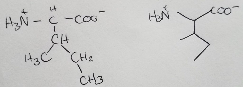
Leucine

Isomer of isoleucine.

Resources.

References.

Bean, J. (2019). Amino.

Bean, J. (2019). Macromolecules.

Bean, J. (2019). Nucleotides and nucleic acids.

Bean, J. (2019). Protein structure.

Ferrier, D. (2017). Biochemistry (7th ed.). Philidelphia, PA: Lippincott Illustrated Reviews.


Bean, J. (2019). Nucleotides and nucleic acids.

Bean, J. (2019). Protein structure.

Ferrier, D. (2017). Biochemistry (7th ed.). Philidelphia, PA: Lippincott Illustrated Reviews.

Lieberman, M., & Peet, A. (2017). Marks’ basic medical biochemistry: A clinical approach(5th ed.). Philadelphia, PA: LWW.
Posted on

Structure-Function relationships in proteins.

[Study guide covering chapter 7, PDF]

Last edited: 08.05.2019

Characteristics of 3D Structure.

  • 3D conformation and the type of amino acid side chains determine the character and function of the protein.
  • Globular proteins usu. water soluble.
  • Fibrous proteins are linear, arranged about a single axis, have repeating units.
  • Transmembrane proteins that have 1+ regions aligned so they cross the lipid membrane.
  • Primary structure. Linear AA residues joined by peptide bonds to form polypeptide chain.
  • Secondary structure. Recurring structures that form in short localized regions. Beta pleats, parallel, antiparallel.
  • Tertiary structure. 3d globular structure.
  • Quatenary structure. 2+ protein structures.

Requirements for 3D Structure.

  • Creating a binding site that’s specific for just one molecule or a group of molecules w/similar structural properities/characteristics. Binding sites on a protein define that protein’s role.
  • Has appropriate rigidity and flexibility allow the protein to fulfill its role.
  • Structural flexibility and mobility so that the protein can fold appropriately.
  • The external surface of the protein must be appropriate for its environment and aid in the protein’s functionality.
  • Conformations must be stable.
  • When the protein is degraded/damaged, it must be able to be broken down and disposed of or recycled.

3D Structure of Peptide Bond.

  • When AA are joined to form a polypeptide chain, the peptide bond takes on a “trans” configuration to minimize steric strain between different alpha carbons and side chains.
  • The backbone is a resonance structure and has very restricted “bends”. The carboxyl and amide groups are planar.
  • Torsion angles, are limited allowed rotation about the alpha carbon and alpha amino group around the bond between the alpha carbon and carbonyl group. Limited by steric constraints and favor the max distance between substituents.

Secondary Structure.

  • Recurring localized structures in polypeptide chains are secondary structures.
  • Alpha helix: secondary structure of proteins; membrane spanning; rigid, stable conformation; maximize H-bond. Peptide backbone formed by H-bonds betw. carbonyl oxygen and amide hydrogen located 4 residues down. Ea. peptide bond is connected to the other peptide bond +4 and -4 amino residues away. Proline is known as the “helix breaker” because it cannot fit in the alpha helix due to proline’s dual attachment points to a backbone.
  • Beta sheets: maximize H-bond betw. peptide backbones; maintain allowed torsional angles; paralllel or antiparallel “weaving”; the carbonyl oxygen of one peptide bond is H-bonded to the amide hydrogen of a peptide bond on adjacent strand. Antiparallel is like hairpin turns or the chain folded backwards on itself. Sheets have hydrophilic and hydrophobic sides.
  • Nonrepetitive secondary structures: bends, loops, and turns that don’t have the same organization as alpha helixes and beta sheets.
  • Patterns of secondary structure.

Tertiary Structure.

  • Secondary elements folded into 3d structure.
  • 3d is dynamic and flexible.
  • Fluctuating movement of the side chains and domains. Don’t require “unfolding”.
  • Maintains appropriate surface resides respective of its environs.
  • Flexibility is a key feature.
  • Forces: ionic bonds, H-bonds, disulfide deformations, London forces, hydrophobia-hydrophilia.
  • Tertiary structural domains: physically indep. regions that fold independently. Domains are fairly obvious to visual inspection.
  • Domain: formed from continuous AA sequence in a polypeptide chain that’s folded into 3d indep. of rest of the protein. 2 domains are connected via a loop or similar simple structure.
  • Folds in globular proteins: relatively large patterns of 3d structure recognizable in many proteins. Each”fold” hosts a certain kind of activity.
  • Actin fold: G-actin.
  • Nucleotide binding fold.
  • Globular proteins solubility in aqueous environment. Most are soluble in the cell. The core is mostly hydrophobic and is densely packed to maximize London forces.
  • Charged/polar parts of the polypeptide chains generally on the surface or close to it.
  • Charged side chains bind to inorganic ions to minimize repulsion betwen like groups.
  • Some charged aminos in the interior usu. serve as binding sites.
  • Polar uncharged: serine, threonine, asparagine, glutamine, tyrosine, tryptophan.
  • Transmembrane proteins have characteristics that allow them to cross membrane structures. They usu. have hydrophobic and hydrophilic components. Transmembrane proteins usu. have many post-translational modifications providing additional chemical groups to interact as needed. Receptors need both flexibility and “rigidity”.
  • Binding domain.

Quaternary Structure.

  • Homo-
  • Hetero-
  • Protomer: an unit made from two identical units.
  • Oligomer: multi subunit proteinmade of identical G-actin subunits.
  • Multimer: a complex w/lots of subunits of 1+ types. “Melting/melding pot”.
  • The larger a complex protein, the more difficult it is to fold and unfold etc. Requires much larger effort. It also means that it’s more stable, too. It has fewer “options”. It can also operate in more of a coordinated effort due to less subunits “going rogue”.

Quantication of Ligand Binding.

  • Ka. Association constant. Binding affinity of a protein for a ligand. The Keq for the binding reaction of P protein and L ligand. Used for comparing proteins made by diff. alleles.
  • Kd. Dissociation constant for the ligand-protein binding. Is the reciprocal of Ka.

Structure-Function in Myoglobin and Hemoglobin.

  • Myoglobin and hemoglobin are oxygen-binding proteins similar in structure.
  • Myoglobin: globular; simple polypeptide that has one oxygen binding site; 8 alpha helices connected via short coils called globin fold; no beta pleated sheets (unusual); helicies create hydrophobic oxygen binding pocket w/tightly bound heme w/an iron atom Fe2+.
  • Hemoglobin: tetramer made up of different subunits (2 alpha and 2 beta polypeptide chains called alpha-beta-protomers). Maximizies oxygen carrying capacity. Planar porphyrin ring of 4 pyrrole rings linked by methenyl bridges lying w/nitrogen atoms in the center, binding Fe(II) in the center. Negatively charged areas interact w/arginine and histidine side chains from hemoglobin; hydrophobic methyl and vinyl groups interact w/hydrophobic amino acid side chains as well that help position the heme group. There are 16 diff. interactions betw. myoglobin amino acids and diff. groups in porphyrin ring.
  • When PO2 (partial pressure of oxygen) is high (in lungs), myoglobin and hemoglobin are oxygen-saturated.
  • When PO2 is lower (e.g. in tissues), hemoglobin can’t bind to oxygen as well as myoglobin can.
  • Myoglobin is in heart and skeletal muscle and is able to bind to (and store) O2 released by hemoglobin.
  • Cytochrome oxidase, a heme-containing enZ in ETC( electron transport chain), has higher affinity than myoglobin.
  • Prosthetic groups: organic ligands tightly bound to proteins. They are a part of the protein and don’t dissociate until the protein is degraded.
  • Holoprotein: a proteinw/prosthetic groups.
  • Apoprotein: a protein w/out its prosthetic group.
  • In the binding pocket of myoglobin and hemoglobin, oxygen binds to the Fe2+. The Fe2+ can chelate (bind to) 6 different ligands (4 ligands are co-planar, 2 ligand positions are pependicular). 4 ligand positons taken by N, 1 of the perpendicular positions is taken by N on histadine called proximal histidine, the other position taken by O2.
  • When O2 binds, conformational changes occur >> Tertiary structure changes from T-state (tense state) with low O2 affinity >> to R (relaxed) state w/high O2 affinity. Binding rate for the first O2 is low but with subsequent O2’s the binding rate is higher. This is known as positive cooperativity. The first O2 is difficult but subsequent binding of O2’s gets easier.
  • HbO2 –> Hb + O2
  • Agents that affect O2 binding: hydrogen ions; 2,3-bisphosphoglycerate; covalent binding of CO2.
  • 2,3-bisphosphoglycerate (2,3-BPG). Formed in red blood cells. 2,3-BPG binds to hemoglobin increasing energy requirements for the conformational changes that facilitate O2 binding. Lowers hemoglobin affinity for O2. RBC can use 2,3-BPG to modulate affinity to O2 binding as needed by changing the rates of synthesis/degradation of 2,3-BPG.
  • Proton binding, (Bohr Effect). When hemoglobin binds protons, it has less affinity for oxygen. pH decreases in tissues (proton concentration is higher) as metabolic CO2 is converted to carbonic acid via carbonic anhydrase in RBC’s. Dissociated protons react w/AAs >> conformational changes >> promote release of O2. In the lungs, this process is reversed. Lungs have high O2 concentration >> O2 binds to hemoglobin causing release of protons >> pH of blood rises >> carbonic anhydrase cleaves carbonic acid to H2O and CO2 >> CO2 exhaled.
  • Carbon dioxide. Most of it is from tissue metabolism. CO2 is carried to lungs as bicarbonate. Some of the CO2 is covalently bonded to hemoglobin.
  • https://www.khanacademy.org/science/health-and-medicine/advanced-hematologic-system/hematologic-system-introduction/v/bohr-effect-vs-haldane-effect
  • https://www.khanacademy.org/science/health-and-medicine/advanced-hematologic-system/hematologic-system-introduction/v/hemoglobin-moves-o2-and-co2
  • http://www.pathwaymedicine.org/bohr-effect
  • https://youtu.be/FtA4Xy-lMSY

Structure-Function Relationships in Immunoglobin.

  • Immunoglobins/antibodies bind to antigens (ligands) on the invaders, like marking them for inactivation/destruction. Marking the invaders as “not self”.
  • Immunoglobins have the same structure: ea. antibody molecule has 2 identical polypeptide chains (L light chains) and 2 identical large polypeptide (H heavy) chains. L and H chains are joined via disulfide bonds.
  • 5 major classes of immunoglobins.
  • IgG gamma (most abundant); 220 aminos in light chain; 440 aminos in heavy chain. L and H chains have immunoglobulin fold (collapsed number of B sheets called Beta-Barrel). Have attached oligosaccharides that help mark “not self”.
  • V = variable regions. VL (variable light chain) and VH (variable heavy chain) interact to make one antigen-binding site at each branch of the Y-shaped molecule. V regions have different AA compositions.
  • C = constant regions. Form the fragment, crystallizable part of the antibody important for the antigen-antibody complex.
  • Antigens bind tightly w/almost no tendency to dissociate. Kd is betw. 10^-7 to 10^-11 M.

Protein Folding.

  • Peptide bonds may be rigid, but other bonds in the molecule can allow some flexibility.
  • Native conformation: every molecule of the same protein has the same stable “native” conformational state.
  • Primary structure (sequence of AA side chains) determines folding and assembly of subunits.
  • Denaturation can affect protein structure. However, under certain conditions, the denaturation may be reversed.
  • Not all proteins fold into their native state by themselves. Sometimes the folding and refolding occurs as the protein searches for its most stable state.
  • Kinetic barriers are the higher energy states and conformation the protein may pass through.
  • These kinetic barriers may be overcome by heat-shock (chaperonins) which use energy from ATP hydrolysis to help in the folding process.
  • Cis-trans isomerase.
  • Protein disulfide isomerase.
  • Collagen: fibrous protein; made mostly by fibroblasts (cells in interstitial connective tissue), muscle cells, and epithelial cells. Type 1 collagen most abundant in mammals and major component in connective tissue. Found in ECM, loose conn. tissue, bone, tendons, skin, blood vessels, and cornea. 33% glycine, 21% proline, and hydroxyproline.
  • Hydroxyproline is an AA made by posttranslational mods of peptidyl proline residues.
  • Procollagen 1 is the precursor of collagen 1; triple helix of 3 pro-alpha polypeptide chains twisted around ea. other (ropelike). Interchain H-bonds. Every 3rd residue is a Glycine.
  • Collagen 1 polymerizes >> collagen fibrils, great tensile strength.
  • Vit C functions as a cofactor of prolyl hydroxylase and lysyl hydroxylase. These hydroxylase aid in H-bond formation >> strength and stability. Vit C deficiency causes the melting point to drop from 42 deg C to 24 deg C.
  • Aldehyde residues, allysine, make covalent crosslinkages betw. collagen and other structures >> improve stability and structural support.
  • Allysine on one collagen molecule + lysine of another molecule >> Schiff base (N=C double bond).
  • Aldol condensation may occur betw. 2 allysine to form lysinonorleucine.
  • Protein denaturation via nonenzymatic modification of proteins.
  • Protein denaturation via temp, pH, and solvent.
  • Protein denaturration via misfolding and prions.

Resources.

References.

Lieberman, M., & Peet, A. (2017). Marks’ basic medical biochemistry: A clinical approach(5th ed.). Philadelphia, PA: LWW.

Posted on

Amino Acids.

[Study guide covering chapter 6, PDF]

Last edited: 08.05.2019

Structure.

  • Phosphate group.
  • Nitrogen/amino group: attached to the alpha carbon of the carboxylate.
  • Carboxylate group.
  • R-side chain.
  • Concept of zwitterion.
  • Amine group pH ~9.5
  • Physiological pH ~7.5
  • Alpha carbon is chiral except in glycine.
  • Glycine, the carbon is achiral (has two H).
  • Mammalian AA are L configuration (as opposed to D).
  • Alpha aminos are AA attached to alpha carbon.
  • Primary structure: sequence of AAs.
  • AA joined via peptide bonds betw. COO- of one AA and the NH3+ of the other.
  • Backbone >> carbolxylate, alpha carbon, and amino group.
  • AA are the side chains.
  • Binding sites.
  • Ligand-receptor pairs.
  • N-terminal (where the amino group is).
  • C-terminal (where the carboxylate is).

Classification of Amino Acids.

  • pKa
  • Hydropathic index: a scale to describe hydrophobicity of the side chain.
  • Glycine is a special case where its R-group is just H. Has the least steric hinderance. Can be found in the “nooks and crannies”.
  • Nonpolar: alanine, valine, leucine, isoleucine; aliphatic; very hydrophobic; typically form the hydrophobic cores.
  • Proline is another special case as it’s attached to the backbone twice. It has both an alpha carbon and alpha amino group. Proline is an imino acid. Rigid and forms kinks. Restricted conformations.
  • Aromatic acids: 6-member ring of C and H; conjugated double bonds. H on the ring doesn’t participate in H-bonding. The substituents on the ring influence side chains characteristics & determine what action takes place (e.g. hydrophilic or hydrophobic).
  • Aliphatic, polar, uncharged AA: has amide group (asparagine, glutamine); or hydroxyl group (serine, threonine); form H-bonds with water. Typically found on surfaces.
  • AA with sulfur: cysteine and methionine. Can form disulfide bridges via oxidation of the sulfhydral groups, but it doesn’t always do that. The action/function depends on it’s surrounding environment and “what else” there is.
  • Acidic AA: carboxylic acid groups; e.g. aspartate and glutamate. Their negative charge can form ionic bonds with cations.
  • Basic AA: have side chains containing N (+) and tends towards basicity; e.g. histidine, lysine, arginine. Their positive charges can form ionic bonds with anions. Lysine and arginine can form bonds to anionic compounds w/protein binding sites (i.e. those protein binding sites can become incorporated into a new compound as well as become altered). Those ligand-receptor sites can become “grandfathered in”.
  • Acidic/basic characteristics can allow AAs to participate in H-bonds and salt bridge formations.
  • Carbon positions can be described using the Greek letters: alpha, beta, gamma, delta, epsilon).
  • If pH < pKa then it favors the protonated form (-COOH, -NH3+). If pH>pKa, then it favors the deprotonated form (-COO- and -NH2).
  • Imidazole ring. C3N2H4 (compound).
  • In proteins, only the side chains, N-terminal, and C-terminal are dissociateable. Other C, N, H that form part of the backbone do not participate in acidic/basic characteristics.
  • Electrophoresis: separate proteins via charge differences. Helps to identify proteins and components.

Variations in Protein Structure.

  • Protein structure and characteristics can vary between different individuals and different ages. This variant nature is called variant regions.
  • Hypervariable describes a situation where variation is tolerable within reason.
  • Invariant regions, in contrast, do NOT vary between individuals, species, etc. Variation is NOT tolerated.
  • Polymorphisms: when allele variations occur with great frequency.
  • Homologous proteins: these belong to the same ancestral proteins.
  • Paralogs: proteins that have similar structure and function that have evolved from the same gene after gene duplication.
  • Divergent evolution: when one gene performs it’s expected function yet a copy mutates into a different function or have different characteristics.
  • Superfamily: large family of homologous proteins.
  • Isoforms: 2+ functionally similar proteins with similar structure (but not identical) or AA sequence; isoforms of a protein have the same function.
  • Isozymes: 2+ enZ w/similar functions but differ in structure; isozymes catalyze the same reactions.
  • Developmental variation: structures and functions differ at different developmental stages.
  • Tissue-specific isoforms: proteins that function “the same” but they vary in structure/characteristics from tissue group to another tissue group.
  • Tissue-specific isozymes: enZ that function “the same” but they vary in structure/function from tissue group to tissue group.
  • Species variation: proteins and enzymes (and their structure and function) can vary from species to species.

Modified Amino Acids.

  • Post-translational modification. Post-protein synthesis, some AA residues in the primary sequence may be modified. These changes may or may not serve/enhance function or characteristics. Usu. occur after protein has already folded into its specific conformation.
  • Glycosylation: the addition of carbohydrates to a molecule.
  • Fatty acylation: the addition of lipid group(s) to a molecule. These types of changes (N- or O-) can enhance barrier/surface protection (e.g. N-linked oligosaccharides). O-linked oligosaccharides can enhance secretions.
  • https://www.ncbi.nlm.nih.gov/pmc/articles/PMC4975971/
  • https://www.nature.com/articles/nrm.2015.11
  • Prenylation: the addition of farnesyl or geranylgeranyl groups via thioether linkate to specific cysteine residues of membrane proteins.
  • Regulatory modifications. Phosphorylation, acetylation, and adenosine diphosphate (ADP)-ribosylation of some AAs can alter bonding characteristics of the AA.
  • Other amino acid posttranslational modifications: can alter the activity of the protein.
  • Selenocysteine: found in a few enZ and is required to activate those enZ.

Resources.

References.

Lieberman, M., & Peet, A. (2017). Marks’ basic medical biochemistry: A clinical approach(5th ed.). Philadelphia, PA: LWW.

Posted on

Tour and quick review of the major compounds important to the human body.

[Study guide covering chapter 5, PDF]

Last edited: 08.04.2019

Important functional groups: those with oxygen, nitrogen, phosphorus, sulfur.

  • Acyl group: the part of the structure that provides the –C=O carbonyl group in an ester/amide linkage. “-yl” ending.
  • Aliphatic: open chains (non-ring).
  • Aromatics/Benzene rings. “Phenyl” if ring is a substituent.

Carbohydrates (sugars).

  • Formula: CnH2nOn. Glucose is C6H12O6.
  • Classified by: carbonyl group (aldose/ketose); # of carbons; positions of the -OH group on the anomeric carbon (D=right/L=left OH position, stereoisomers, epimers); any additional substituents; number of saccharides; how the components are linked (e.g. glycosidic bonds).
  • For n asym centers >> 2^n stereoisomers generally speaking.
  • Humans use D-sugars. “D for Delightful sugars!”
  • Epimer: a pair of stereoisomers that differ only in one position of the OH at a chiral carbon.
  • Epimerases: enzymes that make epimer conversions.
  • Glycoproteins: proteins + sugars.
  • Proteoglycans: proteins that are heavily glycosylated. Many long unbranched polysaccharide chains attached to a protein core. VIP to extracellular matrix, aqueous humor, cells that make mucous secretions, & cartilage.
  • Glycosaminoglycans: polypeptide chains with repeating disaccharide units w/oxidized acid sugars, sulfated sugars, and N-acetylated amino sugars. Structure looks like a bottle brush.
  • https://themedicalbiochemistrypage.org/glycans.php
  • https://www.mdpi.com/1424-8247/11/1/27/pdf
  • Glycosylation: a reaction where a carbohydrate is attached to a hydroxyl or other functional group.
  • Glycolipids: lipids + sugars.
  • In solution, OH on anomeric carbon spontaneously changes (mutarotation) from alpha to beta and back to change from open to ring forms such as chair/boat etc. Chair/boat etc are usually more stable so there’s a greater chance that a compound will be in those configurations.
  • If the anomeric carbon forms a bond with another molecule, those mutarotations cannot happen due to the bond which limits configurational possibilities.
  • Common substituted groups: phosphate, amino, sulfate or N-acetyl.
  • Most free monosaccharides in the body are phosphorylated at the terminal carbons preventing transport out of cell.
  • Galactosamine & glucosamine are examples of an amino group replacing one of the OH groups. Usu. the amino group gets acetylated forming an N-acetylated sugar.
  • Acetylation: adding an acetyl functional group to a compound.
  • Acyl group. http://www.chem.ucla.edu/~harding/IGOC/A/acetyl_group.html
  • https://www.oit.edu/docs/default-source/library-documents/library-publishing/che102-intro-organic-chemistry/chapter-1-7.pdf
  • Sugars can get oxidized at the aldehyde carbon to form “-onic acid” or “-onate”.
  • Uronic (“-uronic acid”) acid forms when the the terminal OH group gets oxidized.
  • Polyol sugar: a sugar where the aldehyde gets reduced where all the carbon atoms have OH. Eg. Sorbitol.
  • Deoxy sugar: a sugar that has reduced such that 1+ carbons contains only H’s. Carbon 2 of deoxyribose.
  • The OH of the anomeric carbon can react with an OH (O glycosidic bonds found in sugar-sugar, sugar-hydroxyl bonds) or NH (N glycosidic bonds found in nucleosides and nucleotides) group to form an alpha/beta glycosidic bond.
  • Alpha glycosidic bond. The Greek alpha looks like a fish which is DOWN in the sea.
  • Beta glycosidic bond. The Greek beta looks like a bird UP in the air.
  • Disaccharide: 2 monosaccharides joined by O-glycosidic bond.
  • Oligosaccharide: 3-12 linked monosaccharides via N or O glycosidic bonds.
  • Polysaccharides: thousands of monosaccharides joined to make chains and/or branches.

Lipids.

  • Hydrophobic.
  • Usu. straight chains, methyl group at one end (w-carbon) and carboxyl at the other end.
  • Most FA in humans have even number of carbons betw. 16-20.
  • Most common FA in cells are stearic and palmitic FAs.
  • *Special notation for FA’s pg. 69-70.
  • http://rogersal.people.cofc.edu/Lipids.pdf
  • https://www.cs.mcgill.ca/~rwest/wikispeedia/wpcd/wp/f/Fatty_acid.htm
  • https://courses.lumenlearning.com/suny-nutrition/chapter/2-33-fatty-acid-naming-food-sources/
  • https://www.ncbi.nlm.nih.gov/pmc/articles/PMC3646453/
  • https://www.uio.no/studier/emner/matnat/farmasi/nedlagte-emner/FRM2041/v06/undervisningsmateriale/fatty_acids.pdf
  • FA also classified by distance from w-carbon to the double bond.
  • Fatty acids. Esterified to glycerol >> triacylglycerols (triglycerides) or phosphoacylglycerols (phosphoglycerols).
  • Tri acyl glyderols: 3 acyl FA groups attached to glycerol; fat stores in the body.
  • Sphingolipids: FA + sphingosine (serine + palmitate FA). No glycerol backbone.
  • Ceramides (type of amides) = sphingosine + FA attached at the amino group.
  • More sphingolipids formed from attaching substituents onto the OH of the ceramide.
  • Cerebrosides + gangliosides = sugars glycosidically bonded to OH of ceramides.
  • Sphingomyelin = phosphorylcholine + ceramide; vip part of cell membranes and myelin sheath.
  • Glycolipids: lipids + sugar hydroxyl group.
  • Polyunsaturated FA: building blocks of eicosanoids.
  • Eicosanoids: signalling molecules via enZ or non-enZ oxidation of arachidonic acid or other polyunsat. FA. Hormone-like compounds. Polyunsat FA with 20 carbons (eicosa) and have 3-4-or-5 double bonds (e.g. prostaglandins, thromboxanes, leukotrienes).
  • Naturally occurring FA typically cis.
  • There are also trans.
  • Cholesterol: formed from isoprene units.
  • Bile salts.
  • Steroid hormones.
  • Isoprenyl unit: combined in long chains to make structures such as side chains of Coenzyme Q in humans and Vit A in plants.
  • Geranyl groups = 10 carbons & polymers of isoprenyl units.
  • Farnesyl groups = 15 carbons + isoprenyl units.
  • Geranyl and farnesyl groups often get attached to proteins so that proteins can interact w/other cellular structures.
  • Acylglycerols: glycerol with 1+ FAs (acyls via ester linkages). Mono- di- and tri-acylglycerols contain 1, 2, and 3 FA esterified to glycerol. Triacylglycerols don’t usu. have the same FA at all 3 positions (usu. mixed).
  • Phosphoacylglycerols: FA at positions 1 and 2; phosphate group (or substituent attached to phosphate group) at position 3. If it’s only the phosphate group and NO other substituents at position 3, then it’s a phosphatidic acid.
  • Phosphatidylcholine (lecithin) found in membranes. Has polar and nonpolar duality.
  • Lysolipid = phosphoacylglycerols – fatty acyl group
  • Steroids: 4-ring steroid nucleus; cholesterol precursor; diff species made by modifying ring or C20-side chains.
  • Cholesterol hydrophobic can convert to hydrophilic bile salt (eg. cholic acid). Branched 5-carbon units w/1 double bond (isoprenyl unit)
  • Bile salts are on micelles survaces in the intestinal lumen.

Nitrogen Compounds.

Free Radicals.

  • Compounds w/single electron in outer shell.
  • Extremely reactive and unstable.
  • Usually formed as intermediates.
  • Usu. negative effects.

Oxidation/Reduction.

  • Carbon-carbon or carbon-oxygen bonds said to be oxidized or reduced depending on # of electrons around the carbon.
  • LEO: lose electrons (lose H atoms) oxidation.
  • GER: gain (gain H or lose O) electrons reduction.
  • More oxidized from alcohol to aldehyde/ketone to carboxyl.

Acid/Base.

  • Cations are catfabulous (+)! Anions (onions) make you cry (-).
  • Common anionic groups: carboxylate; phosphates (P); sulfates.
  • Common cationic groups: N, amines.

Bond Polarity & Partial Charges.

  • Carboxylate.
  • Phosphate.
  • Sulfate.
  • Ester = carboxylic acid + alcohol – water
  • Thioester = acid + sulfhydryl
  • Amide = acid + amine
  • Phosphoester = phosphoric acid + alcohol
  • Anhydride = acid1 + acid2

Resources.

References.

Lieberman, M., & Peet, A. (2017). Marks’ basic medical biochemistry: A clinical approach(5th ed.). Philadelphia, PA: LWW.

Posted on

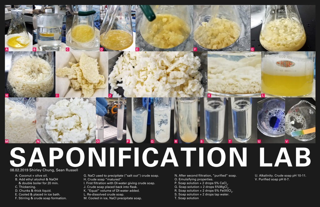
Saponification Lab Poster

Copyright 2019 Shirley Chung. All rights reserved.

Articles of interest.

Chagrin Valley Soap & Salve Company. (2014, June 16). The Origin and History of Soap. Retrieved from https://www.chagrinvalleysoapandsalve.com/blog/posts/what-is-the-origin-of-soap/

Wallace, T., Gibbons, D., O’Dwyer, M., & Curran, T. P. (2017). International evolution of fat, oil and grease (FOG) waste management–A review. Journal of Environmental Management, 187, 424-435.

Zhang, L., Wang, S., Yang, R., Mao, J., Jiang, J., Wang, X., … & Li, P. (2019). Simultaneous determination of tocopherols, carotenoids and phytosterols in edible vegetable oil by ultrasound-assisted saponification, LLE and LC-MS/MS. Food chemistry, 289, 313-319.

Zhu, X. N., Nie, C. C., Zhang, H., Lyu, X. J., Qiu, J., & Li, L. (2019). Recovery of metals in waste printed circuit boards by flotation technology with soap collector prepared by waste oil through saponification. Waste Management, 89, 21-26.

Posted on

Carbohydrate Notes

Carbohydrates have the general formula of Cn H2n On, where the “n” is a number and subscripted. They hydrolyze to a polyhydroxy aldehyde or a polyhydroxy ketone. At the top of the Haworth projection

  • Carbohydrates have the general formula of Cn H2n On, where the “n” is a number and subscripted.
  • They hydrolyze to a polyhydroxy aldehyde or a polyhydroxy ketone.
  • At the top of the Haworth projection, there’s either an aldehyde group for an aldose or a ketone group for an aldose.
  • Monosaccharides have 3-6 carbons in the parent chain with a carbonyl group.
  • If the carbonyl is at the C1 position, it’s an aldose. If the carbonyl is at the C2 position, it’s a ketose.
  • In a Fischer projection, the vertical lines/backbone “recede and go away and into the page like a dashed-wedge” while the horizontal lines “come out like a wedge”.
  • If there are n carbons, then look closely at the n-1 carbon position. If the H–OH with the OH on the right side, then the compound is labeled “D”. If the HO–H at the n-1 carbonposition, then the compound is designated “L”.
  • A chiral carbon is one where the carbon is attached to four unique bonding groups. A carbon participating in a double or triple bond cannot be a chiral carbon.
  • The human body uses “D” sugars.
  • To draw the enantiomer, swap the position of “H” and “OH” down the backbone (i.e. just make them reverse of what they currently are).

Fischer to Haworth Projections (straight chain to cyclic form)

For a 6-member ring.

  • Turn the Fischer diagram 90 degrees clockwise (lay it on it’s side).
  • Substituents below the carbon backbone get the downward position. Substituents above the carbon backbone get the up position.
  • Twist carbon-5 so that the OH group is near the carbon-1 putting carbon-6 (CH2OH) sticking up above the ring.
  • The -OH group on carbon-5 reacts with the aldehyde group on carbon-1 thereby closing the ring. An oxygen takes the position between carbon-5 and carbon-1.
  • The OH-H group on carbon-1 can have the OH in the “up” position (beta form, b like a bird). The OH-H group on carbon-1 can also have the OH in the “down” position (alpha form, Greek A looking like a fish in the sea).
  • Carbon-1 is called the anomeric carbon where linkages are possible.

For a 5-member ring.

  • A ketone at the second carbon position reacts with the OH at the n-1 carbon position to form a 5-member ring instead of a 6-member ring.

More to be continued. Work in progress.

Posted on

Naming/ IUPAC Nomenclature 2

Now that you have the basics of naming, here are some tips on prioritization rules (i.e. giving functional groups priority or the lowest position number).

  • Double bond “-enes” have priority over triple bond “-ynes”.
  • Triple bond -ynes get priority over over phenyl.
  • Halides/halogens have no special priority over alkyl groups.
  • In general, you can expect any functional group containing an oxygen will have priority over functional groups without the oxygen.
  • Alcohol has priority over alkyl, amines, halides, ethers, phenyl, and thiols.
  • Thiols -SH get priority over alkyl and halides.
  • If both thiols and alcohols are present, then alcohol gets priority.
  • Aldehyde gets priority over double bond -enes and alcohol.
  • Carboxyl group gets priority over aldehydes.
  • Ketones get priority over -enes, phenyls, and alcohols.
  • Carboxylic acids are highest priority and are terminal; there can only be a maximum of two carboxylic acids. Carboxylic acids get priority over alcohols, amines, ketones and aldehydes.
  • Non carboxylic acid carbonyls (acting as substituents) get the “oxo” name.
  • Esters have priority over carboxylic acids and the nitrile group.
  • Groups with oxygen have priority over amines.
  • Amides get priority over amines.
Posted on

Naming/IUPAC Nomeclature

  • Identify the longest carbon chain.
  • When naming, identify different substituents and functional groups. Keep in mind that at some point, you’ll put these into alpha order unless otherwise noted.
  • X, Y, Z-name. Position numbers are separated by commas. The substituent(s) is separated from those position numbers by a dash.
  • Ignore di-, tri-, etc. when naming (they don’t “count” when putting substituents/groups into alpha order).
  • Double and triple bonds. Find the longest chain that includes as many of these bonds as possible. Give the first carbon of the multiple bond the lower position number. Double bonds “-enes” get priority over triple bonds “-ynes”.
  • Alkyl groups have the same level of priority as halogens.
  • Double and triple bonds get priority (lower position numbers) over alkyls and halogens.
  • Alcohols get priority over: alkenes; alkynes; alkyl; halogens. Prefix “-ol”. If there’s double bonds “-enol”.
  • Benzene: ortho (o) is the 1,2 position; meta (m) is the 1,3 position; para (p) is the 1,4 position. Memorize as OMP-benzenes!!
  • Ether are R-OR. Alkoxy is the -OR group. Hydroxy is -OH. Methoxy is -OCH3. Ethoxy is -OCH2CH3.
  • Thiols are -SH and named like alcohols. Drop the “e” of the parent chain and add -thiol.
  • Aldehydes. Change -ane to -anal. They’re at ends of chains so the max you can have on one chain is two. For double bonds use “-enal”.
  • Ketones. Change “e” to “one”. The position number of the carbonyl carbon gets to be the lowest number (or highest priority). If an alcohol exists, “hydroxy-“. If NH2 exists, “amine-“.
  • Work in progress.

Some helpful resources.

Posted on

Aldol Panic Attack

Oh! So you’re like me and you’re cramming to understand the Aldol set of reactions and enols? My secret resource is Leah4Sci (basically anything you need that’s chem, ochem, or MCAT oriented). GO SEE LEAH4SCI.

Here’s a link to the Aldol page http://leah4sci.com/aldol-reaction-shortcut/

Here is a link to Aldol addition/condensation reaction trick https://www.youtube.com/playlist?list=PLaySzQJTCO1mXW0zCC3CbRK6sCYvP9FOy

Here is the link to Leah’s entire playlist relating to enols and aldols https://www.youtube.com/playlist?list=PLaySzQJTCO1mXW0zCC3CbRK6sCYvP9FOy