Posted on

Microbiology: Oxidase test.

Oxidase enzymes (e.g. cytochrome oxidase) are important (as electron transporters) to aerobes, facultative anaerobes, and microaerophiles in aerobic respiration. The oxidase test helps to differentiate between generas Neisseria and Pseudomonas (both of which are oxidase-positive) and Enterobacteriaceae (oxidase-negative).

After inoculation and incubation for 12-48 hours, 2-3 drops of reagent p-aminodimethylaniline oxalate is added to the medium. If the medium turns pink, then maroon, and then dark purple, the test is positive for cytochrome oxidase production. If there is no color change, the test is negative.

Clinical significance. The oxidase test helps to differentiate between generas Neisseria and Pseudomonas (both of which are oxidase-positive) and Enterobacteriaceae (oxidase-negative). The oxidase test helps to identify Neisseria meningitis, and also separate yeast Candida from Saccharomyces and Torulopsis.

Reference

Cappuccino, J. G., & Welsh, C. (2018). Microbiology: A laboratory manual.

Posted on

Microbiology: Catalase test.

Aerobes, facultative anaerobes, and microaerophiles may produce hydrogen peroxide and/or superoxide as a result of aerobic respiration (oxygen is final electron acceptor). These organisms must be able to break down hydrogen peroxide (and/or superoxide) via production of: catalase enzyme degrades hydrogen peroxide into water and free oxygen gas; superoxide dismutase enzyme degrades superoxide into hydrogen peroxide.

A trypticase soy agar (TSA) broth/slant/dish is inoculated and incubated. Add 3-4 drops of hydrogen peroxide and observe for bubble formation (positive test result for catalase).

Clinical significance. The catalase test helps to differentiate between Staphylococci (catalase positive), Streptococci (catalase negative), and members of Enterobacteriaceae.

Reference

Cappuccino, J. G., & Welsh, C. (2018). Microbiology: A laboratory manual.

Posted on

Microbiology: Nitrate Reduction test.

Nitrate reduction is an anearobic process and can occur with some aerobes and facultative anaerobes. Anaerobic respiration (an oxidative process) uses nitrates (NO3-) or sulfates (SO4-2) as an oxygen resource, because oxygen is the final electron acceptor.

A nitrate broth (standard broth medium with 0.1% potassium nitrate) with 0.1% agar (makes the medium a semisolid promoting an anaerobic environment due to lack of free gas exchange) is inoculated and incubated for 12-48 hours (this test must be done in this time window).

Five drops of reagent A (sulfanilic acid) is added followed by five drops of reagent B (alpha-naphthylamine). A cherry red color change indicates a positive result (the organism can metabolize nitrates).

Other possible situations may be:

A. If there’s no color change after the additions of reagents A and B, the organism was not able to reduce the nitrates. To test this possibility, add a small amount of zinc powder to the test tube (which already has reagents A and B). If nitrates exist, the zinc will reduce the nitrates to nitrites and the medium will turn into a cherry red color (positive test indicates that the organism was not able to metabolize nitrates). IF upon the addition of zinc there was NO color change…then you have situation B (see below).

B. If there’s no apparent color change, the organism may have metabolized and broken down the nitrates (to ammonia or molecular nitrogen) too quickly for testing. This is why it is a good idea to inoculate a few test tubes, and to perform this test at the 12-hour mark and 48-hour mark.


Clinical significance. A patient with tuberculosis-like symptoms should provide a sputum sample in order to check for Mycobacterium tuberculosis. M. tuberculosis is the only Mycobacterium species with the ability to metabolize nitrates.


Reference

Cappuccino, J. G., & Welsh, C. (2018). Microbiology: A laboratory manual.

Posted on

Microbiology: Litmus-Milk reactions.

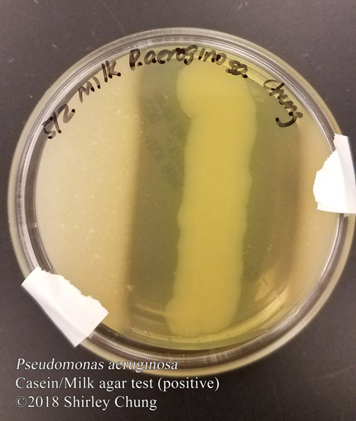
The important components of milk are the milk-sugar lactose and the proteins casein, lactalbumin, and lactoglobulin. These may be incorporated into a standard agar medium along with litmus (redox pH indicator) in order to observe microorganisms that may possess the ability to metabolize lactose and the milk-proteins.

Using the litmus-milk medium, it is possible to test for: lactose fermentation; gas production; litmus reduction; curd formation; proteolysis; and alkaline reaction.

Lactose fermentation. Lactose may be used as an alternative carbon source by organisms capable of producing beta-galactosidase. Beta-galactosidase helps to break down lactose into glucose and galactose which can then enter the glycolytic (Embden-Meyerhof) pathway. The end product, lactic acid, may then be detected by the pH indicator: litmus is purple when there is no change (negative result); litmus turns pink when the medium is acidic up to pH 4.

Gas formation. Gases likely to be produced are carbon dioxide and hydrogen gas. Tiny fissures/breaks/bubbles in the agar medium may indicate such gas formation.

Litmus reduction. Lactose oxidation produces carbon dioxide and hydrogen gas. Litmus acts as an electron acceptor as indicated by a white or milk-ish color.

Curd formation. It’s possible that curds/clots may form as a byproduct. Curds are acidic or rennet. Acid curds: are precipitates (calcium caseinate) of lactic acid or other organic acid; hard and usually sticks firmly to sides of a test tube (especially if tube is inverted). Rennet curds: are precipitates that from when rennin (enzyme) acts on casein forming paracasein; paracasein plus calcium ions converts to calcium paracaseinate, an insoluble semisolid curd/clot which will flow slowly when test tube is inverted.

Proteolysis (peptonization). Some organisms cannot metabolize lactose for energy, but they may be able to metabolize milk-proteins for energy instead. As milk-proteins (usually casein) are broken down into their component amino acids, the byproduct ammonia causes the pH of the medium to turn alkaline. Litmus will turn deep purple at the upper part of the tube, and the medium may turn translucent brown or become whey-like.

Alkaline reaction. When casein is only partially metabolized into short polypeptide chains, the medium turns alkaline (alkaline reaction). The medium’s color remains unchanged or it may change to a deep blue.

To summarize…

Litmus milk results:

A. Pink—lactose fermentation (acid)

  • Pink band up top + white color medium below—acid followed by reduction.
  • Pink band up top + white color medium below + solids—acid, reduction, and curd.
  • Pink band up top + white color medium below + solids + fissues/cracks/bubbling—acid, reduction, curd, and gas.

B. White + purple band up top—litmus reduction.

C. Deep puple band up top + translucent whey-looking brown medium—proteolysis.

D. Medium unchanged or is a deep blue color—alkaline reaction.


Clinical significance. Litmus-milk is a differential medium for Enterobacteriacaeae and other gram-negative bacilli.

Reference

Cappuccino, J. G., & Welsh, C. (2018). Microbiology: A laboratory manual.

Posted on

Microbiology: Urease test.

Urease is an enzyme made by some microorganisms (e.g. Proteus vulgaris) to breakdown/hydrolyze urea into carbon dioxide, water, and ammonia. P. vulgaris happens to be able to do this rather quickly as compared to other Proteus species.

An urea broth with pH indicator phenol red is used. The presence of ammonia will cause the phenol red to change color to a deep pink (positive result). If no deep pink color is detected, the result is negative.

Clinical significance. There are only a few enterics classified as “rapid urease positive” organisms due to their ability to hydrolyze urea.

Reference

Cappuccino, J. G., & Welsh, C. (2018). Microbiology: A laboratory manual.

Posted on

Microbiology: Hydrogen Sulfide test.

Hydrogen sulfide producers may be categorized by their fermentation pathways.

Pathway 1. Hydrogen sulfide (g) is produced via hydrogenation (reduction) of sulfur in cysteine (amino acid) which is part of the medium. Cysteine desulfurase breaks cysteine into pyruvic acid, hydrogen sulfide gas, and ammonia.

Pathway 2. Hydrogen sulfide (g) is produced when thiosulfate reductase breaks down thiosulfate into sulfite and hydrogen sulfide gas.

A SIM medium containing peptone and sodium thiosulfate is used. A positive result is indicated by the ferrous sulfate trail/precipitate. Also, the motility of an organism may also be observed via the “blackened trail”.

Clinical significance. This test helps to separate and identify Shigella dysentariae (does not produce hydrogen sulfide) from Proteus or Salmonella.

Reference

Cappuccino, J. G., & Welsh, C. (2018). Microbiology: A laboratory manual.

Posted on

Microbiology: IMViC Test.

Enterobacteriaceae, found in the intestinal tract of humans and some animals, are short, gram-negative, and non-spore producing. The IMViC (indole, methyl red, Voges-Proskauer, and citrate utilization) test can help differentiate groups of Enterobacteriaceae.

Indole production test. Indole (C8H7N) is a 6-benzene ring fused to a 5-benzene ring, and is a foul smelling crystalline structure. The essential amino acid, tryptophan, may be hydrolyzed via tryptophanase into indole, pyruvic acid, and ammonia. SIM agar deep tubes with tryptophan are inoculated. After inoculation and incubation, 10 drops of Kovac’s reagent is added. A cherry-red top-layer indicates a positive test for indole production.

Methyl red test. Glucose fermentors often produce quite acidic by-products detectable using the methyl red pH indicator. E. coli will produce and maintain a very acidic environment. E. aerogenes will start producing some acidic by-products but then converts these into nonacidic products (e.g. 2,3-butanediol and acetoin/acetylmethylcarbinol) making the environment more basic (pH 6). Methyl red indicator turns red for an acidic environment (positive), and is yellow for a negative result.

Voges-Proskauer test. Some organisms produce nonacidic (or even more “neutral” types) products (e.g. acetylmethylcarbinol)  like the aforementioned E. aerogenes. Barrit’s reagent (alpha-naphthol and 40% potassium hydroxide) oxidizes acetylmethylcarbinol into diacetyl and Guanidine. A deep rose color after the addition of Barrit’s indicates a positive result (presence of acetylmethylcarbinol).

Citrate test. Some organisms can use citrate as a carbon source instead of glucose/lactose. These organisms can make citrate permease which breaks down citrate into oxaloacetic acid and acetate which get converted to pyruvic acid and carbon dioxide. The carbon dioxide combines with sodium and water to for sodium carbonate which is alkaline and changes the bromthymol blue indicator in the medium from green (negative) to deep blue (positive).

Clinical significance.


Reference

Cappuccino, J. G., & Welsh, C. (2018). Microbiology: A laboratory manual.

Posted on

Microbiology: Triple Sugar-Iron Test.

The triple sugar-iron (TSI) agar slants are 1% lactose, 1% sucrose, 0.1% glucose, and phenol red to detect carbohydrate fermentation (medium changes from orange-reddish to yellow, positive). In addition, TSI slants contain sodium thiosulfate and ferrous sulfate for hydrogen sulfide production.

Inoculated slants should be observed between 18-24 hours.

Glucose fermentation is indicated when: slant is red; butt (bottom) of tube is yellow; with or without gas production (which would’ve appeared as tiny breaks/bubbles in the agar).

Lactose and/or sucrose fermentation is indicated when: slant is yellow; butt is yellow; with or without gas production (which would’ve appeared as tiny breaks/bubbles in the agar).

No carbohydrate fermentation when: slant is red (or no change); butt is red (or no change).

Hydrogen sulfide production is indicated by a black “trail”/blackening.

Clinical significance. The TSI test differentiates among types of Enterobacteriaceae (all are gram-negative, glucose fermenters w/acid production) and other gram-negative enteric bacilli based on the type of carbohydrate fermentors and hydrogen sulfide producers. TSI can separate out Proteus vulgarisProteus mirabilis (causes urinary tract infections, and is sensitive to ampicillin and cephalosporins), and Proteus penneri.


Reference

Cappuccino, J. G., & Welsh, C. (2018). Microbiology: A laboratory manual.

Posted on

Microbiology: Carbohydrate Fermentation.

Carbohydrates may be metabolized in many different ways: aerobically, anaerobically, both or neither.

In carbohydrate (or alcohols) fermentation, anaerobic dissimilation produces an organic acid (e.g. lactic, formic, acetic) with a gas (hydrogen or carbon dioxide) as a by-product. Facultative anaerobes are usually carbohydrate fermentors.

A carbohydrate fermentation medium involves: nutrient broth containing a specific type of carbohydrate (e.g. glucose, sucrose); Durham tube (inverted vial which can detect gas indicative of fermentative processes); phenol red (pH indicator) which is red at neutral pH 7 and yellow in an acidic environment such as pH 6.8.

Inoculated broth tubes should be observed in 48 hours.  If carbohydrate fermentation occurs, tubes will change color to yellow (acidic) and a gas bubble may/may not be observed.

Clinical significance. Lactose fermentation helps to differentiate between enteric and non-enteric bacteria. Dextrose fermentation helps differentiate between Vibrio (+) and Pseudomonads species in septicemia after consumption of fish.


Reference

Cappuccino, J. G., & Welsh, C. (2018). Microbiology: A laboratory manual.

Posted on

Microbiology: Extracellular Enzymes.

Large and/or complex molecules/substances/nutrients (e.g. polysaccharides, lipids, proteins) need to be broken down into simpler parts before they are able to pass through into the cell membrane. Extracellular enzymes help hydrolyze and break down these macromolecules.

Starch hydrolysis. Starch is polymer of glucose units linked via glycosidic bonds. Amylase helps break starch down into dextrins (smaller polysaccharide units), and maltose (disaccharide). Maltase helps break down the maltose into glucose. Starch agar is a nutrient medium with starch added. When drops of iodine are added, remaining starch will turn blue-black. If exoenzymes have broken down the starch (i.e. if the organism is capable of starch hydrolysis), a clear zone should be evident around the growth. Presence of a clear zone indicates positive for starch hydrolysis.

Lipid hydrolysis. Lipases (esterases) help breakdown ester bonds of lipids into glycerol and fatty acids. Agar is supplemented with tributyrin (triglyceride) which will appear cloudy/milky. Presence of a clear zone (positive result) around the specimen growth indicate that the specimen is able to break down lipids.

Casein hydrolysis. Casein is a protein in milk. It’s made of amino acids units held together by peptide bonds (CO-NH). Proteases help to break down casein via a process called peptonization or proteolysis: peptones, polypeptides, dipeptides, and finally into amino acids. Agar is supplemented with milk (containing casein). A positive result is indicated by a clear zone around the growth.


Reference

Cappuccino, J. G., & Welsh, C. (2018). Microbiology: A laboratory manual.