Posted on

Microbiology: Biochemicals and Enzymes.

Microorganisms may be described and characterized by their biochemical/metabolic functions, including their use of enzymes. Here are some terms to know.

Extracellular enzymes (exoenzymes). As the name suggests, exoenzymes act on substances outside of the cell (or extracellular). Keep in mind these simple organisms may not be able to have complex materials pass through the membrane/wall and into the cell itself. Instead, enzymes are excreted so that the break-down can occur outside of the cell. Then the more basic/simpler material building-blocks may then be transported into the cell. Exoenzymes are mostly hydrolytic (reduce complex materials into much simpler building blocks via water “hydro” molecule and “lysis” which means break apart).

  • Starch hydrolysis.
  • Lipid hydrolysis.
  • Casein hydrolysis.
  • Gelatin hydrolysis.

Intracellular enzymes (endoenzymes). These enzymes act within the cell. Mostly, they aid in taking the simple building blocks and synthesizing those into useful products.

  • Carbohydrate fermentation.
  • Litmus milk reactions.
  • Hydrogen sulfide production.
  • Nitrate reduction.
  • Catalase reactions.
  • Urease test.
  • Oxidase test.
  • IMViC (Indole, Methyl red, Voges-Proskauer, Citrate utilization).
  • Triple sugar-iron test.


Reference

Cappuccino, J. G., & Welsh, C. (2018). Microbiology: A laboratory manual.

Posted on

Microbiology: Bacterial growth phases.

The general bacterial growth curve has 4 phases: lag, logarithmic (log), stationary, and decline/death. You can remember this by “LLSD” (yea, like the illegal drug).

Lag phase. After an inoculation, the bacterial cells need time to acclimate to their new environment such as a petri dish with nutrient agar. It takes some time for the cells to adjust. Metabolism is in overdrive—especially the production of necessary enzymes. Cells increase in size, but not in numbers (no cell division yet).

Log phase. In favorable environmental condtions (e.g. abundance of nutrients), cells reproduce via binary fission at an uniform rate. The number of cells increase exponentially. The generation time is the time needed for the number of cells to double.

Stationary phase. Think of it like this: the number of births = the number of deaths. The number of cells undergoing binary fission = the number of cell deaths. Therefore, there is no appreciable increase in population size. The environmental nutrient levels may not support the population (nutrition getting depleted). There may be a build-up of metabolic by-products that could be toxic to the population. The “real estate” or space (e.g. petri dish size) may not support a population growth spurt.

Decline/Death phase. Usually this is due to limited and depletion of nutrients; build-up of toxins; and limited space. The decline “curve” is very close to the log phase curve.

Reference

Cappuccino, J. G., & Welsh, C. (2018). Microbiology: A laboratory manual.

Posted on

Microbiology: Serial dilutions.

Dilutions serve to reduce the concentration of something. In microbiology, dilutions reduce the concentration of an organism per unit volume. For example, one may need to reduce the microbial concentration in order to be “countable” or run a test/diagnostic (e.g. efficacy of an antimicrobial drug).

A [single] log dilution means the concentration is decreased by a factor of 10 or 10^1 (10 raised to the power of 1) or 1:10. The “1” is one unit volume of solute, and the “10” is the total unit volume (which in this case means that there are 9 unit volumes of solvent). Refer to Figure 1.

In serial dilutions, each dilution decreases the concentration of the solute by a factor of 10 by repeatedly taking 1/10th of the previous diluted solution and adding it to 9/10ths of solvent (also called diluant). Refer to Figure 2.

A great video I found is (click here) by Microbiologics.

Reference

Cappuccino, J. G., & Welsh, C. (2018). Microbiology: A laboratory manual.

Posted on

Microbiology: Oxygen requirements.

Oxygen requirements vary per organism.

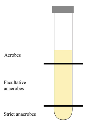
Aerobes. Need oxygen for growth. O2 is the final electron acceptor in their metabolic pathway. You may notice growth at the top of a broth tube or agar deep tube (after stab inoculation). If someone has an open wound, aerobes may be found on (or close to) the wound’s surface.

Microaerophiles. As the name suggests (“micro” small), these organisms need a little oxygen for their growth. Too much oxygen inhibits growth or kills these types of organisms. You may notice growth towards the middle of a broth tube or agar deep tube (after stab inoculation). If someone has an open wound, microaerophiles may be found a bit below the wound’s surface.

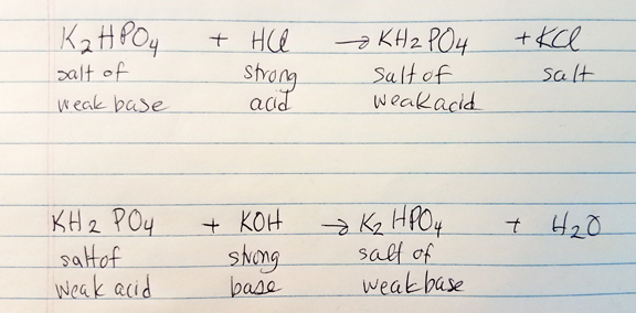
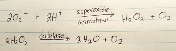
Obligate/strict anaerobes. These organisms growth without oxygen, and the presence of oxygen would be destructive (via build-up of the free radical superoxide). O2 is not the final electron acceptor. Obligate anaerobes lack the enzymes superoxide dismutase and catalase which function to breakdown superoxide to oxygen and water. You may observe growth at the bottom of a broth tube of agar deep tube when placed in a GasPak system. These organisms may reside/penetrate deep in a wound, away from the surface.

Aerotolerant anaerobes. These are fermentors and O2 isn’t the final electron acceptor. They do produce superoxide dismutase and catalase which help them “tolerate” an environment with oxygen. You may find growth below the surface of a broth tube or agar deep tube. These organisms may reside below a wound’s surface.

Facultative anaerobes. These organisms can grow with or without the presence of oxygen. Oxygen may be the final electron acceptor, but nitrate or sulfate compounds may be utilized in the absence of oxygen.

Clinical significance. It helps to narrow down the possibilities if one can determine the oxygen requirements of an unknown organism.

Cultivating anaerobes. There are several ways to cultivate anaerobes, but you can break these methods down into two groups: methods that require a sealed container (e.g. sealed jar), and methods that do not require a sealed container.

Methods using a sealed container. The container must be air-tight and there must be some way to get rid of any oxygen trapped inside the container.

Brewer jar. Uses a pump to evacuate the oxygen and replace it with 95% nitrogen and 5% carbon dioxide.

GasPak. No pump or evacuation is required. A foil package inside the jar generates hydrogen and carbon dioxide when water is added. In the lid, there’s a palladium catalyst to help hydrogen to combine with residual oxygen making the end-product, water. There’s usually a methylene blue indicator strip which is blue in the presence of oxygen and is colorless in the absence of oxygen.

Chromium-sulfuric acid. This requires the production of hydrogen via 15% H2SO4 with Cr+2. The hydrogen is evolved and O2 is evacuated.

Other methods for culturing anaerobes do not require the use of a sealed container.

Shake-culture (solid medium). While agar is in the liquid phase, it is inoculated with a loopful of organism. The tube is then shaken/agitated and rapidly cooled to a solid form, then incubated. Strict anaerobes are likely to grow at the bottom of the tube.

Pyrogallic acid (solid medium). Agar slants are inoculated. A cotton plug is placed at the upper portion of the tube (almost touching the top of the agar). Pyrogallic acid crystals are placed on top of the cotton plug, and sodium hydroxide is added. These act to pull oxygen out of the area below the cotton plug.

Paraffin plug (broth medium). A liquid reducing substance is used. Heat is applied to drive off the oxygen gas, cooled rapidly and inoculated. The tube is sealed with 1/2 inch of melted paraffin, then incubated.

Fluid thioglycollate (broth medium). Sodium thioglycollate binds to oxygen gas. A redox indicator (e.g. resazurin) may be used as a visual aid.


Reference

Cappuccino, J. G., & Welsh, C. (2018). Microbiology: A laboratory manual.

Posted on

Microbiology: Effects of pH.

The environment’s pH affects the growth of organisms. Generally, bacteria grows between pH 4-9 and optimally between pH 6.5-7.5. Fungi may be found in more acidic environments between a pH of 4-6.

Acidophiles: grow between pH 0-7 and optimally around pH 3.5.

Neutrophiles: grow between pH 3.5-10.5 and optimally around pH 7.

Alkalophiles: grow between pH 7-14 and optimally around pH 10.5.

The by-products of an organism’s metabolism may cause pH of the environment to shift (e.g. more acidic or more basic). A build-up of by-products could inhibit or be detrimental to the organism’s growth.

A buffer system (salt of a weak acid and salt of a weak base) may be added to the growth medium to help counter slight shifts in pH.

Clinical significance. Organisms can grow at human body temperature. Many organisms cannot tolerate the acidity of the stomach (pH 2.0). The skin is also a barrier as its pH is around 4-7. The varying pH of different systems in the body act as a stopgap to help prevent infections.

Reference

Cappuccino, J. G., & Welsh, C. (2018). Microbiology: A laboratory manual.

Posted on

Microbiology: Effects of temperature.

Different organisms grow best at different temperature ranges. Temperature ranges (cardinal temperature points) may be described by three points (the names are self-descriptive): minimum growth temperature, maximum growth temperature, and optimum growth temperature.

Psychrophiles: grow well at the lower end roughly between -5-20 °C with the optimum temperature around 12.5 °C.

Mesophiles: may be found “in the middle” of the temperature scale, roughly between 20-45 °C. They can grow at body temperature, 37 °C. Plant saprophytes grow between 20-30 °C.

Thermophiles: may be found at the higher end, roughly between 40-70 °C (but may exceed 70 °C). Facultative thermophiles can grow at 37 °C with optimal temperatures between 45-60 °C. Obligate thermophiles grow above 50 °C with optimum temperatures above 50 °C.

Clinical significance. Cold temperature (such as in refrigerators or freezers) help to decelerate growth (or even inhibit growth via enzymatic metabolism disruption) of bacteria. However, some bacteria can survive such as Listeria monocytogenes which causes flu-like symptoms.

Reference

Cappuccino, J. G., & Welsh, C. (2018). Microbiology: A laboratory manual.

Posted on

Microbiology: Capsule staining, part 7.

Capsule staining (Anthony method): is atypical and cells that have a gelatinous capsule (complex polysaccharides, levans, dextrans, cellulose) are often by nature virulent. Capsule staining is difficult because the capsule is water-soluble—easy to washout stain or destroy the capsule. There are 2 reagents.

The primary stain is crystal violet (stains cell and capsule).

The decolorizing agent is copper sulfate (20%) which takes out the primary stain from the capsule but does not remove color bound to the cell wall. Copper sulfate will also bind to the capsular material.

Clinical significance. Helps to identify capsule-forming Gram-negative bacteria such as Haemophilus influenzae and Klebsiella pneumoniae. Capsule-forming Gram-positive bacteria include Bacillus anthracis and Streptococcus pneumoniae.

Summary of procedure.

  1. On a clean slide, put several drops of crystal violet.
  2. Inoculate the slide (with crystal violet) with 3 loopfuls of a culture.
  3. Using another clean slide, smear the first slide such that the inoculated crystal violet spreads thinly across.
  4. Allow this to stand for 5-7 minutes such that it is dry.
  5. Do not heatfix.
  6. Gently flush smear with copper sulfate solution.
  7. Gently blot with bibulous paper.

Reference

Cappuccino, J. G., & Welsh, C. (2018). Microbiology: A laboratory manual.

Posted on

Microbiology: Spore staining, part 6.

Spore staining (Schaeffer-Fulton method): is helpful when other conventional methods won’t work. Some organisms (members of Cloistridium, Desulfotomaculum, and Bacillus) may exist as vegetative cells or spores in order to survive a harsh environment.

Sporogenesis allows an intracellular endospore to develop which is surrounded by layers of spore coats (very tough layers to protect endospore). As the external conditions worsen, the vegetative cell may release the endospore which allows the endospore to become an independent free spore. When external conditions become more favorable, the free spore may revert back (germination) to a vegetative cell.

Spore staining uses 2 reagents

The primary stain is malachite green (with heat). This will color the spores.

The decolorizing agent is water. This will decolorize the vegetative cells.

The counterstain is Safranin which will colorize vegetative cells red.

Clinical significance. Helps to identify members of Cloistridium, Desulfotomaculum, and Bacillus such as Bacillus anthracis (anthrax) and Cloistridia bacteria (tetanus, gas gangrene, food poisoning and pseudomembranous colitis).

Summary of procedure.

  1. Prepare a smear and heatfix.
  2. Flood slide with malachite green and place over a beaker of warm water (on a hotplate).
  3. Steam for 2-3 minutes.
  4. Cool slide then gently flush with deionized water.
  5. Flood slide with Safranin for 30 seconds.
  6. Gently flush slide with deionized water.
  7. Blot dry with bibulous paper.

Reference

Cappuccino, J. G., & Welsh, C. (2018). Microbiology: A laboratory manual.

Posted on

Microbiology: Acid-Fast stains, part 5.

Acid-Fast stains: are particularly helpful when the organism’s cell wall is thick and waxy (lipoidal)—resisting the other (more conventional) staining methods. Acid-Fast staining requires 3 reagents: the primary stain; the decolorizing agent; and the counterstain.

The primary stain is carbol fuchsin— dark red, 5% phenol. Carbol fuchsin is lipid-soluble which allows it to penetrate the thick lipoidal cell wall. Heat (e.g. steam from water bath) helps to facilitate penetration (Ziehl-Neelson method). Instead of heat, one may use a wetting agent (Tergitol) in the Kinyoun method.

The decolorizing agent is an acid-alcohol of 3% HCl and 95% ethanol. Before using the decolorizing agent, the slide must cool in order for the waxy lipoidal wall to harden and retain the primary stain better. When the decolorizing agent is applied, the color is removed from those non-lipoidal structures or those structures that did not retain the primary stain well.

The counterstain is methylene blue which stains all of the decolorized structures (non acid-fast) blue.

Clinical significance. Helpful in detecting members of the Mycobacterium which are especially pathogenic such as M. tuberculosis and M. leprae.

Summary of procedure.

  1. Prepare a smear and heatfix.
  2. Warm a beaker of water on a hotplate.
  3. Flood the slide with carbol fuchsin and place slide over the beaker. Alternatively, flood smear with Tergitol and let it stand for 5-10 minutes (then skip to step 6).
  4. Steam for 5 minutes. Do not let stain evaporate—you may add more carbol fuchsin during this process. Do not let water boil or get too hot (you will blow your slide).
  5. Remove slide from the steam, and let slide cool.
  6. Gently flush slide with deionized water.
  7. Decolorize by sparingly adding the decolorizing agent drop by drop until the run-off is a pale tinge of red (almost clear looking). Do not overdo this!
  8. Gently flush slide with deionized water.
  9. Counterstain using methylene blue for 2 minutes.
  10. Gently flush slide with deionized water.
  11. Blot with bibulous paper.

Reference

Cappuccino, J. G., & Welsh, C. (2018). Microbiology: A laboratory manual.

Posted on

Microbiology: Differential stains, part 4.

Differential stains: are more complex, using 4 reagents (the order is important), and help to separate organisms/cells; and/or help to visualize structures (organelles) such as flagella, capsule, spore, or nucleus.

The primary stain is the first reagent and its purpose is to colorize all cells.

The mordant is the second reagent which helps to increase the contrast and color intensity of the primary stain.

The decolorizing agent is the third reagent. Depending on what you’re trying to stain, the decolorizing agent selectively (e.g. the cell or cellular structures/organelles) removes the color from the primary stain.

The counterstain is the fourth reagent and is a contrasting stain to the primary stain. The counterstain will add color to that which the decolorizing agent removed. The counterstain will not bind to those cells/structures that retained the primary stain).

Clinical significance. The Gram stain is the predominant differential stain—especially used in the clinical setting. It quickly assesses organisms that fall into one of two categories—Gram positive (purple) and Gram negative (pink/reddish). Please visit this link for more details on the Gram stain.

Summary of procedure.

  1. Primary stain.
  2. Mordant.
  3. Decolorizing agent.
  4. Counterstain.

Please visit this link for more details on the Gram stain.

Reference

Cappuccino, J. G., & Welsh, C. (2018). Microbiology: A laboratory manual.