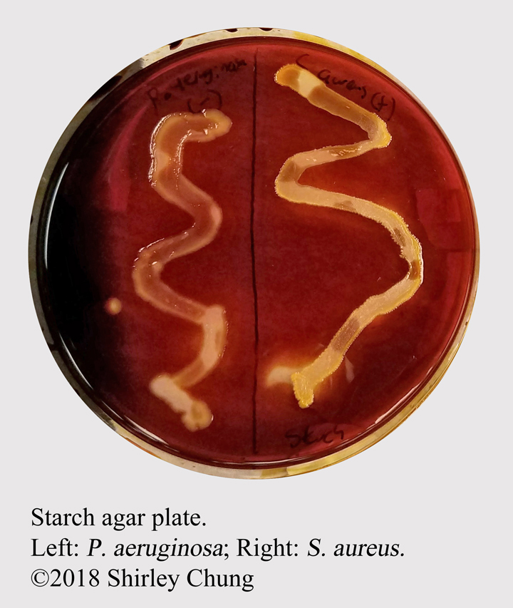
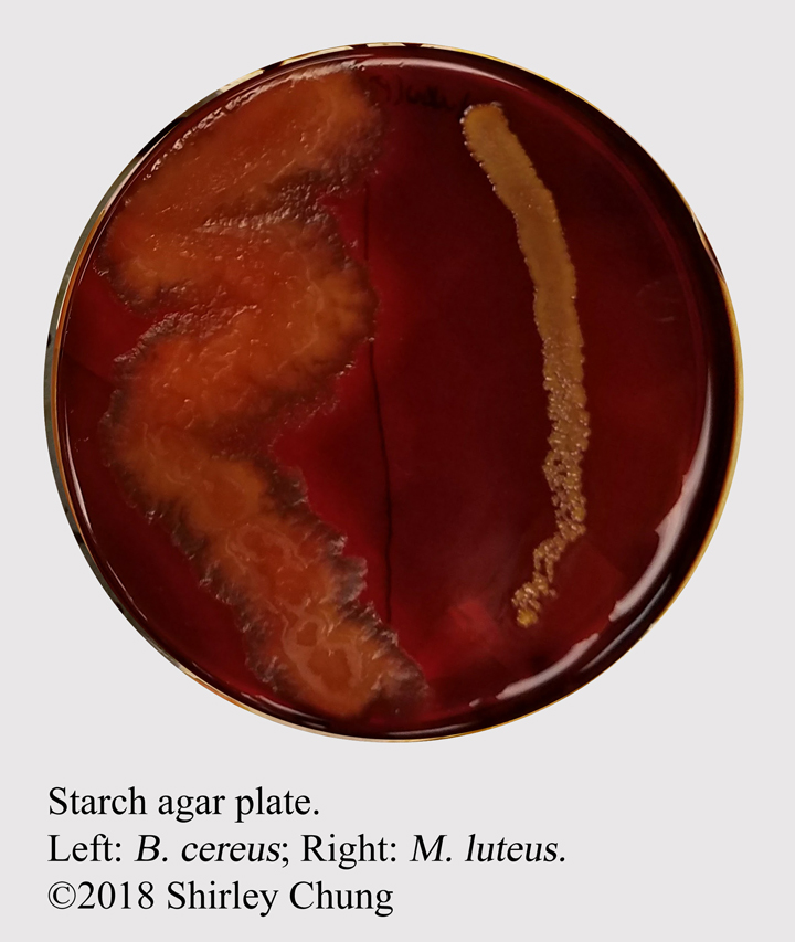
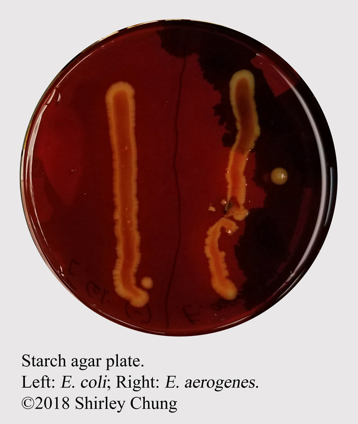
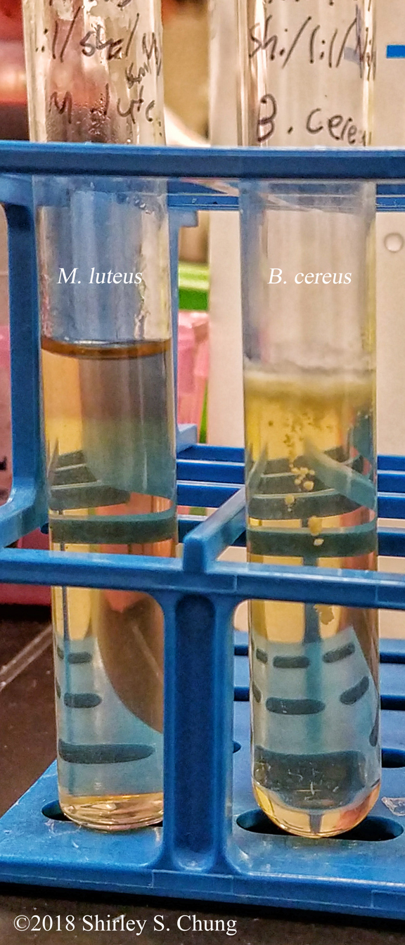
Starch hydrolysis.
| Starch Hydrolysis | |
|---|---|
| E. coli | - |
| E. aerogenes | - |
| P. aeruginosa | - |
| S. aureus | - |
| M. luteus | - |
| B. cereus | + |
Reference
Cappuccino, J. G., & Welsh, C. (2018). Microbiology: A laboratory manual.
| Starch Hydrolysis | |
|---|---|
| E. coli | - |
| E. aerogenes | - |
| P. aeruginosa | - |
| S. aureus | - |
| M. luteus | - |
| B. cereus | + |
Reference
Cappuccino, J. G., & Welsh, C. (2018). Microbiology: A laboratory manual.
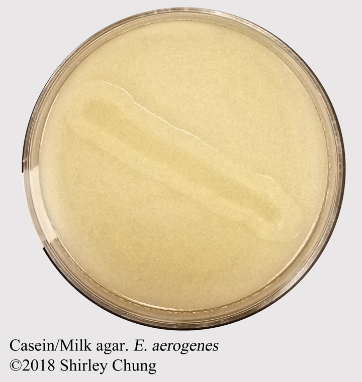
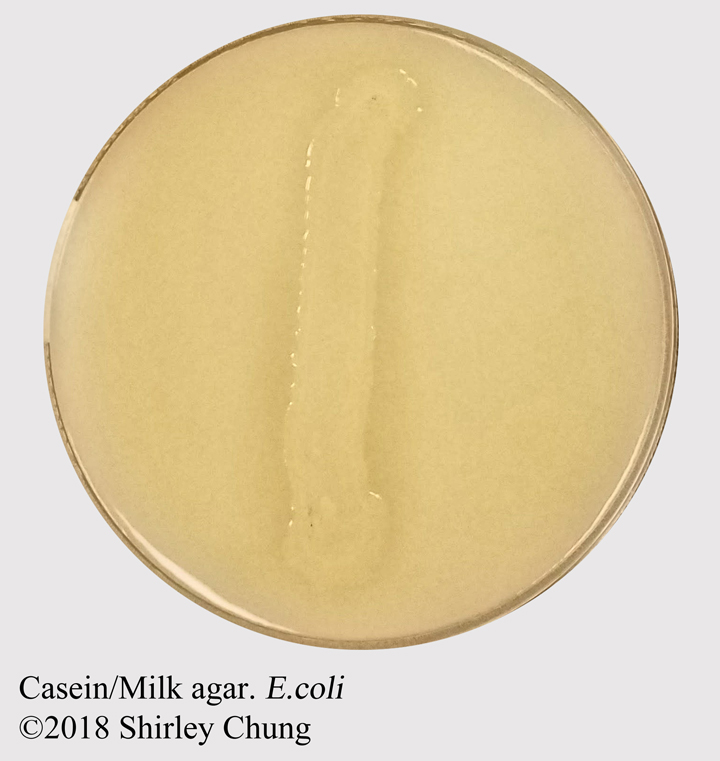
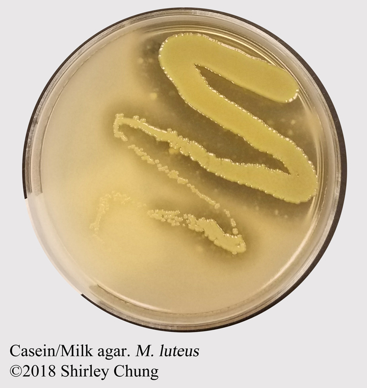
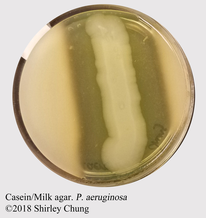
| Milk Agar | |
|---|---|
| E. coli | - |
| E. aerogenes | - |
| P. aeruginosa | + |
| S. aureus | - |
| M. luteus | + |
| B. cereus | + |
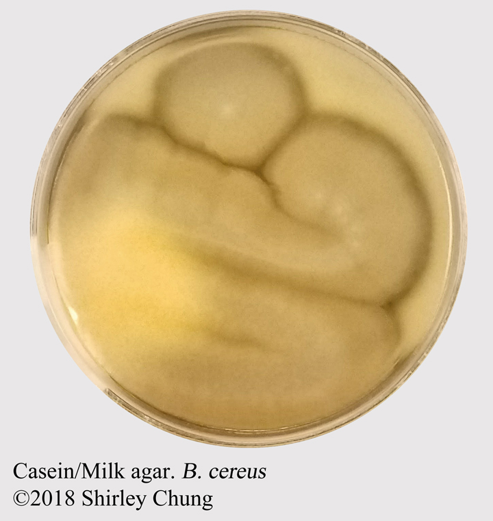

Reference
Cappuccino, J. G., & Welsh, C. (2018). Microbiology: A laboratory manual.
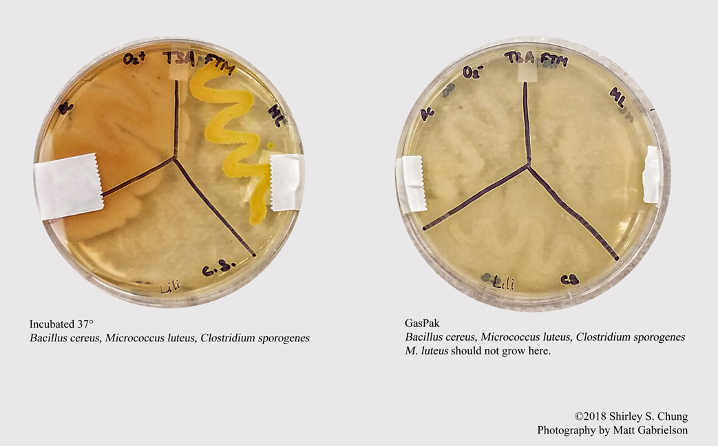
| FTM | -O2 | +O2 | Oxygen Requirements |
|
|---|---|---|---|---|
| M. luteus | Top | - | + | Strict aerobe |
| B. cereus | Throughout | + | ++ | Facultative aerobe |
| C. sporogenes | Below O2 layer | + | - | Strict anaerobe |

Reference
Cappuccino, J. G., & Welsh, C. (2018). Microbiology: A laboratory manual.
Full-size poster download here.
Isolates or is “selective for” certain groups/types of bacteria by incorporating chemical agents that inhibit the growth of certain organisms and promote the growth of other organisms. Selective media include (but not limited to):
Phenylethyl alcohol agar. Isolates Gram-positive organisms. Phenylethyl alcohol partially inhibits Gram-negative organisms.
Crystal violet agar. Selective for most Gram-negative organisms. Inhibits most Gram-positive organisms.
7.5% sodium chloride agar. Promotes halophilic organisms such as Staphylococcus, and is inhibitory for most others.
These types of growth media incorporate materials that aid in selection (promote/inhibit growth) and morphological differentiation. Examples are (but not limited to) MacConkey agar, Mannitol salt agar, and Eosin-methylene blue agar.
MacConkey agar. Contains bile salts and crystal violet which inhibit Gram-positive organisms, but promote the growth of Gram-negative organisms. MacConkey also contains lactose and pH indicator neutral red which distinguishes between lactose-fermentors (red) and non-lactose-fermentors (translucent). Enteric bacteria may be separated into lactose-fermentors and non-lactose-fermentors.
Mannitol salt agar. Promotes halophilic organisms (e.g. staphylococci) as this medium contains 7.5% NaCl (and inhibits most but not other organisms). The differential components are: mannitol which some staphylococci can ferment; pH indicator phenol red which detects acid produced from mannitol fermentation (creates a yellow-zone).
Eosin-methylene blue agar (Levine). Helps distinguish between enteric lactose-fermentors and non-lactose-fermentors and colon bacillus (E. coli). E. coli will appear blue-black with a green metallic sheen due to large amounts of acid by-products. E. aerogenes will make a thick mucous-looking pink colonies. Non-lactose-fermentors will appear transparent and unremarkable.
Enriched media contains generous amounts of certain types of nutrients. For example, blood agar can contain 5% sheep blood to promote growth of fastidious organisms such as Streptococcus spp. Organisms that favor blood agar demonstrate hemolysis (breakdown of heme, blood).
Gamma hemolysis. No lysis of blood; no visible change in the medium.
Alpha hemolysis. Incomplete hemolysis resulting in a greenish halo surrounding the colonies.
Beta hemolysis. Complete hemolysis resulting in a clear zone around the colonies. Streptolysin O produces hemolysis by an antigenic, oxygenlabile enzyme. Streptolysin S is a nonantigenic oxygen-stable lysin.
TSA—tryptic soy agar; MSA—mannitol soy agar; MAC—MacConkey agar; PEA—phenylethyl alcohol agar; EMB—eosin methylene blue agar; BDA—blood agar.

Reference
Cappuccino, J. G., & Welsh, C. (2018). Microbiology: A laboratory manual.
Bacillus subtilis at 1000x
Shaeffer-Fulton Method, infrared contrast filtered. Primary malachite green; secondary safranin.

Reference
Cappuccino, J. G., & Welsh, C. (2018). Microbiology: A laboratory manual.
Serratia marcescens
25 °C —Pigment prodigiosin.
37 °C —No pigmentation.

Reference
Cappuccino, J. G., & Welsh, C. (2018). Microbiology: A laboratory manual.
When designing a dichotomous key, each level represents 1 step in your logic/questioning. The “di” in the word dichotomous means “two”: yes or no. The key is like an upside down tree. At each level, ask a yes/no question. The “yes” should lead the questioning/logic in one direction/branch; the “no” should lead the questioning/logic towards another direction.
See this example.
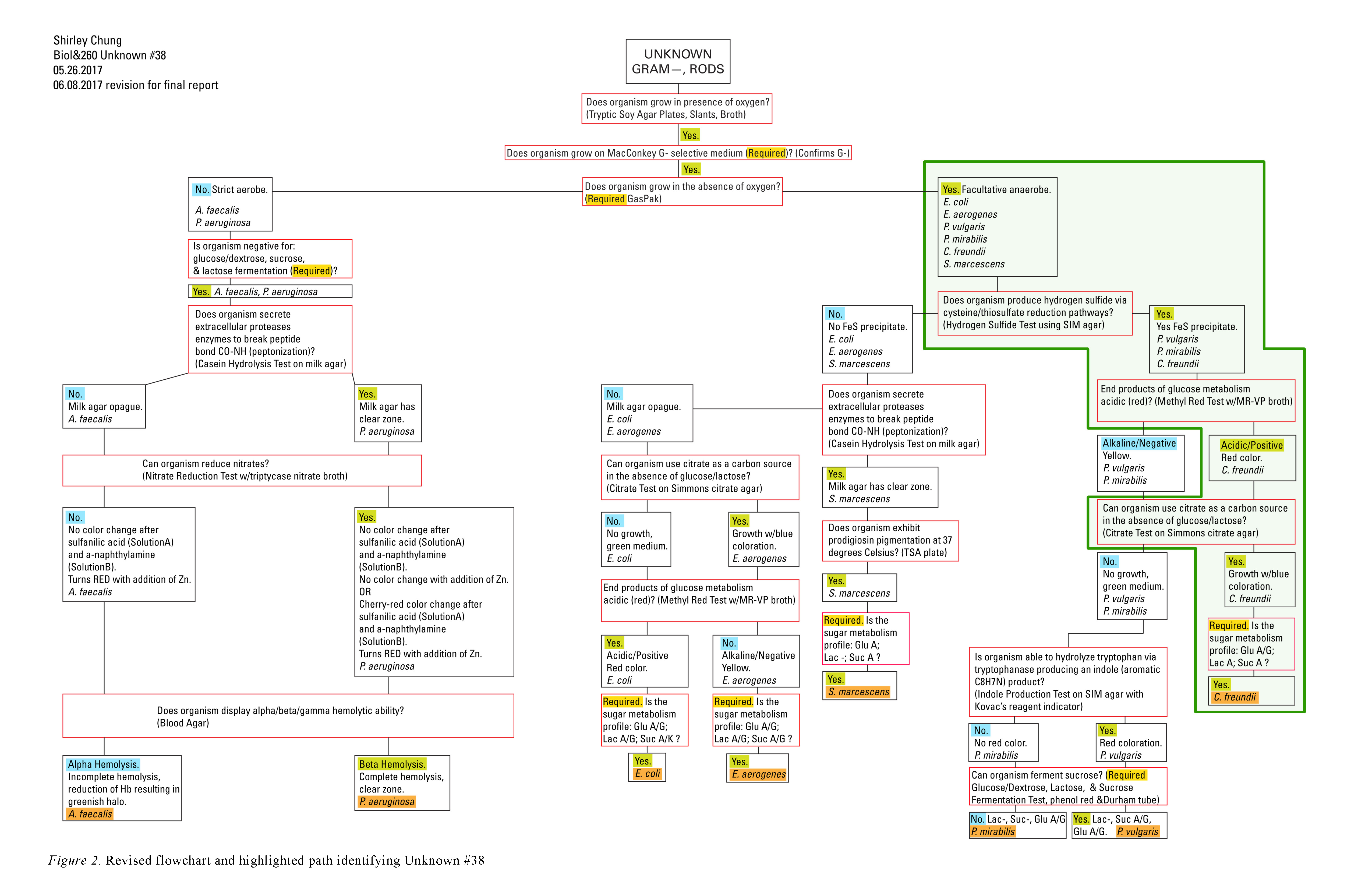
The most common microbiology project is the correct identification of a bacterium using as few tests/diagnostics as possible. This is usually an “end of quarter” project. Here are some tips (in no particular order):
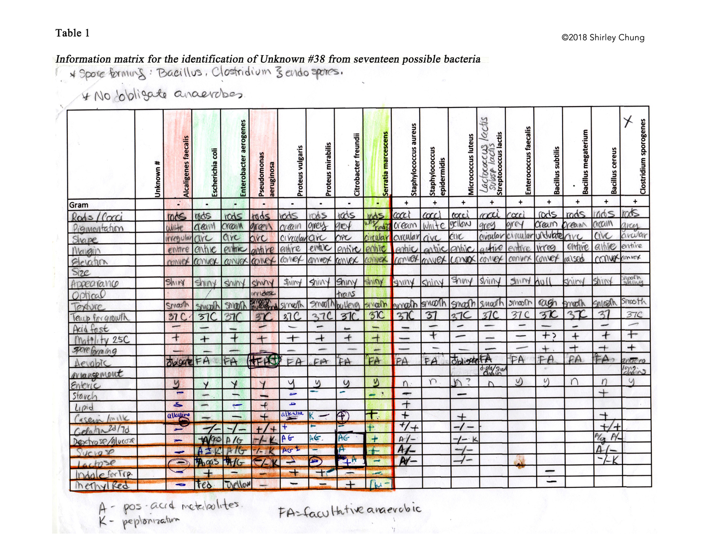


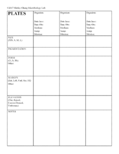
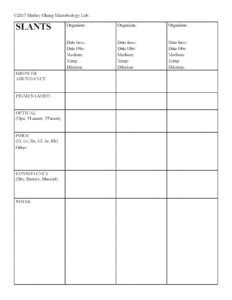
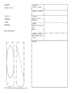
Greetings! Feel free to download (click on the thumbnails to get the PDFs) these lab forms if you find them to be helpful! I designed these while I was taking microbiology class at Green River Community College. ©2018 Shirley S. Chung


Microbiology Lab Procedure Notes By ©2018 Shirley S. Chung, Green River Community College
Carbol Fuchsin, 15-30 sec
Crystal Violet, 20-60 sec
Meth. Blue, 1-2 min
OR
Flood smear with carbol fuchsin WITH TERGITOL for 5-10 minutes
Microbiology Lab Procedure Notes By ©2018 Shirley S. Chung, Green River Community College