Posted on

Joints

Download these notes.

Anatomy & Physiology: Joints.


Structure.

  • Structural classification: based on whether or not there is space tween the articulating bones; type of connective tissue that holds the bones together.
    • Fibrous joints: no synovial cavity; dense collagen-fiber-rich connective tissue.
      • 3 types of fibrous joints: sutures, syndesmoses, & interosseous membranes.
      • Sutures: fibrous joint made of thin layer of dense connective tissue. Only occurs in skull bones. Strength from irregular and interlocking edges. In infants and young children the sutures are amphiarthrotic; in adults the sutures are fused and immoveable (synarthrotic).
        • Synostosis: suture present in infants/children but ossified in adults. Synarthrotic.
        • Frontal/metopic suture: if the suture exists past 6 yrs old.
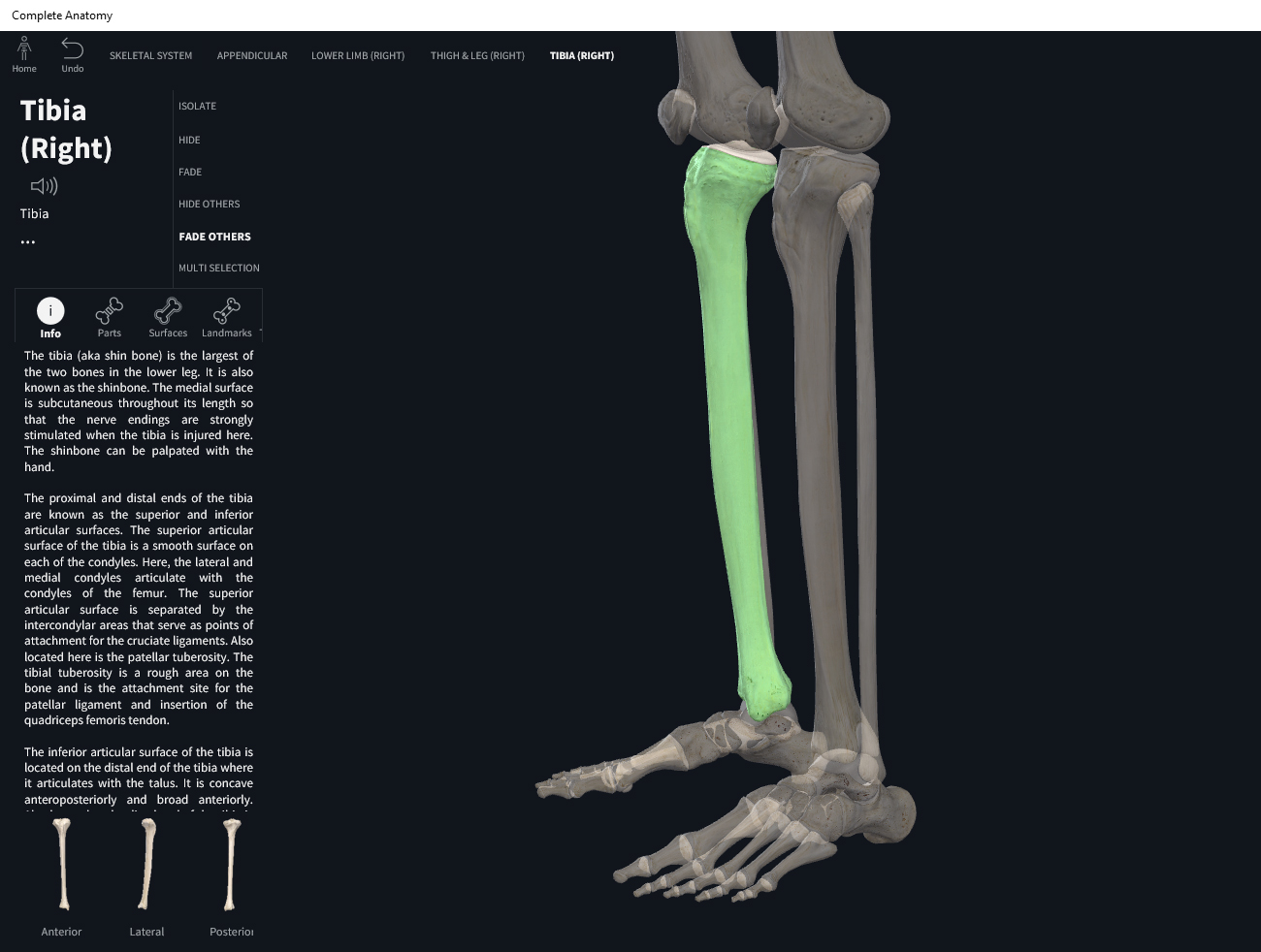
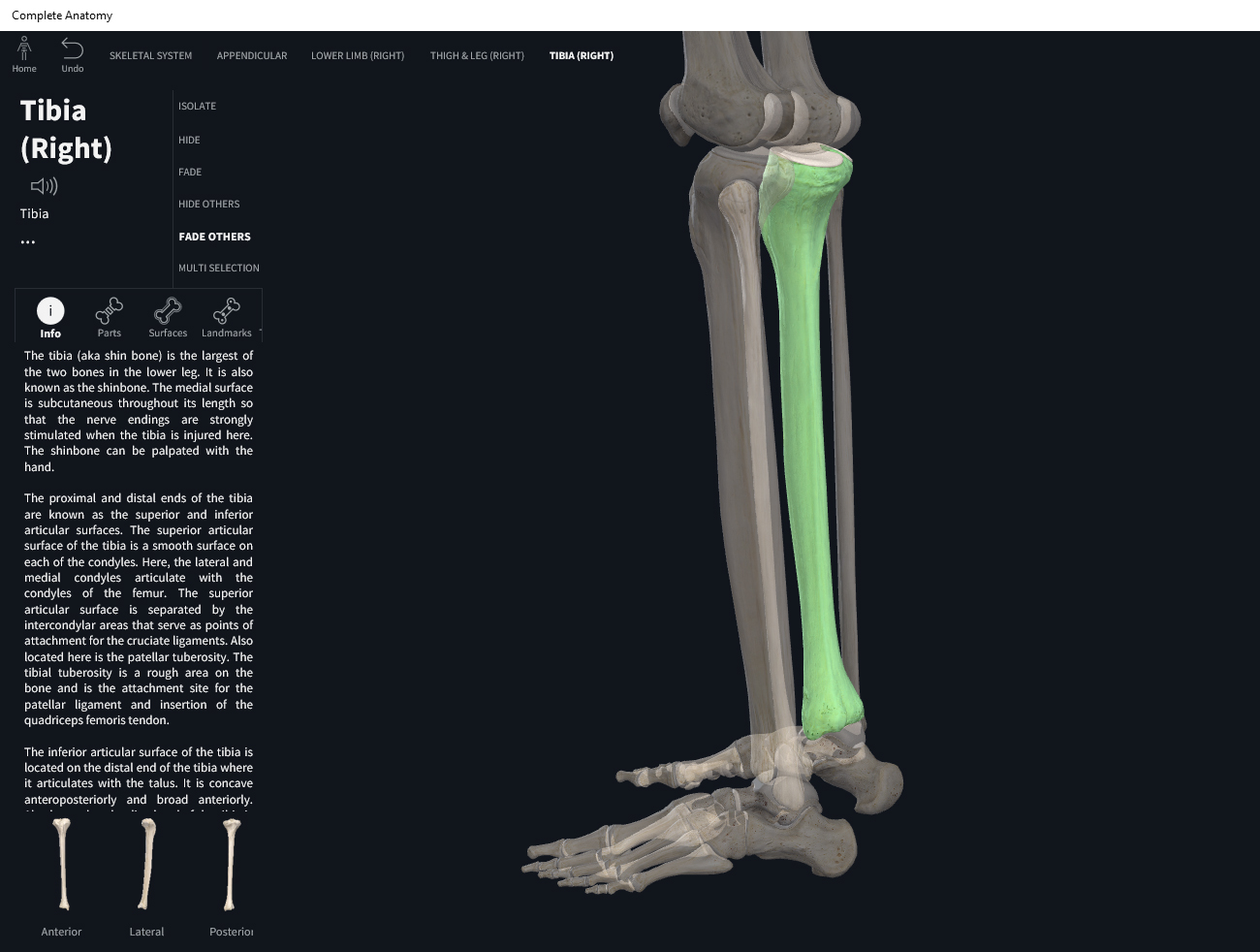
      • Syndesmoses: fibrous joint with some distance between the two bones. Dense connective tissue arranged in bundles (ligamentous) limiting the joint movement. E.g. distal tibiofibular joint.
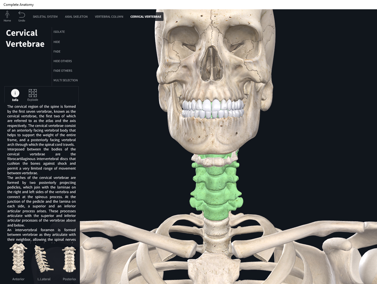
        • Gomphosis: peg fitting into a socket. E.g. teeth and teeth sockets (alveoli).
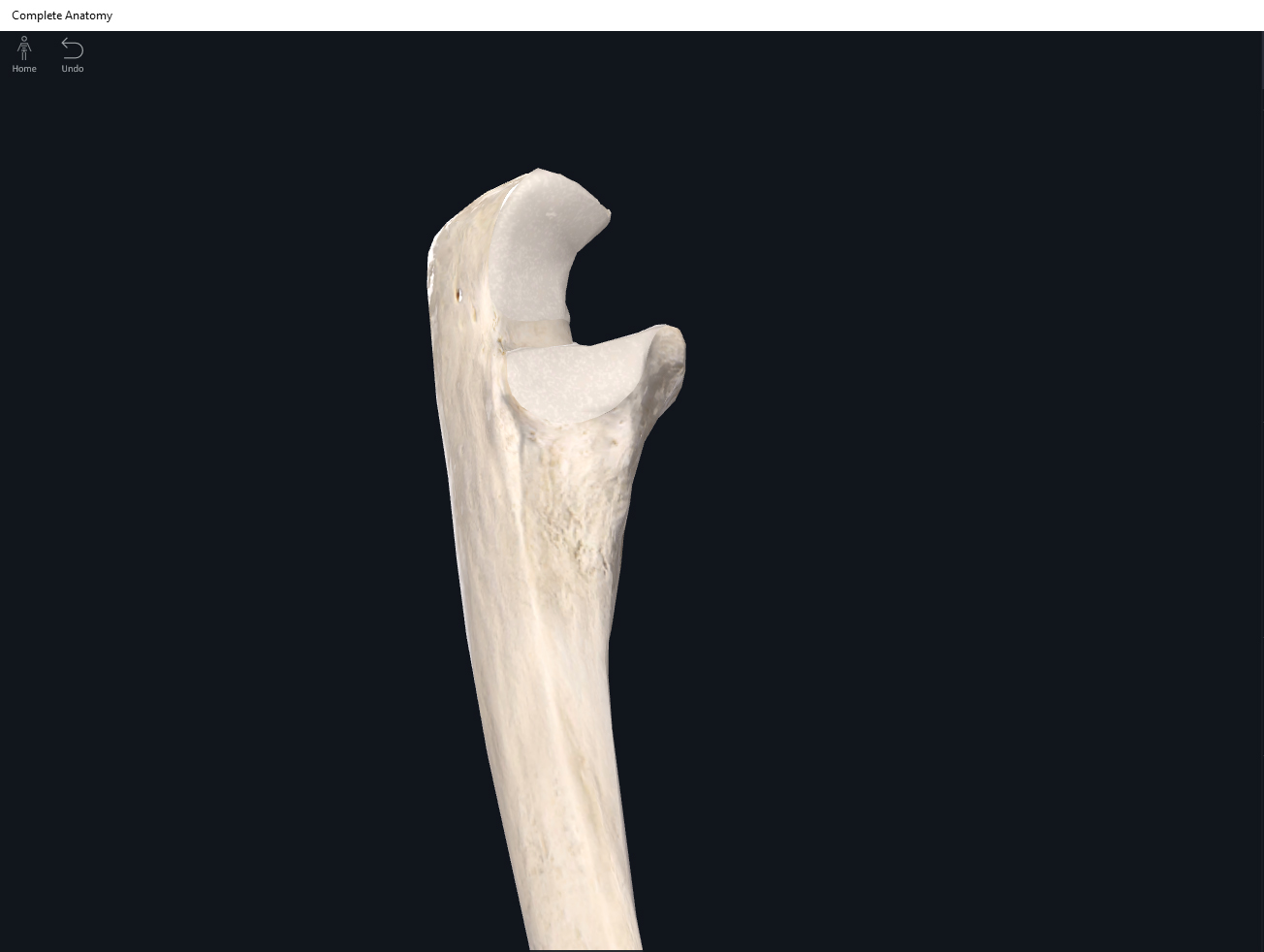
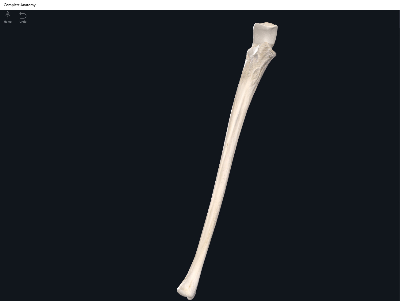
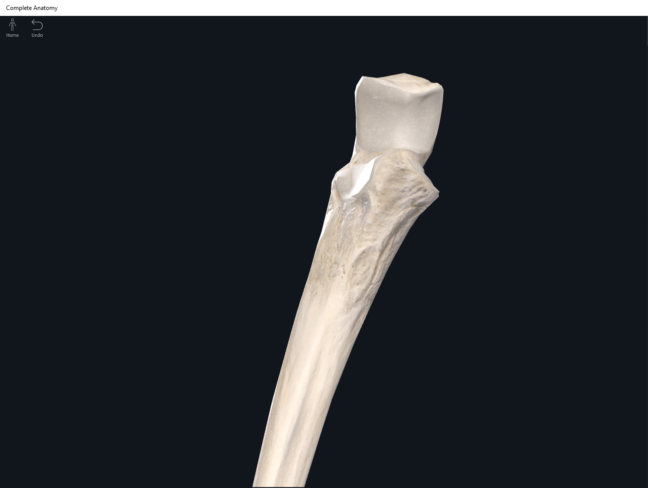
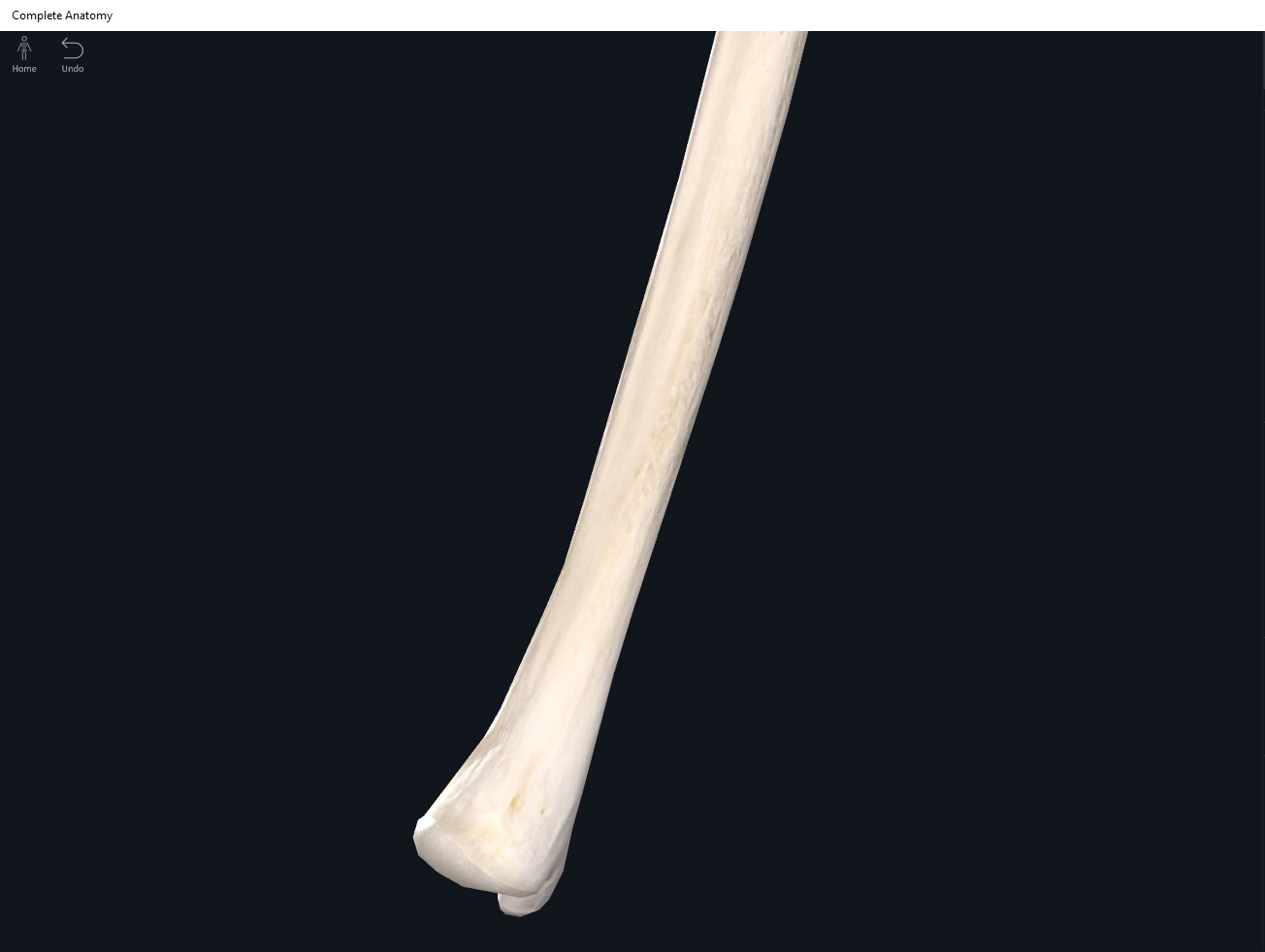
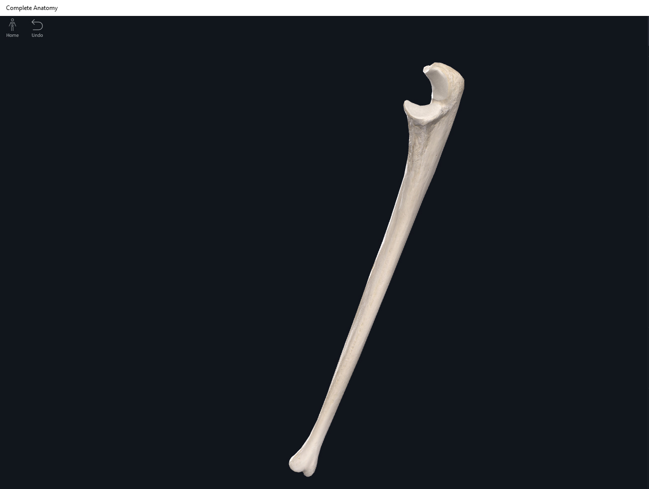
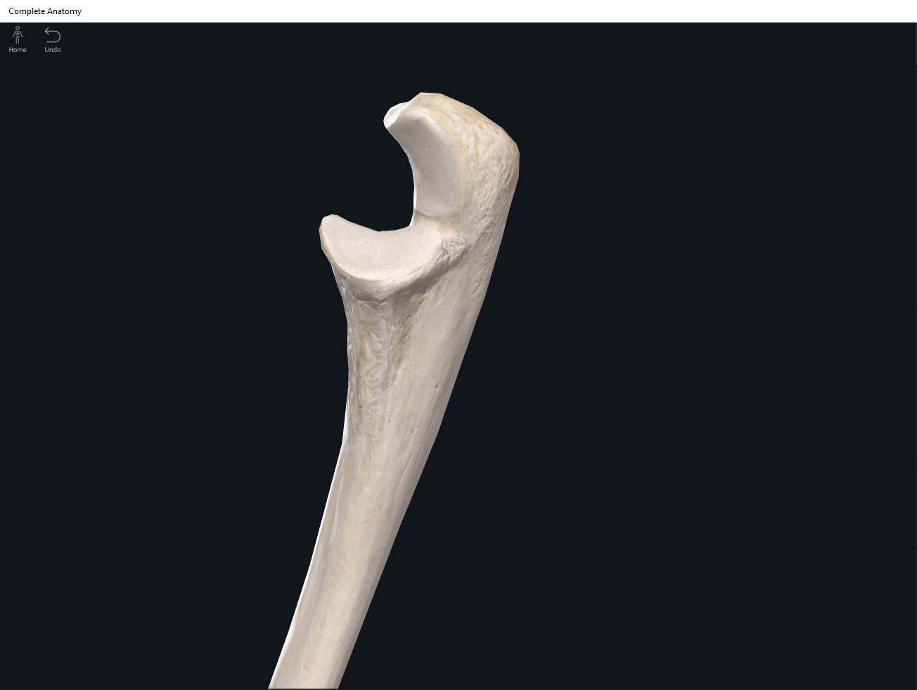
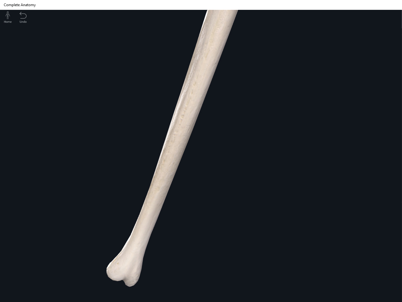
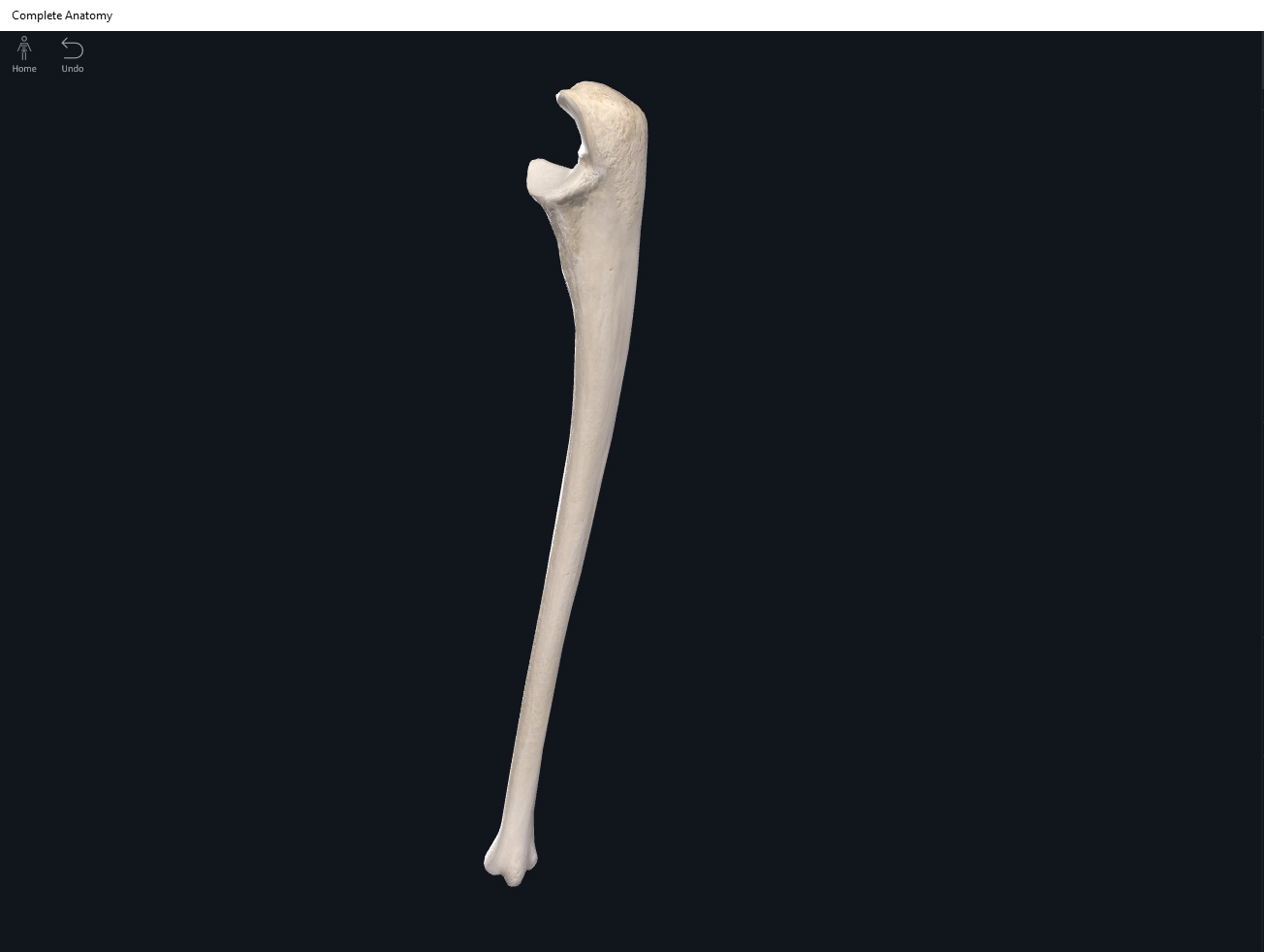
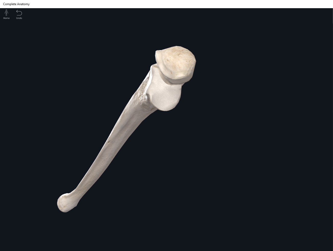
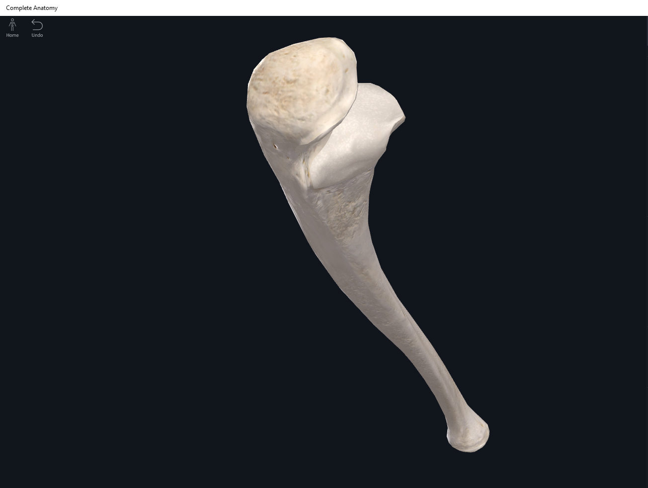
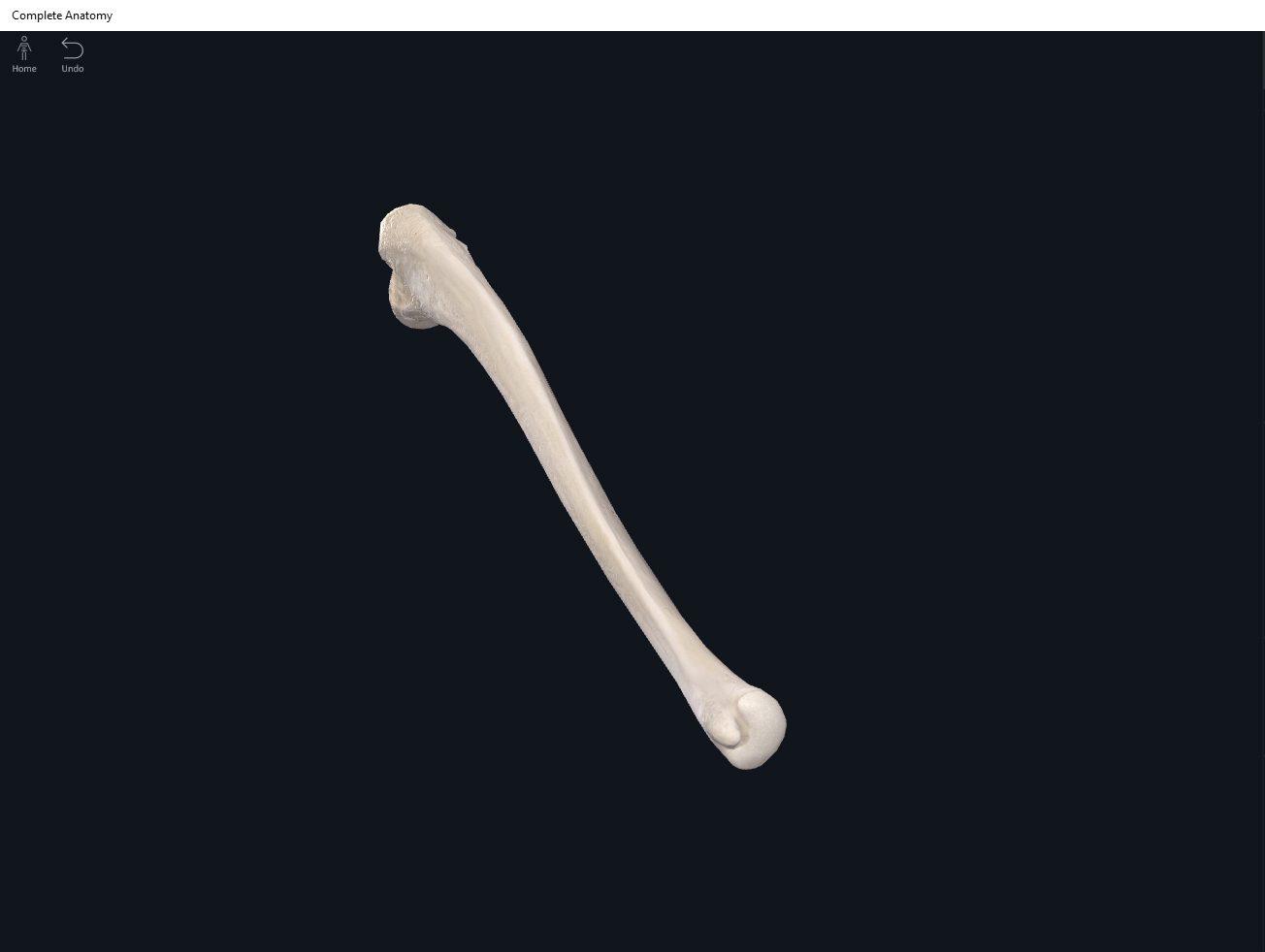
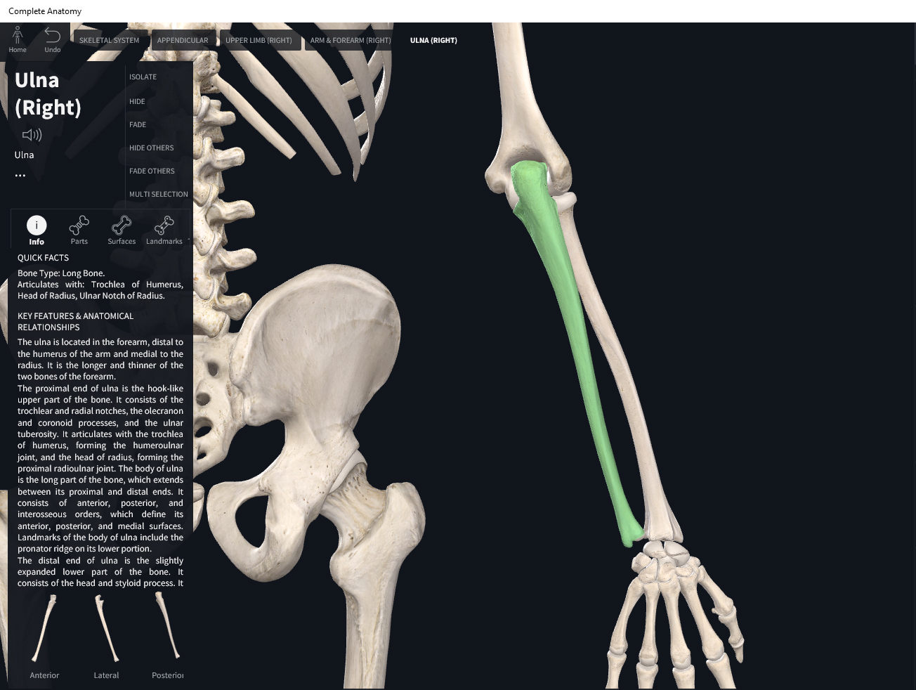
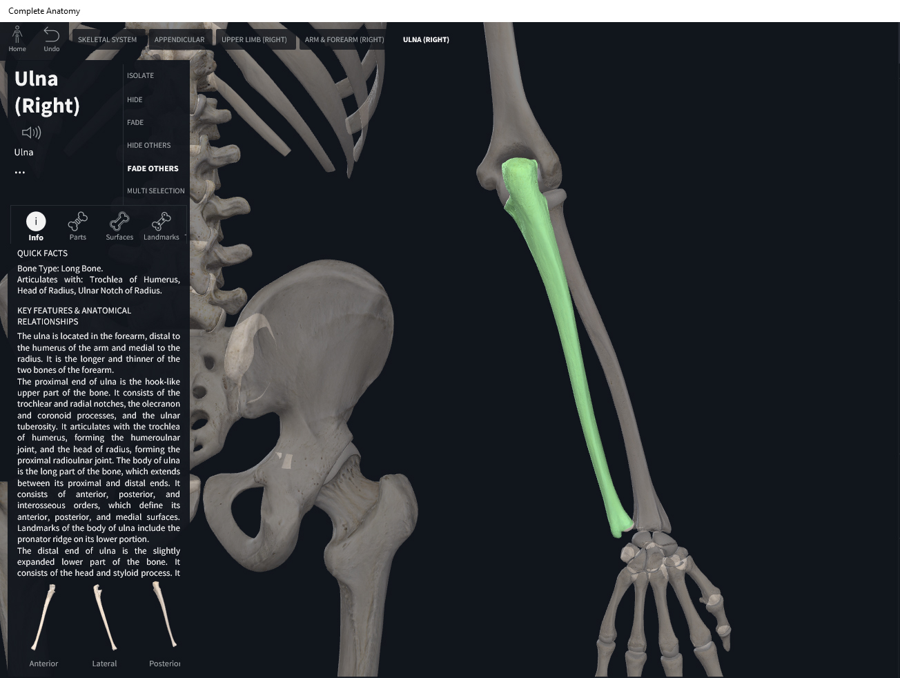
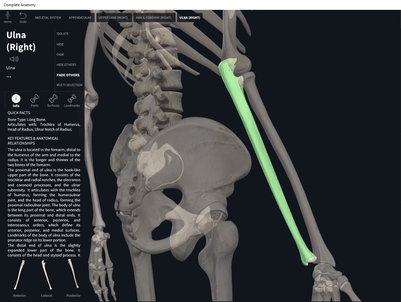
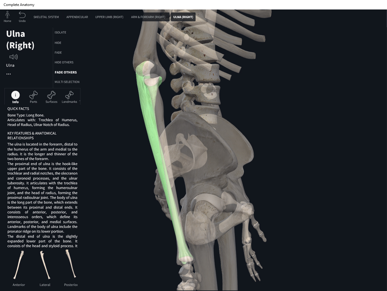
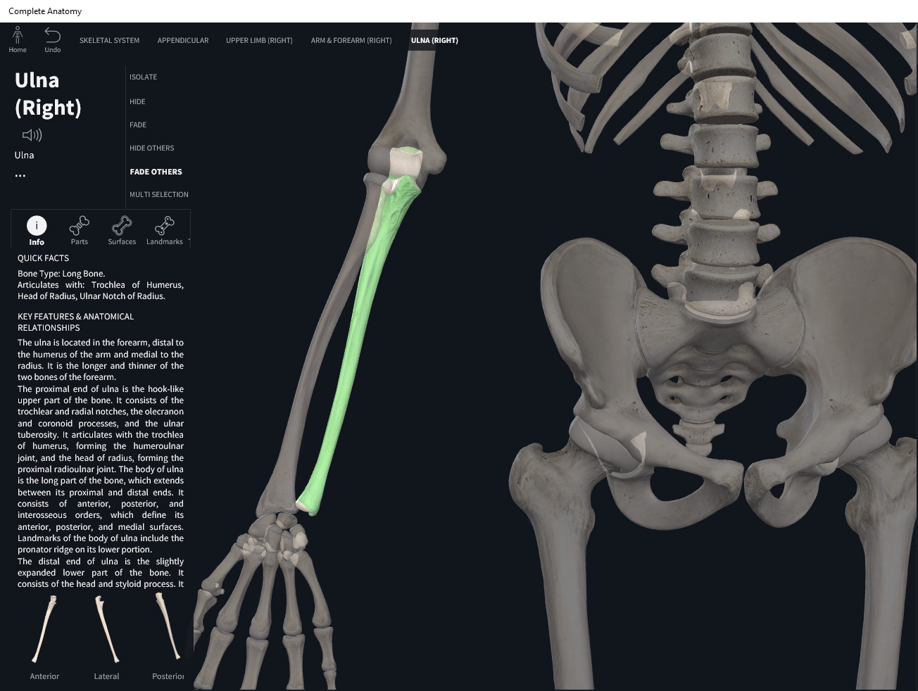
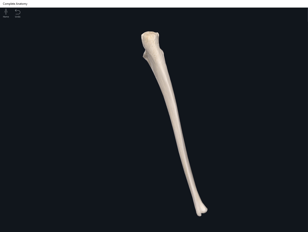
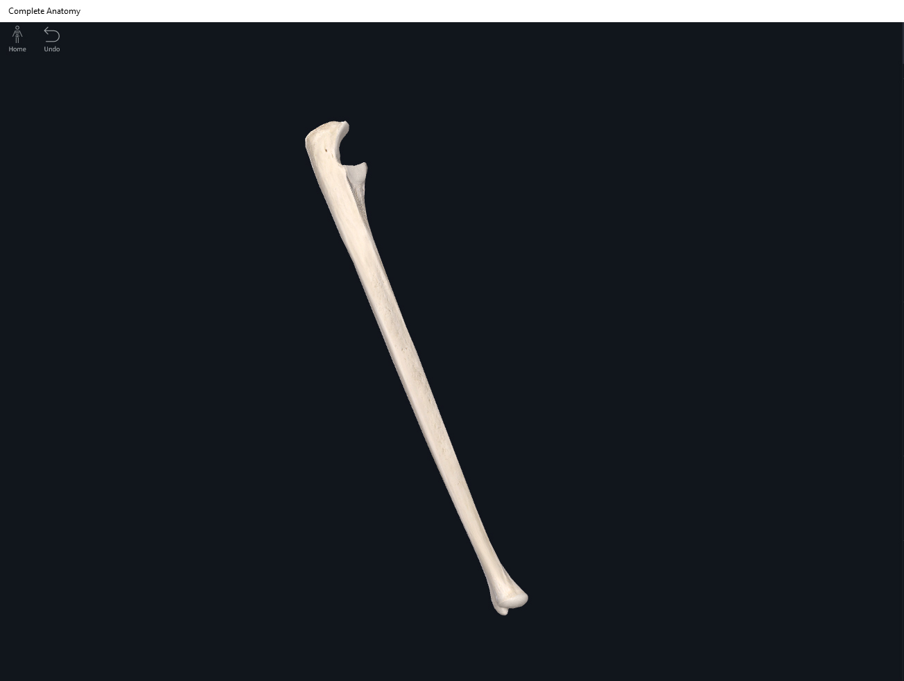
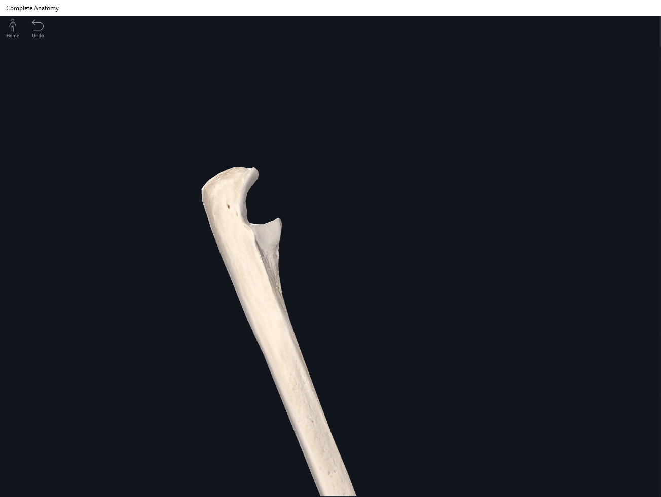
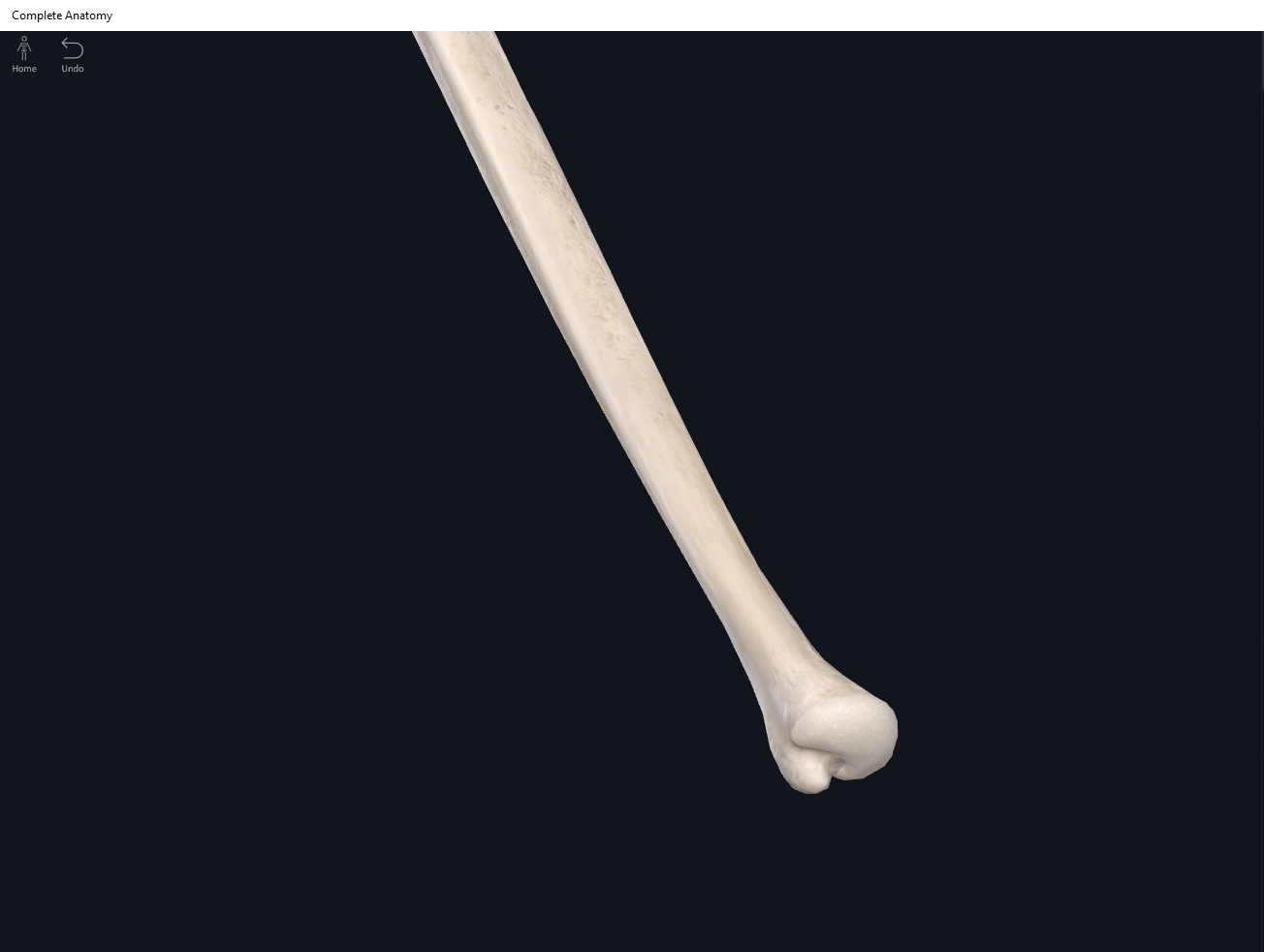
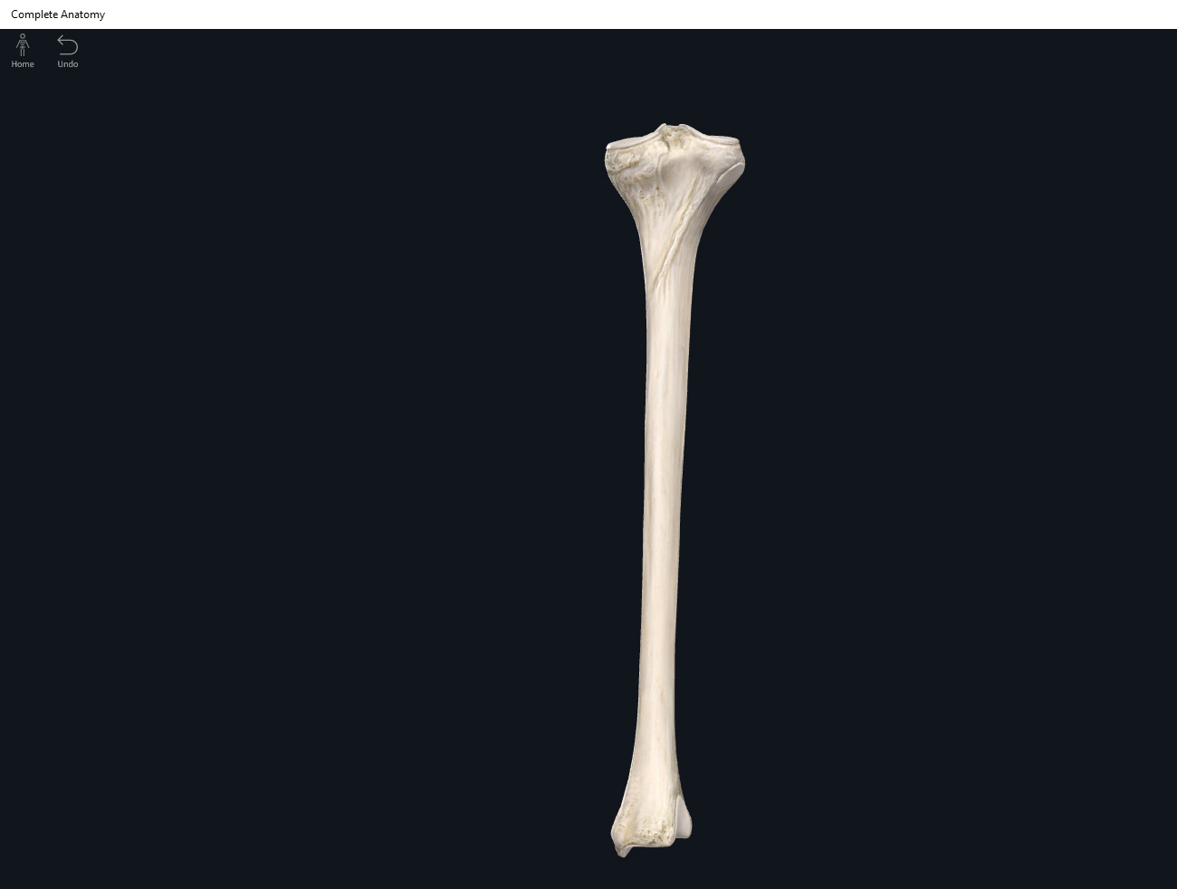
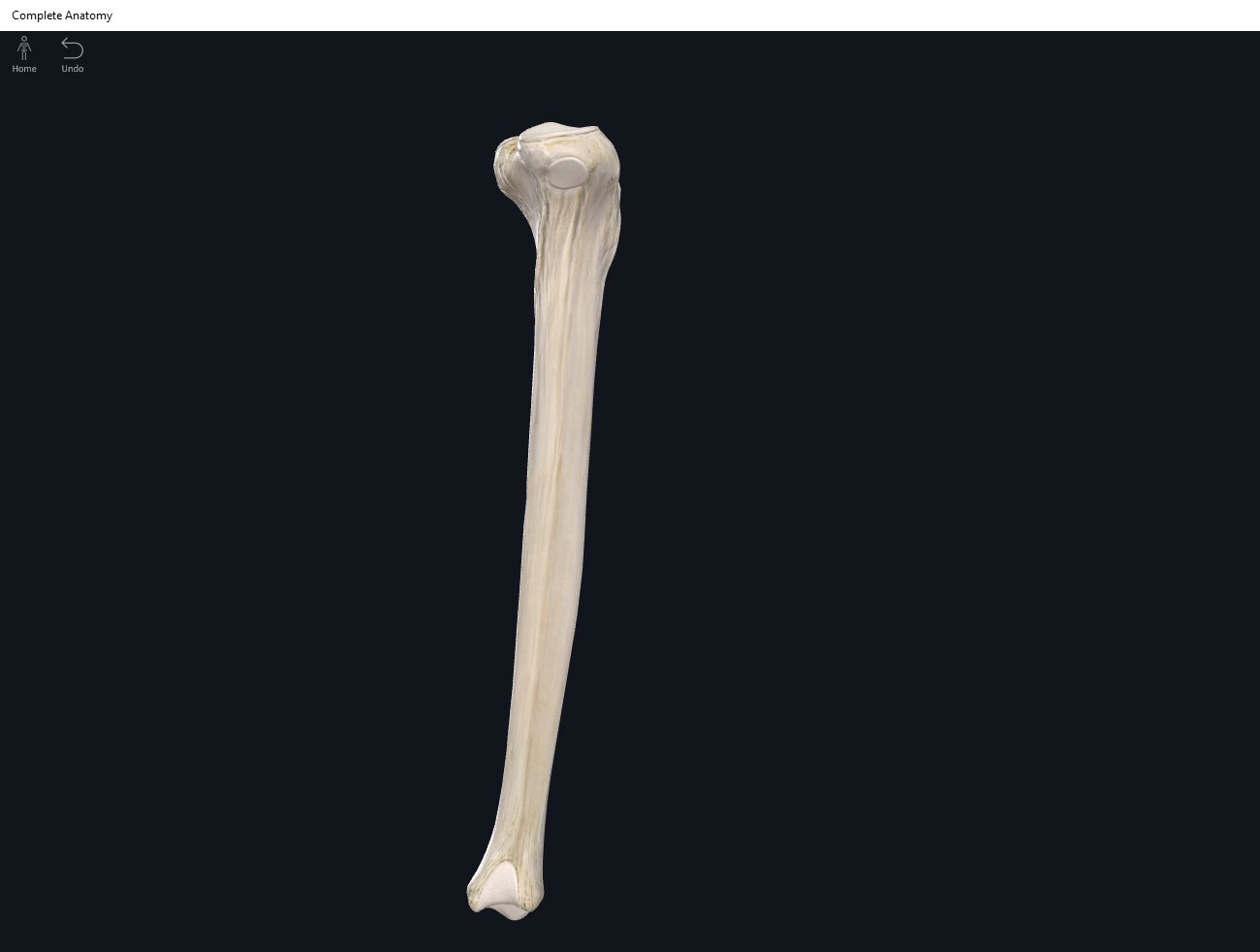
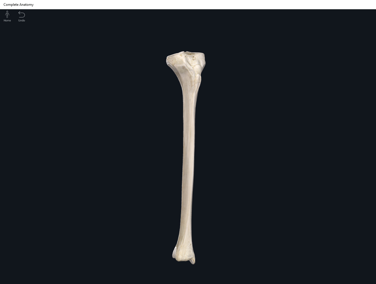
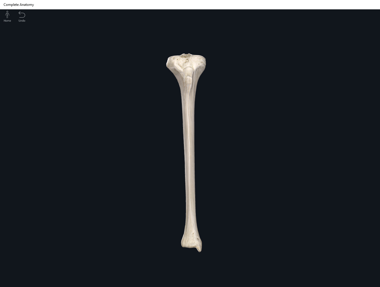
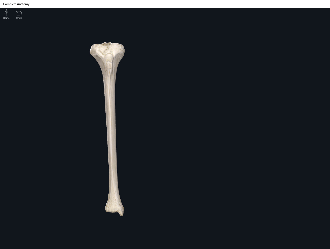
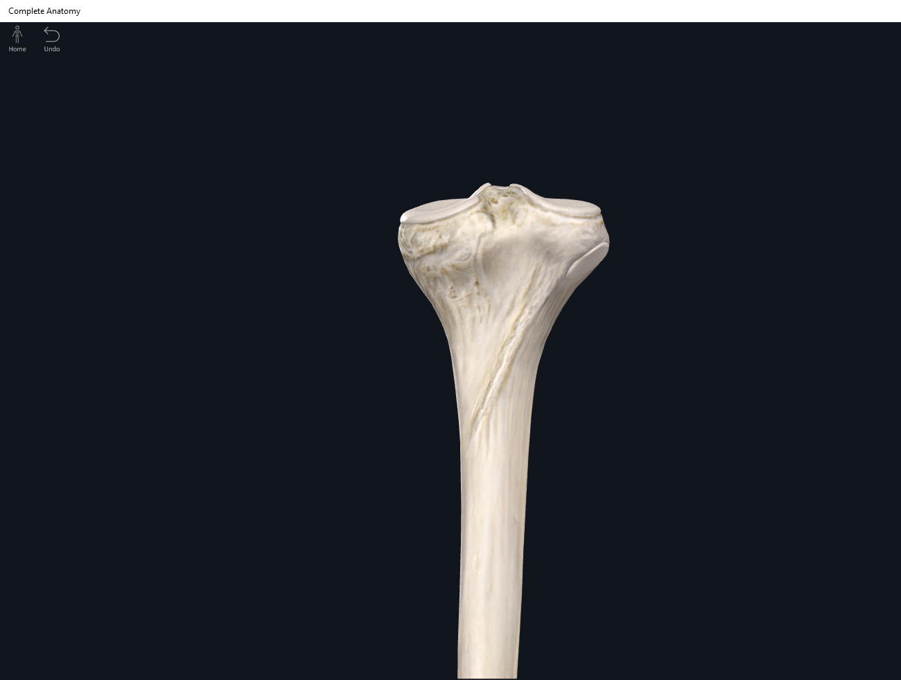
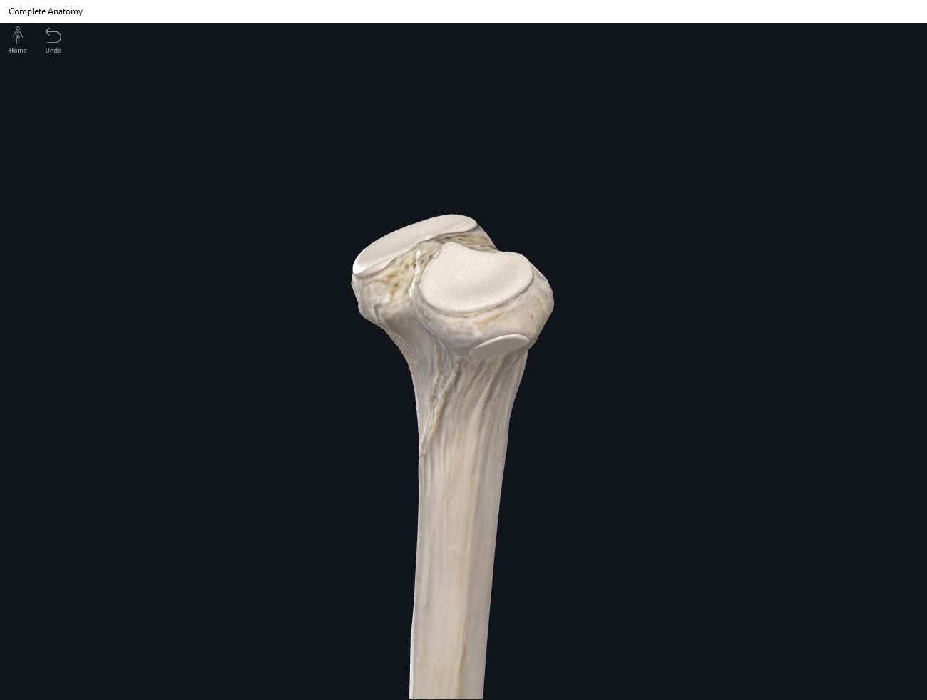
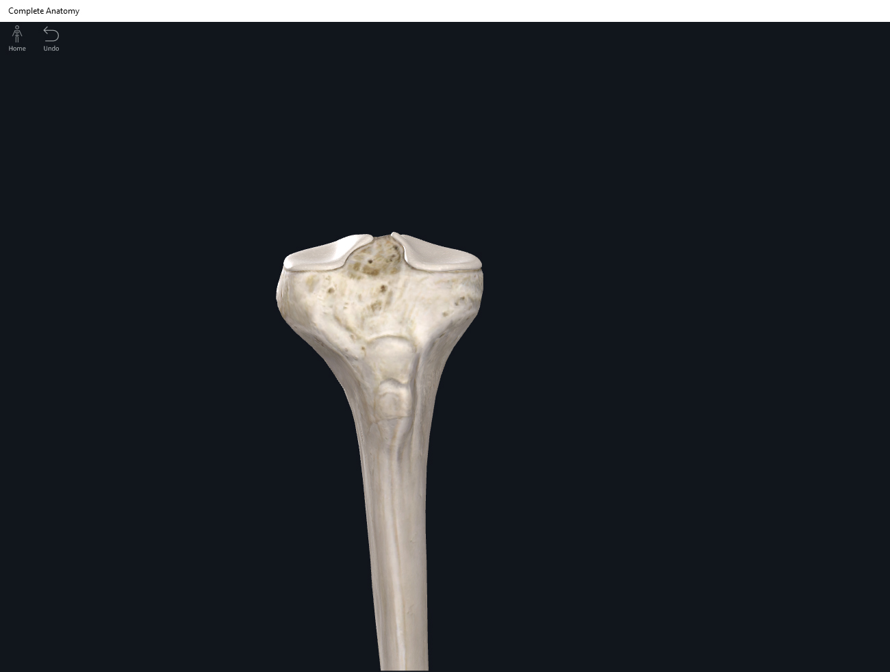
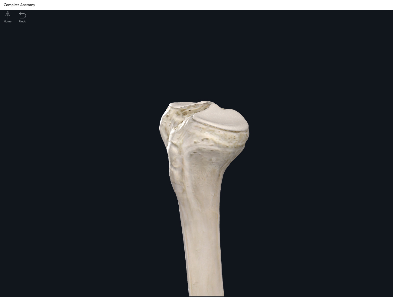
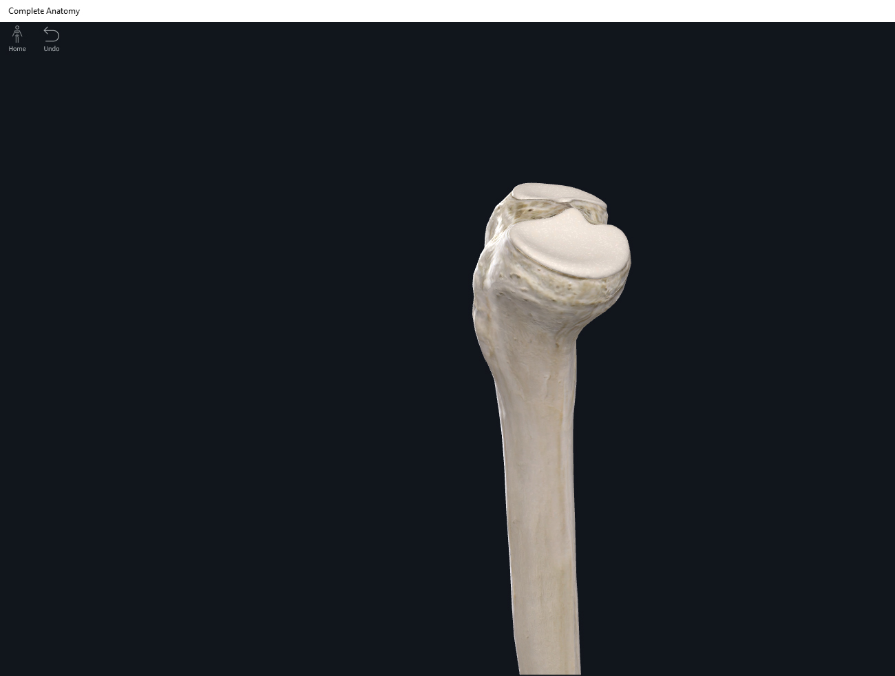
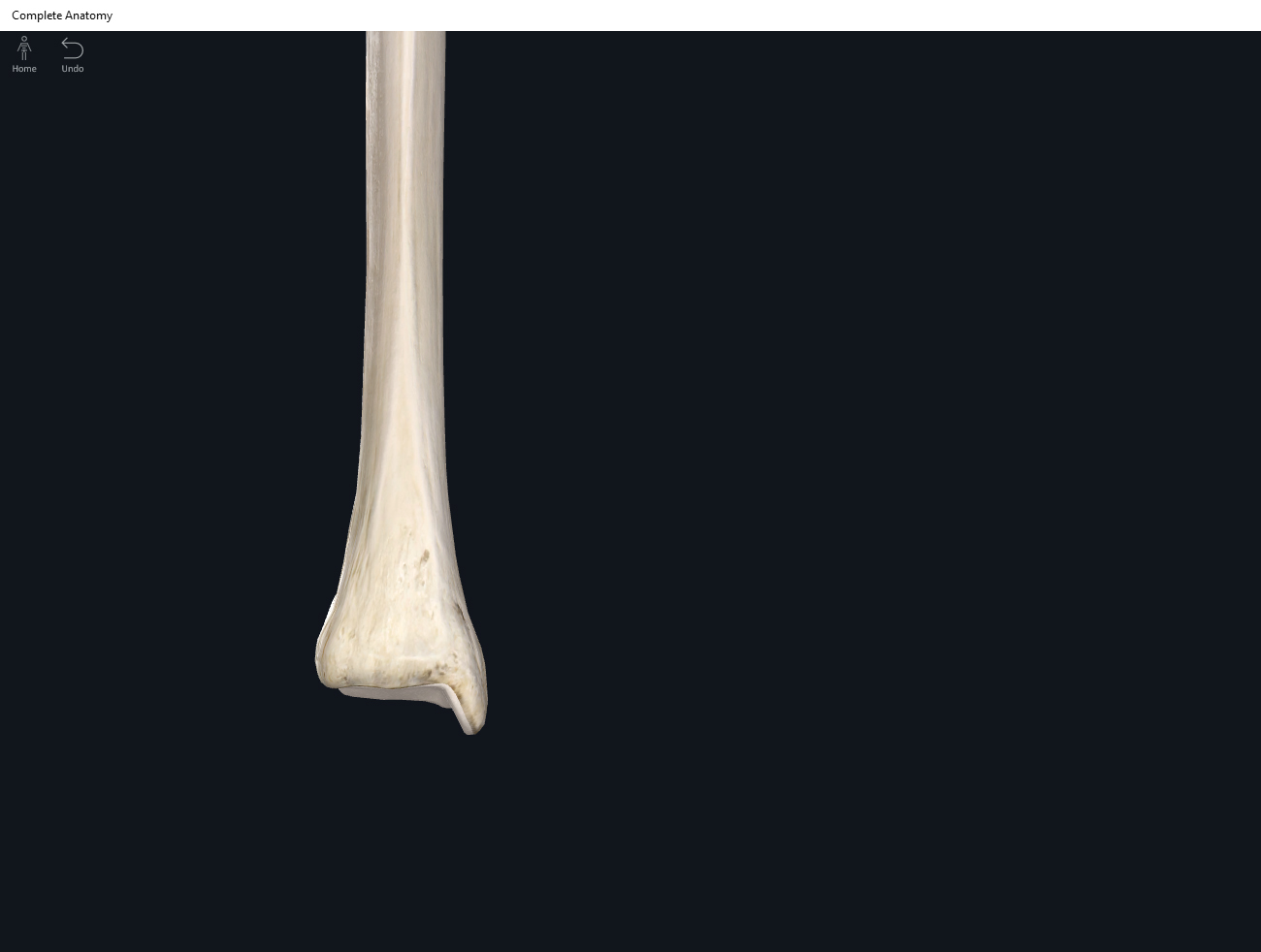
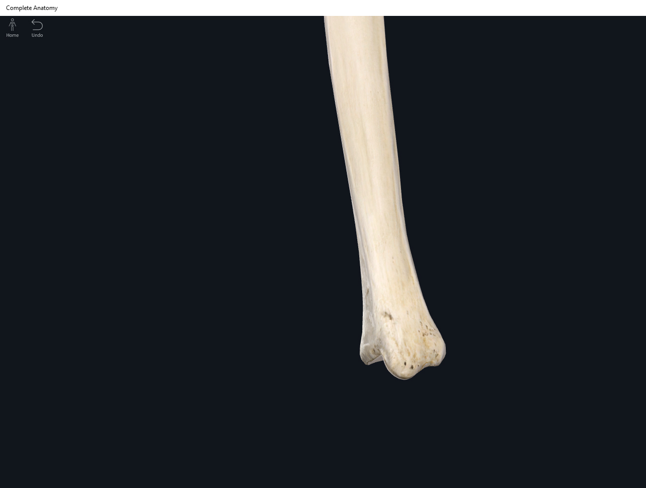
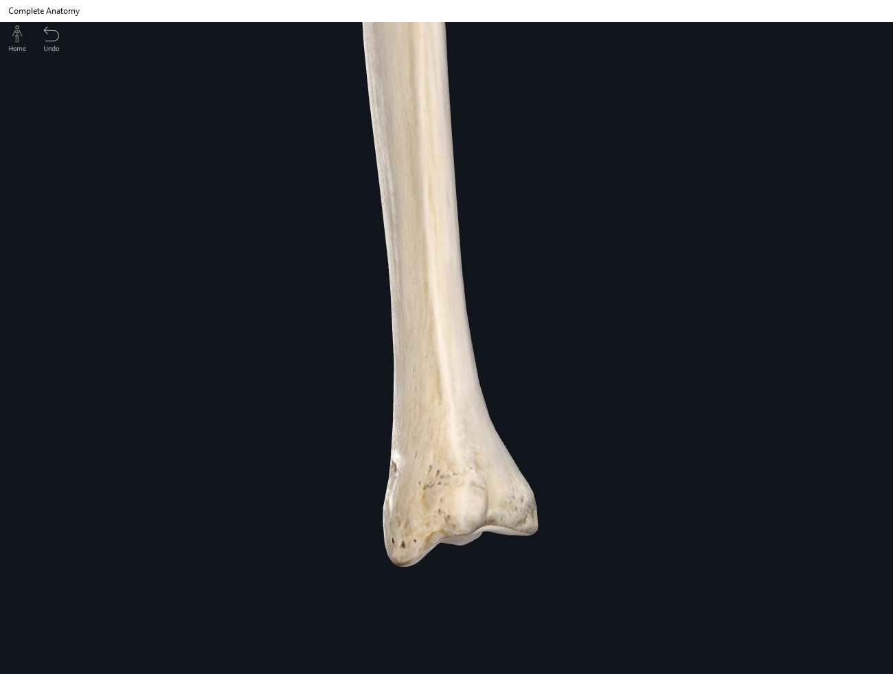
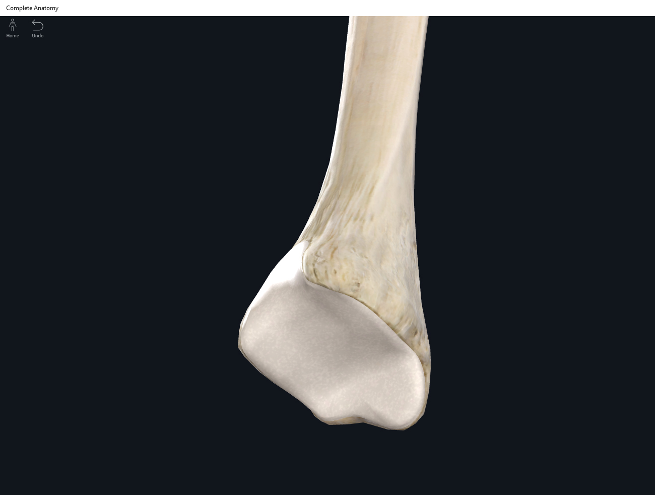
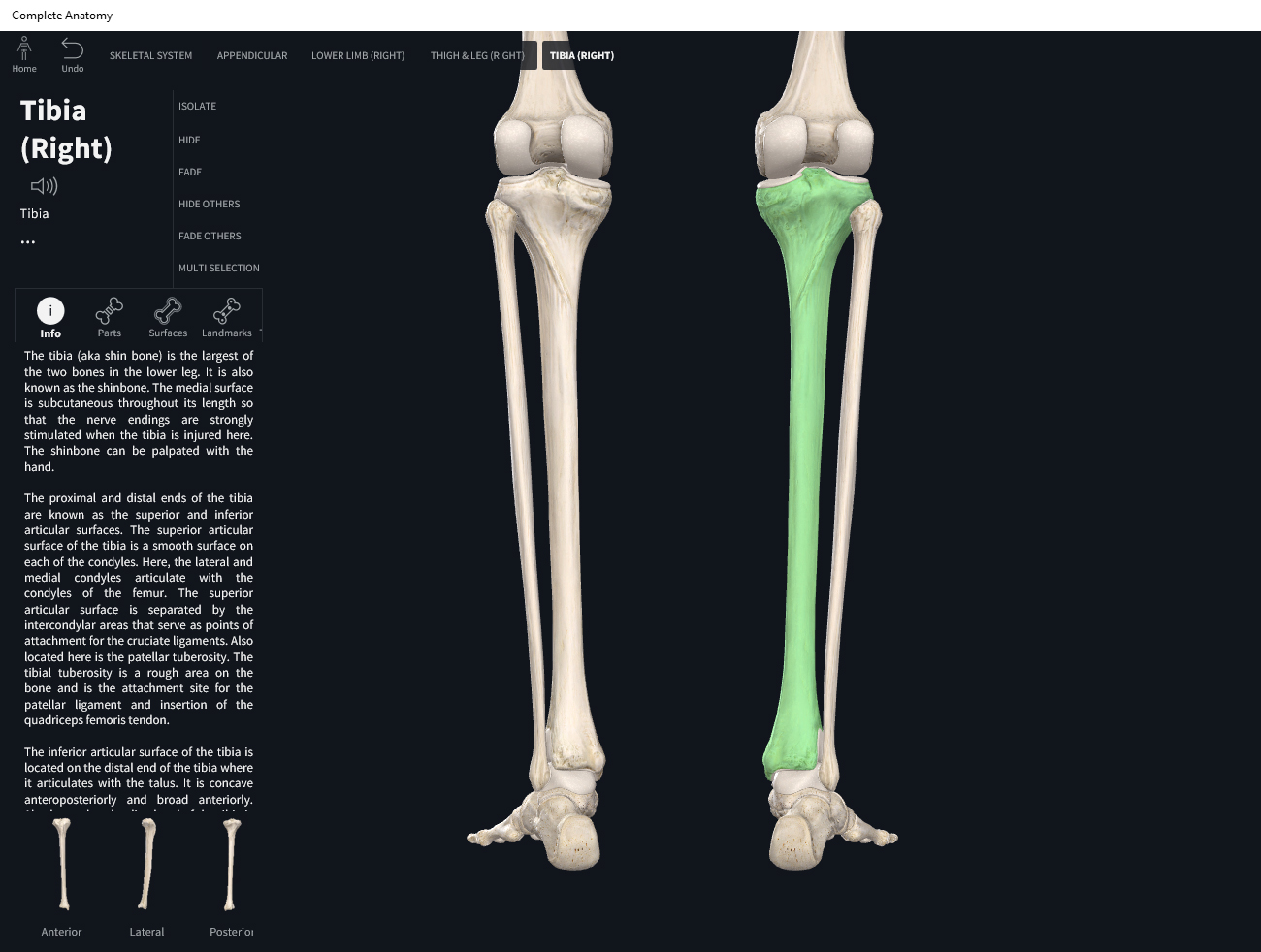
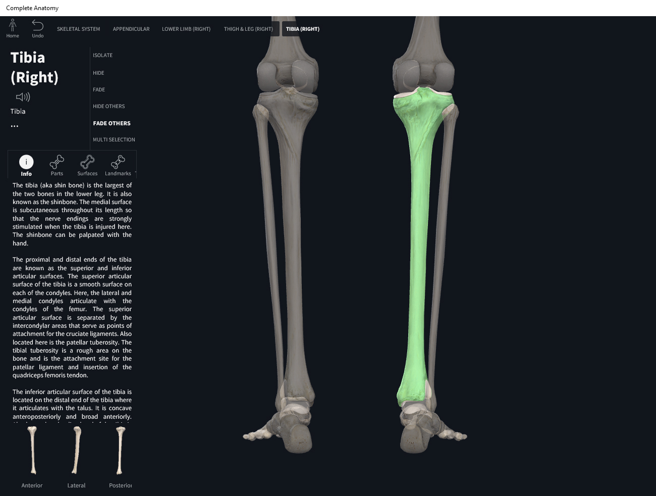
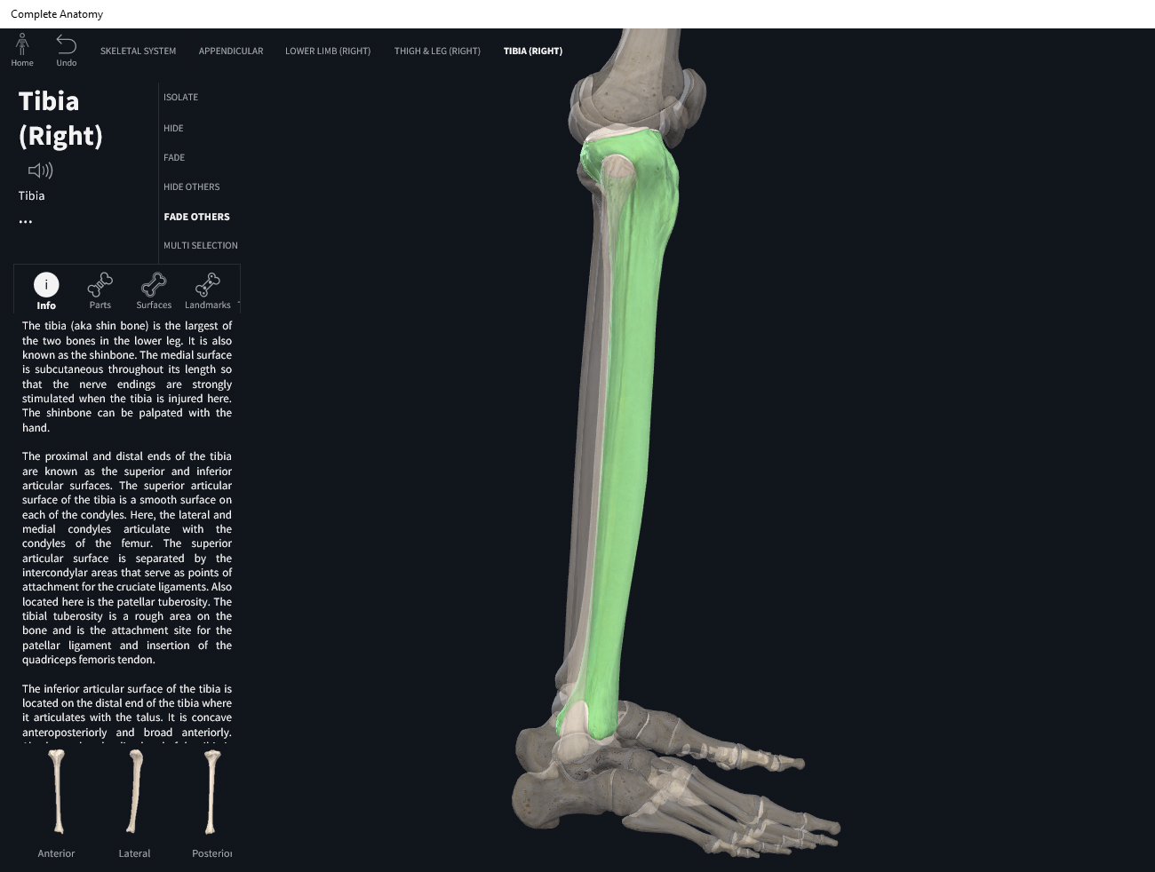
      • Interosseous membrane: sheet-like dense connective tissue between long bones; amphiarthrotic. E.g. between ulna and radius; between tibia and fibula.
    • Cartilaginous joints: no synovial cavity; cartilaginous connective tissue (hyaline or fibrocartilage).
      • Synchondroses: hyaline connective tissue. E.g. epiphyseal plate. Synarthrotic (immoveable).
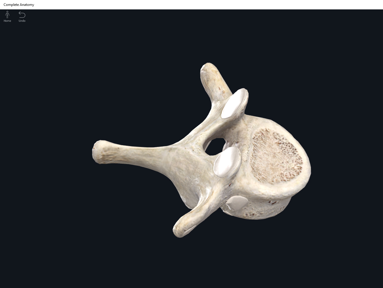
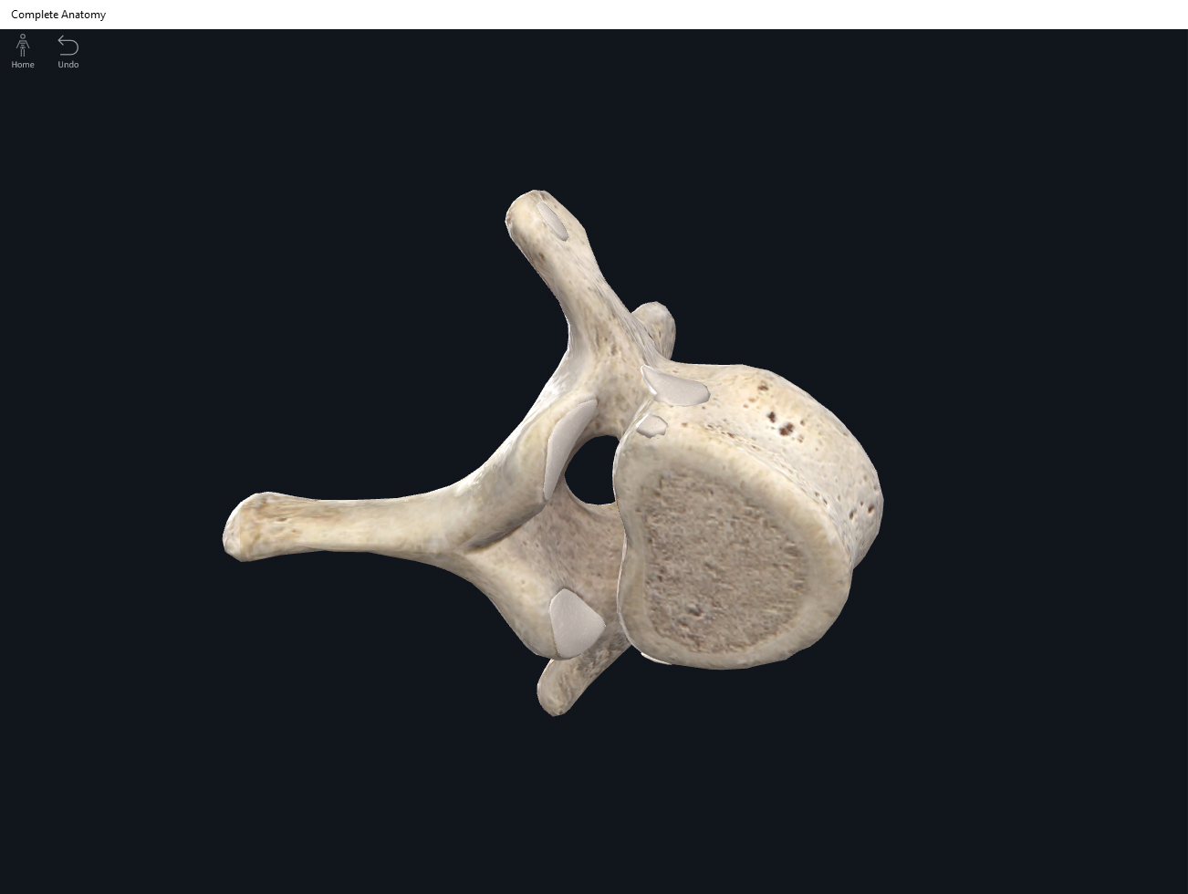
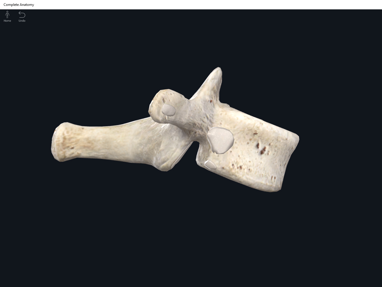
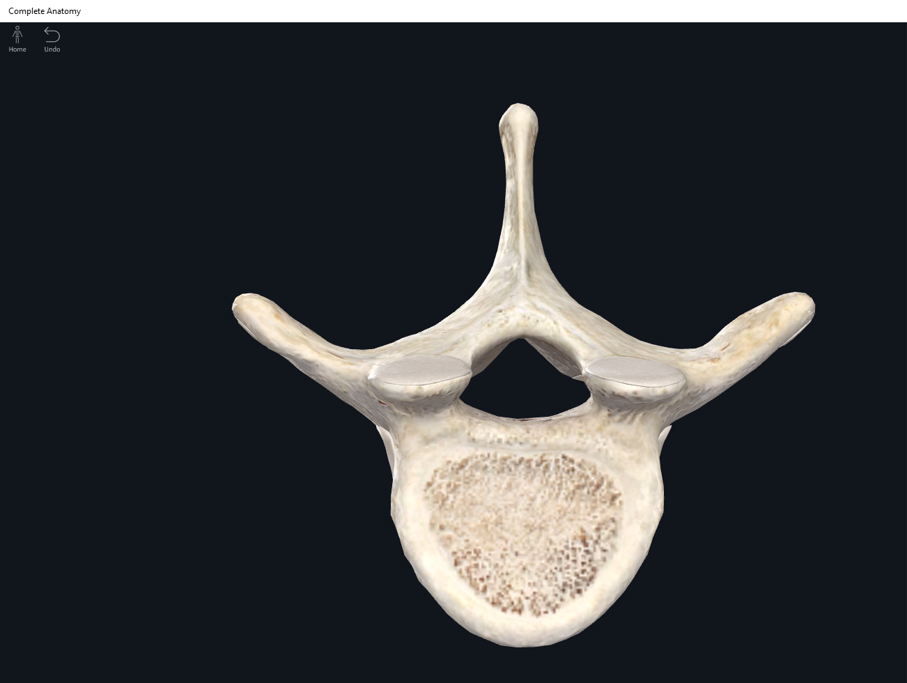
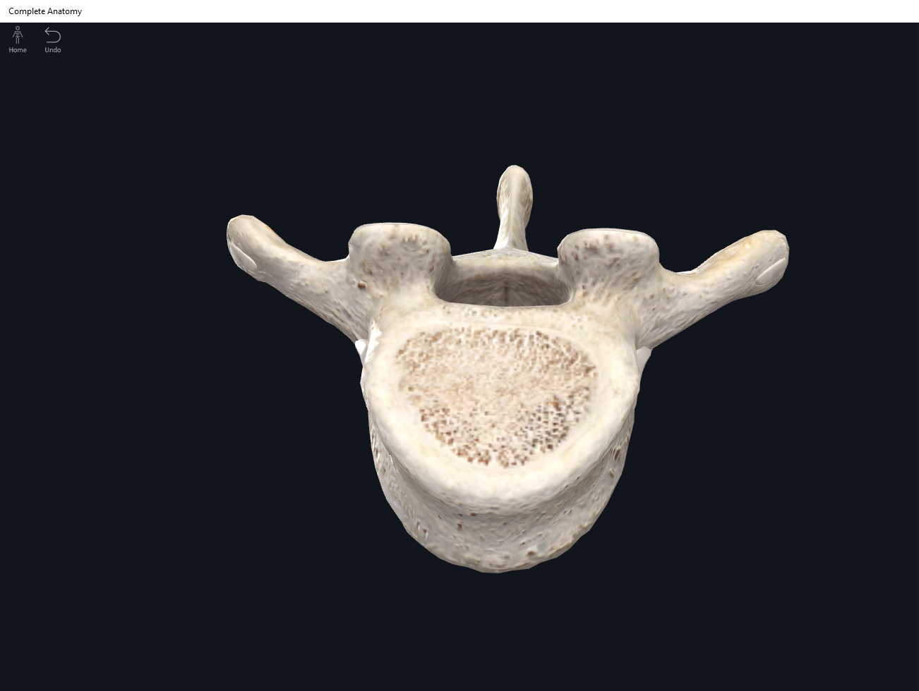
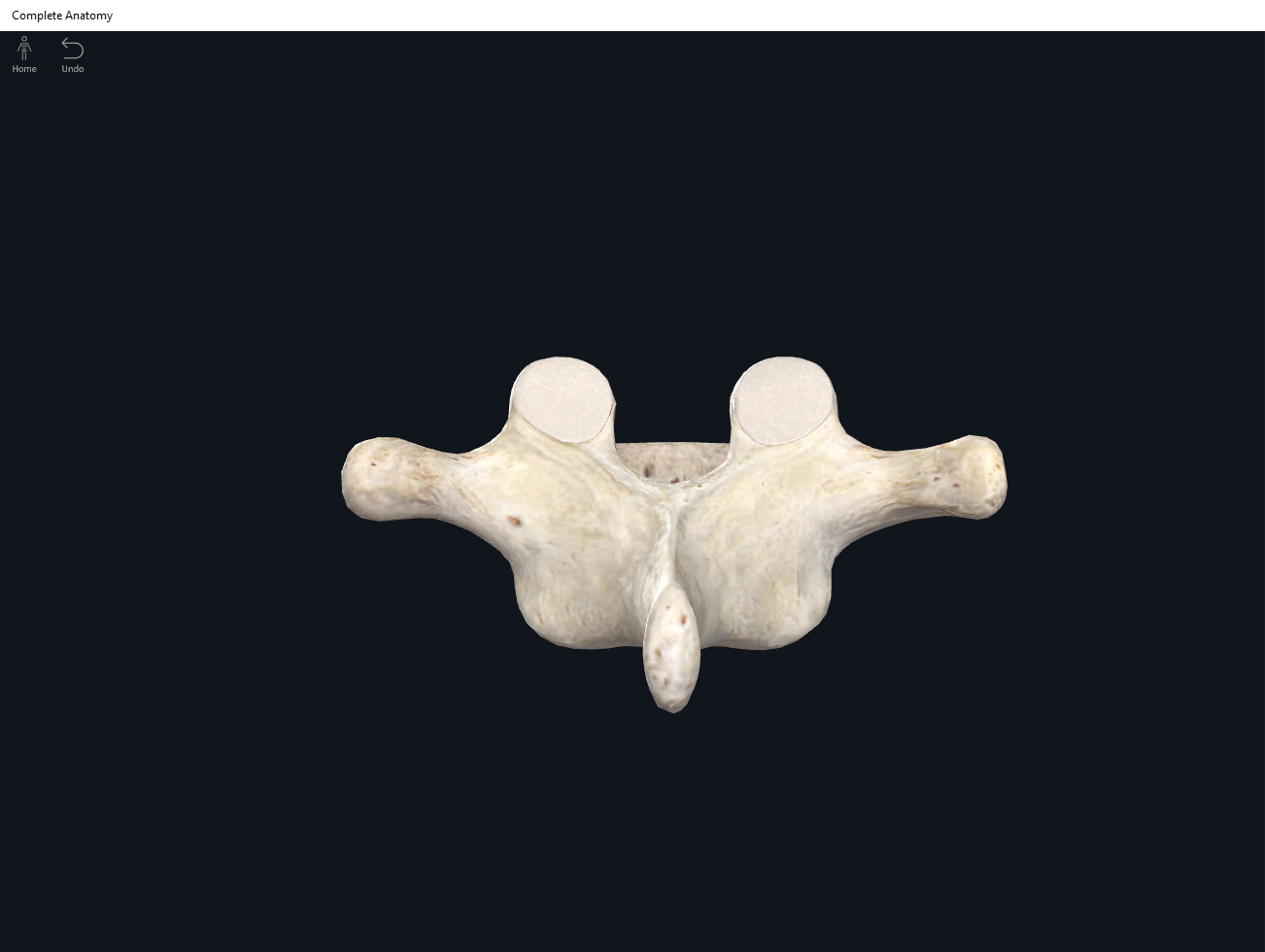
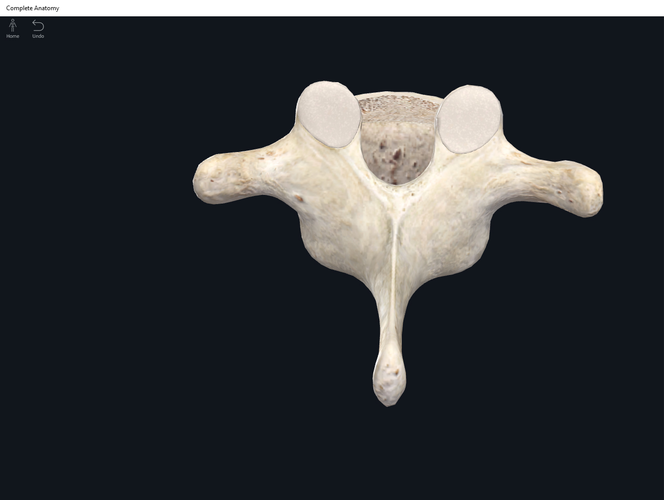
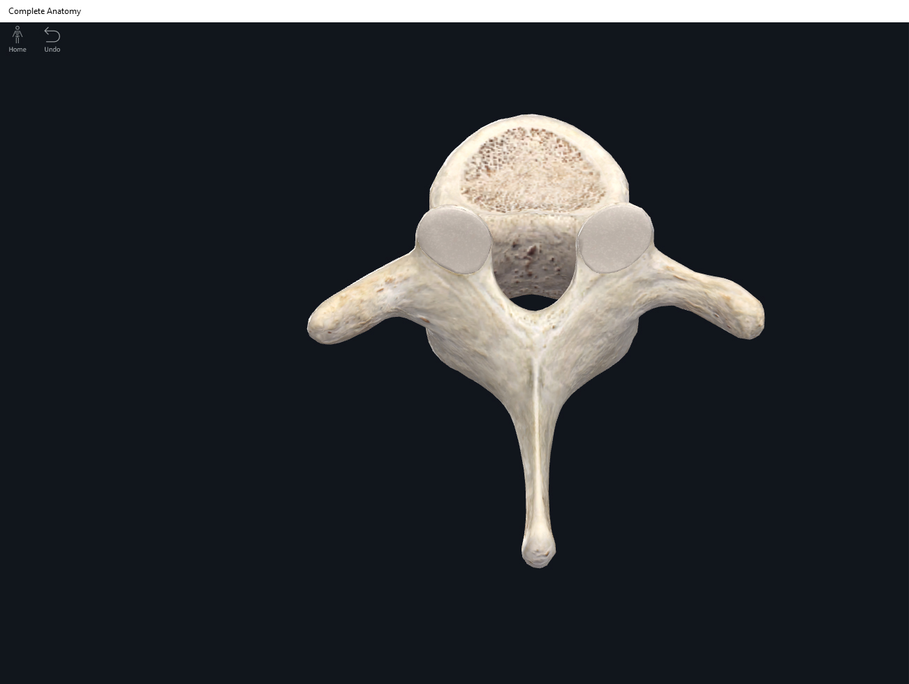
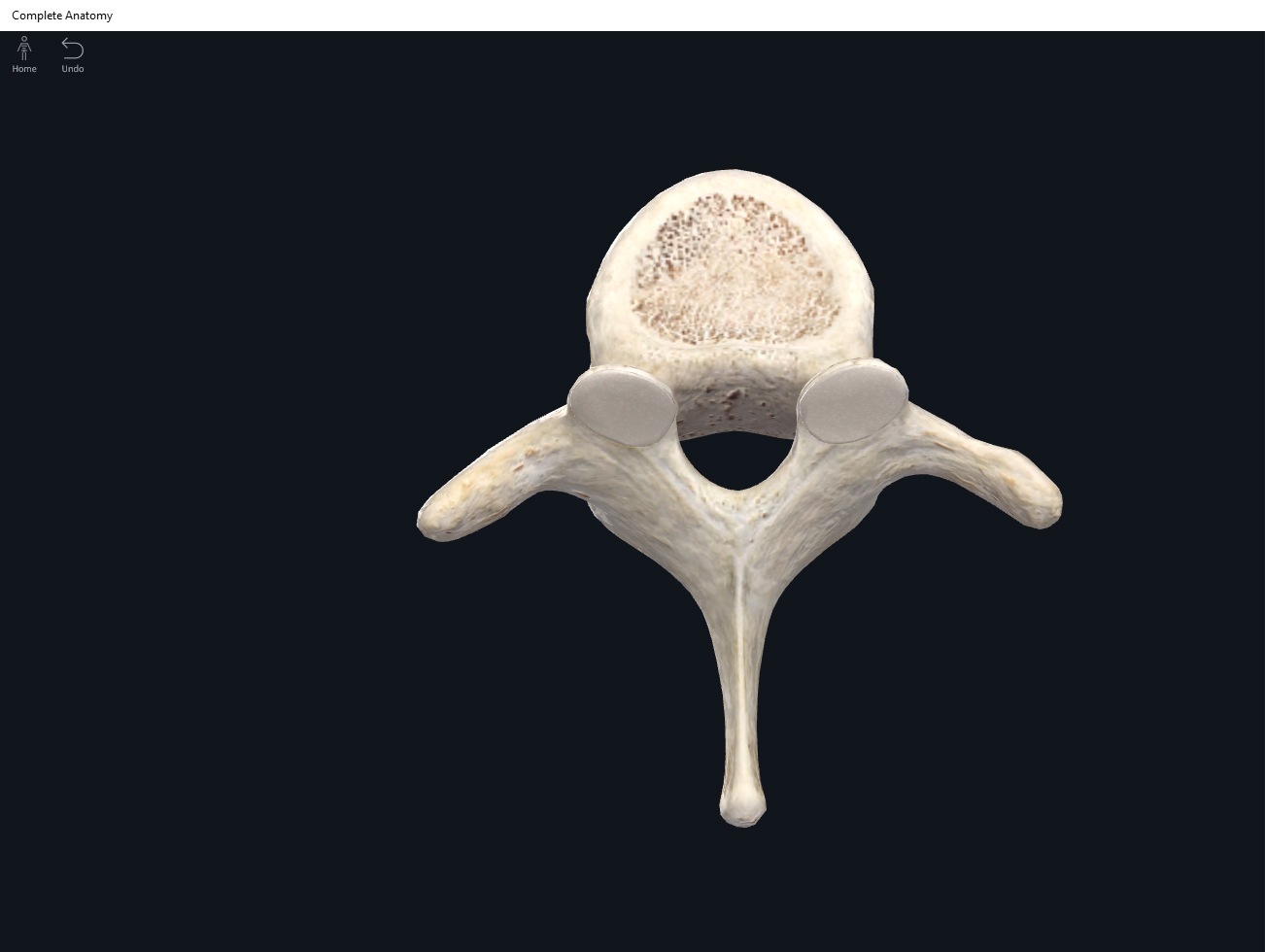
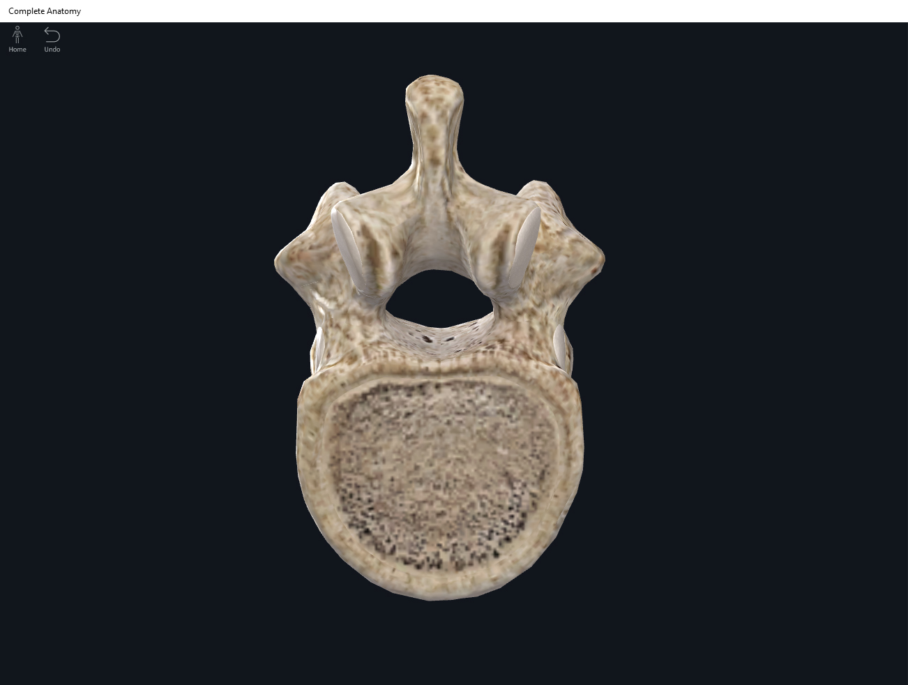
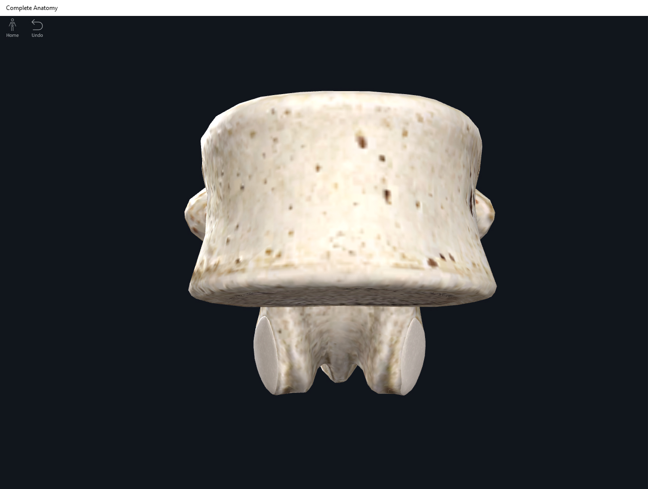
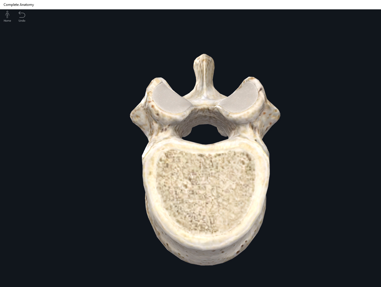
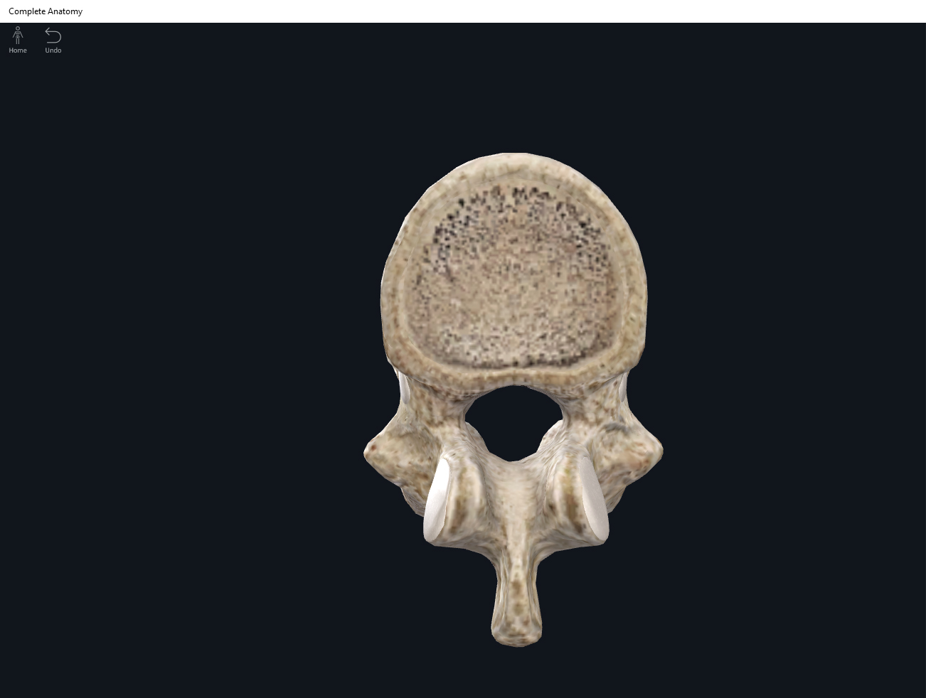
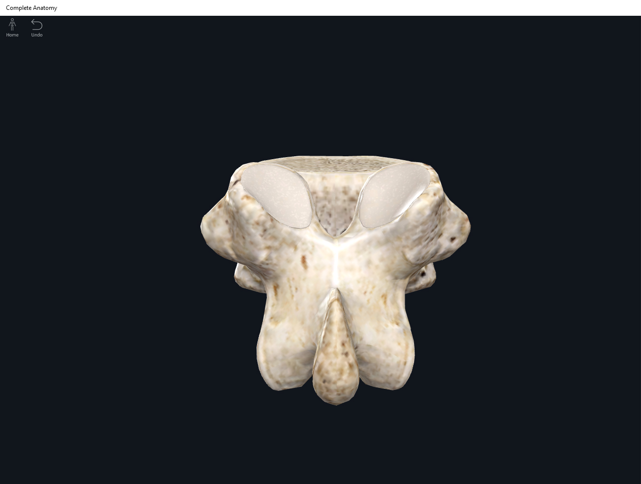
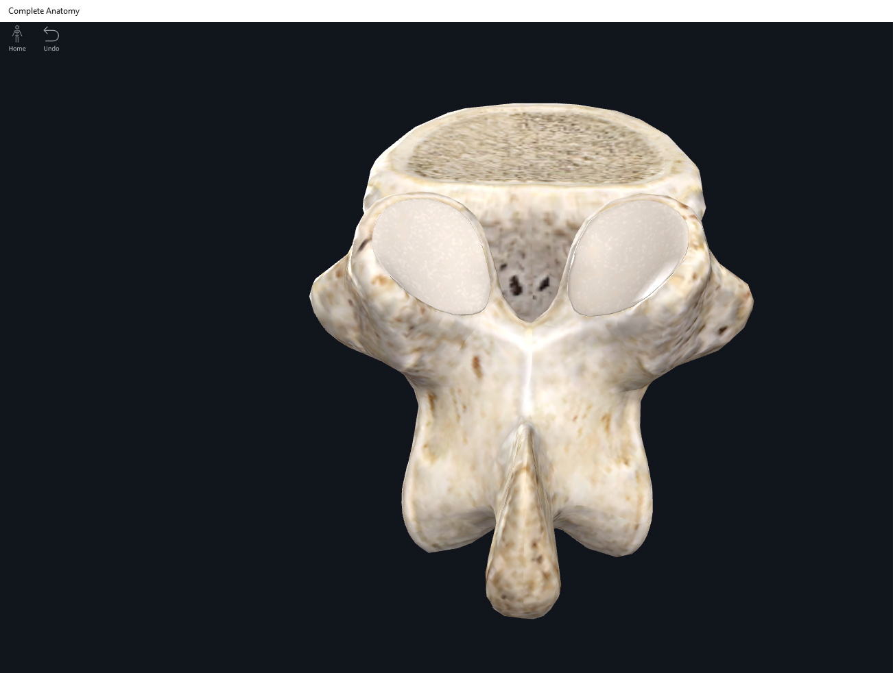
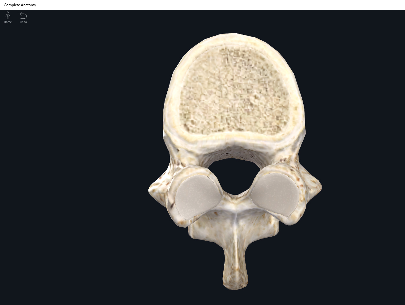
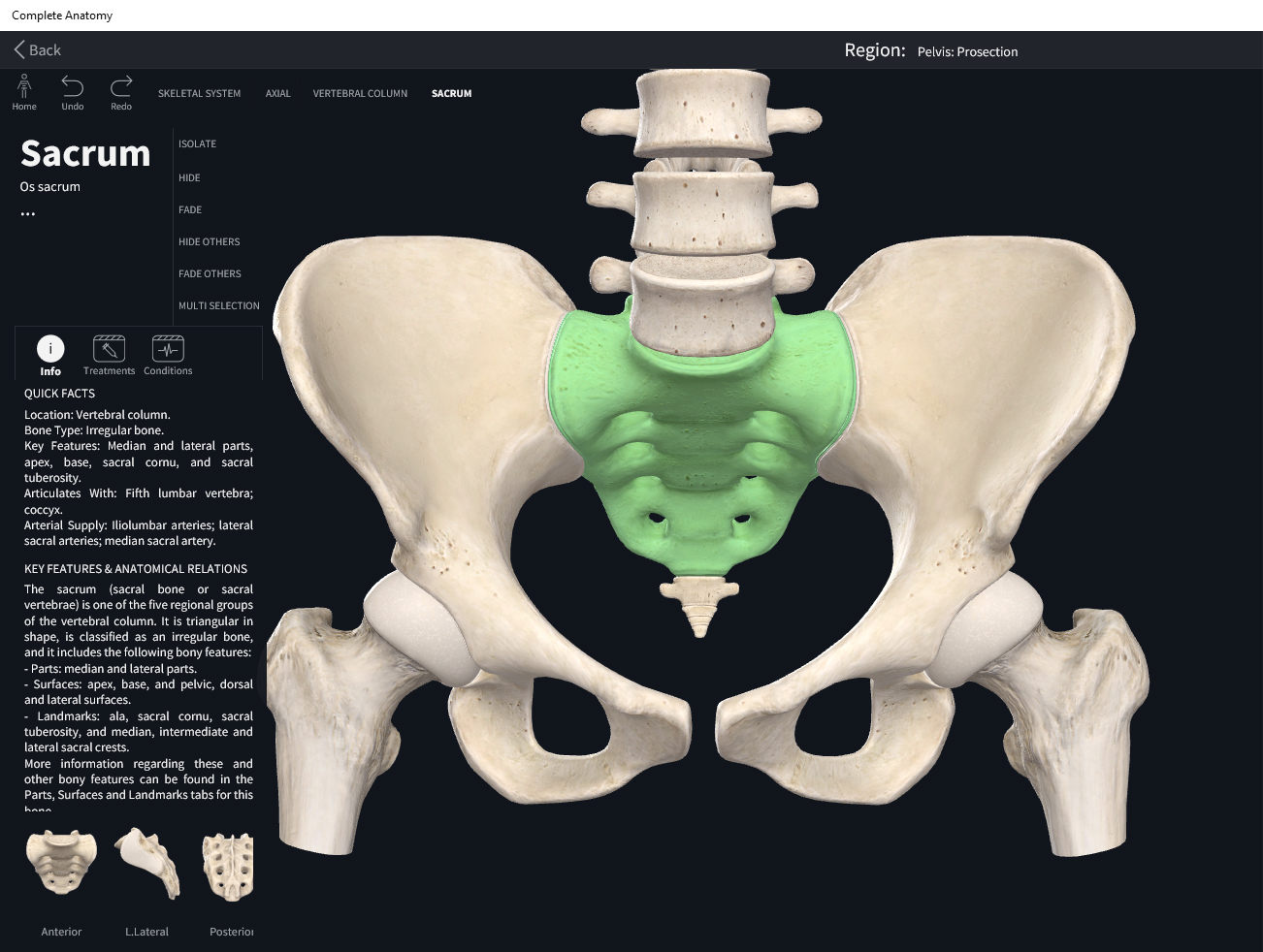
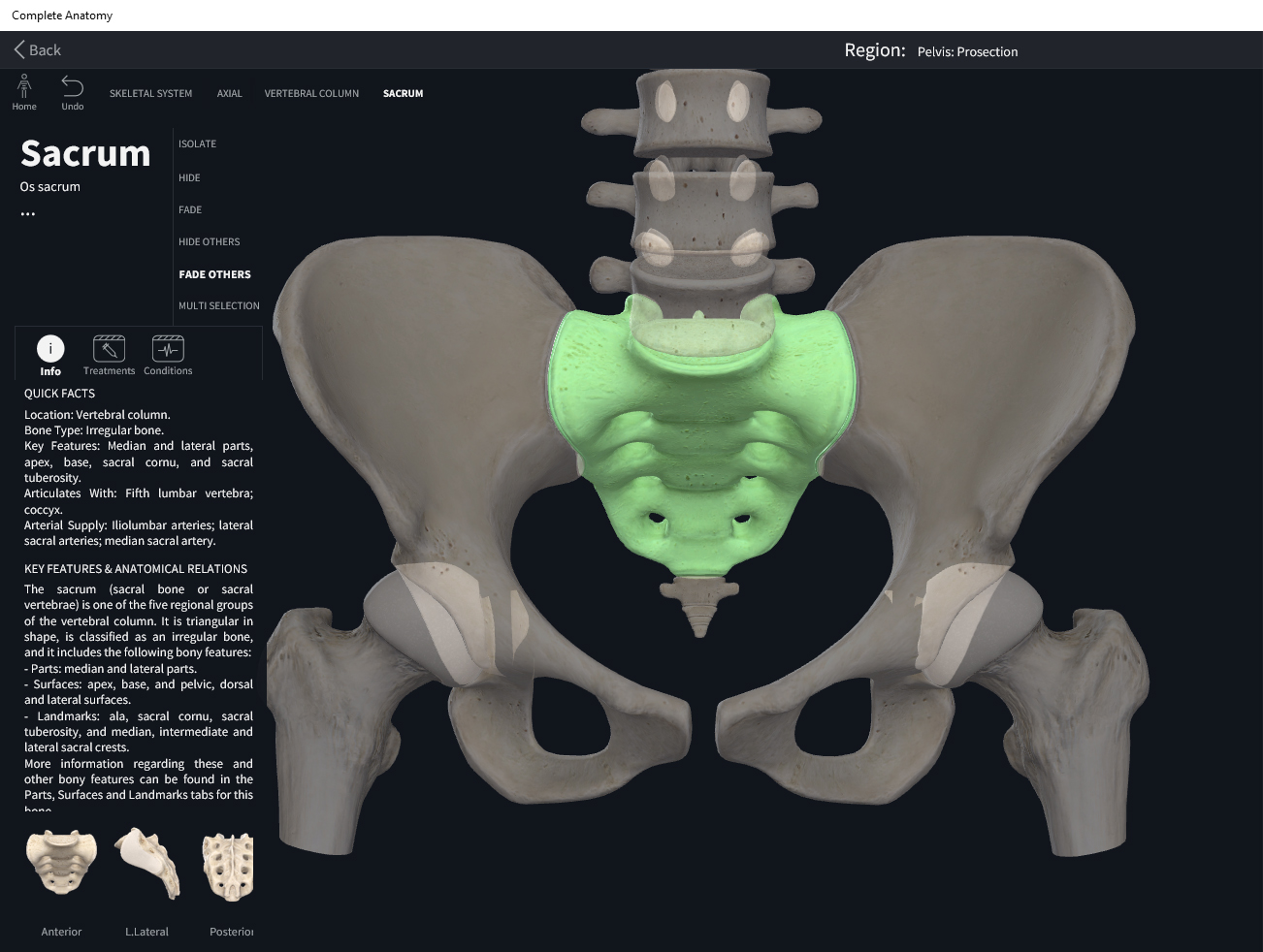
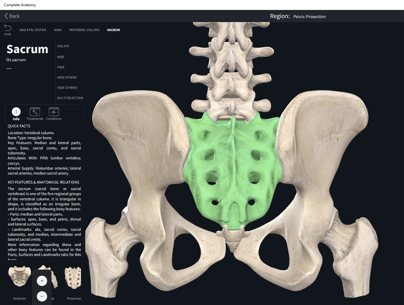
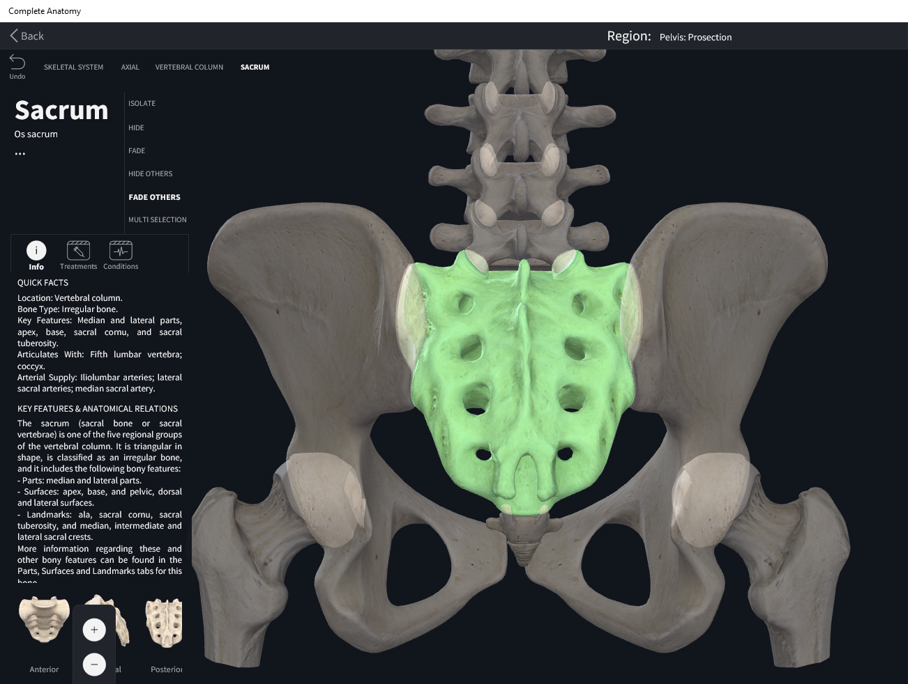
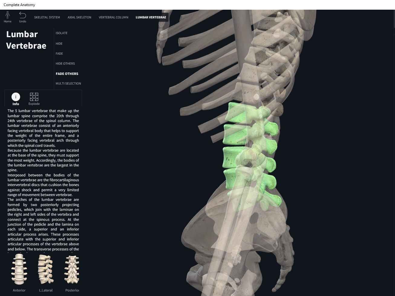
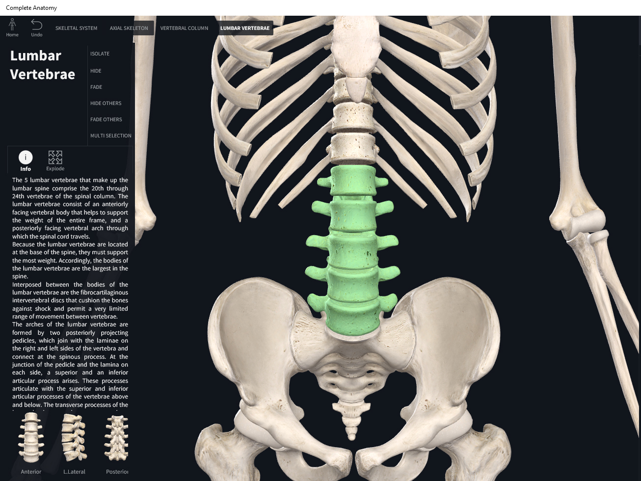
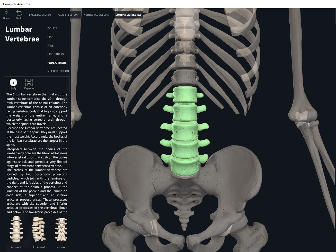
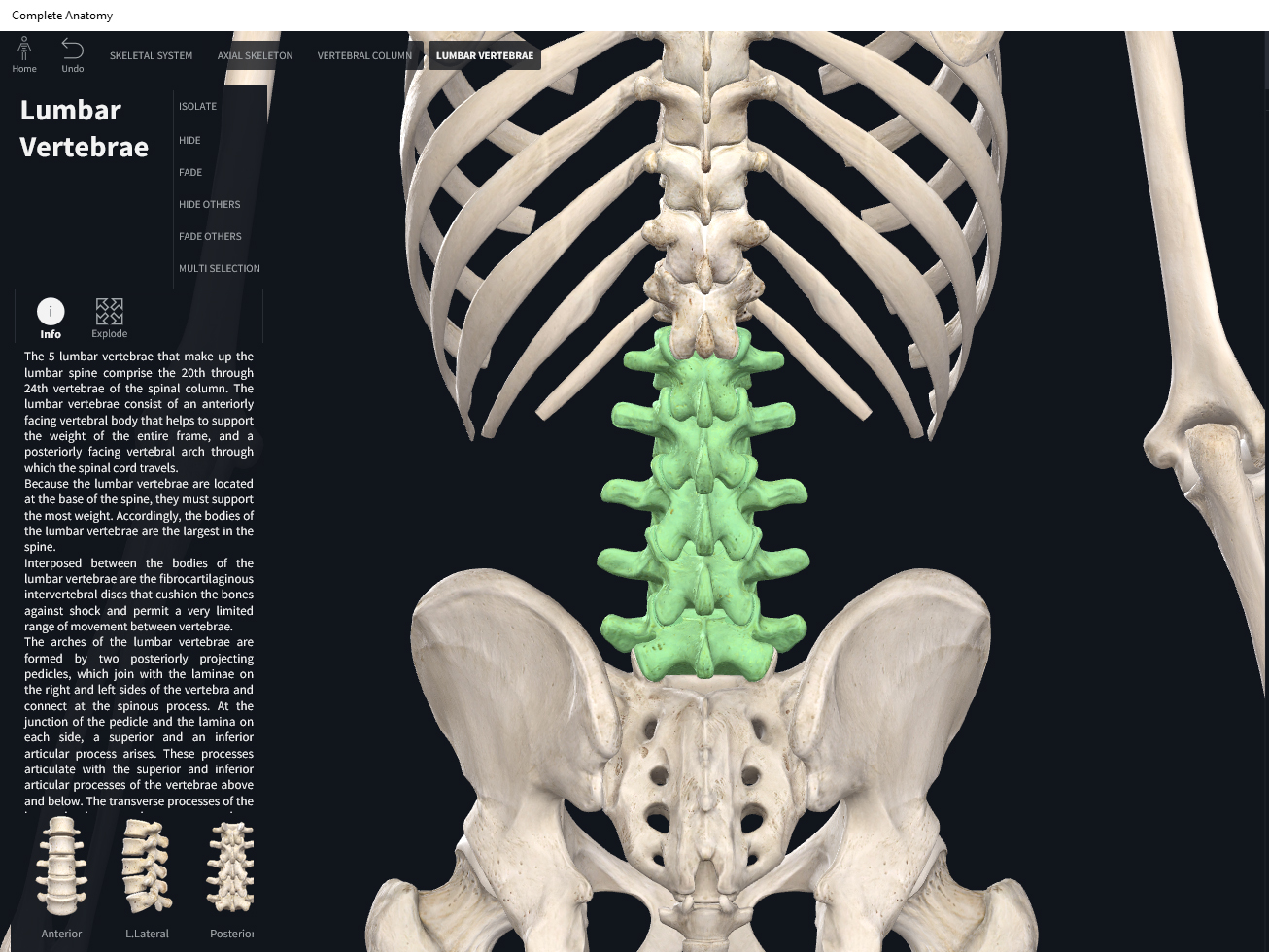
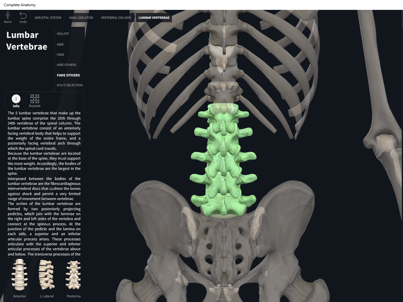
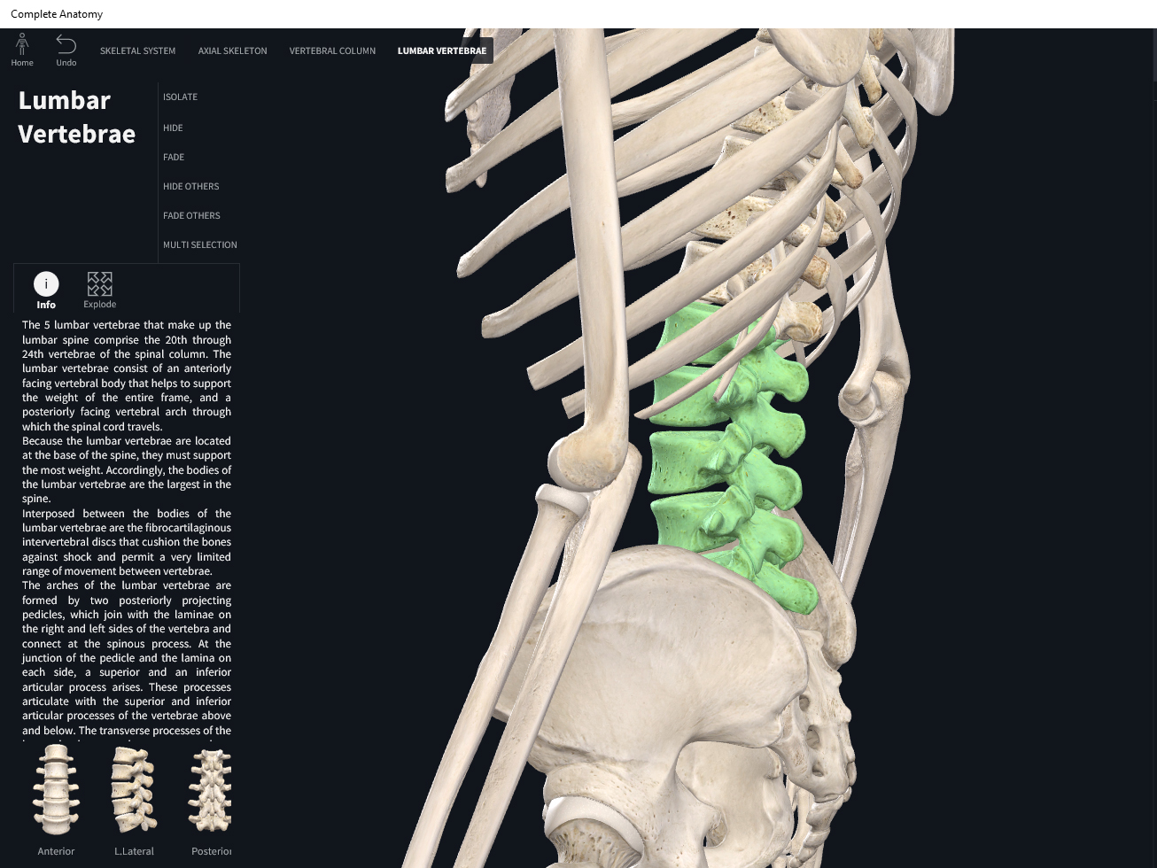
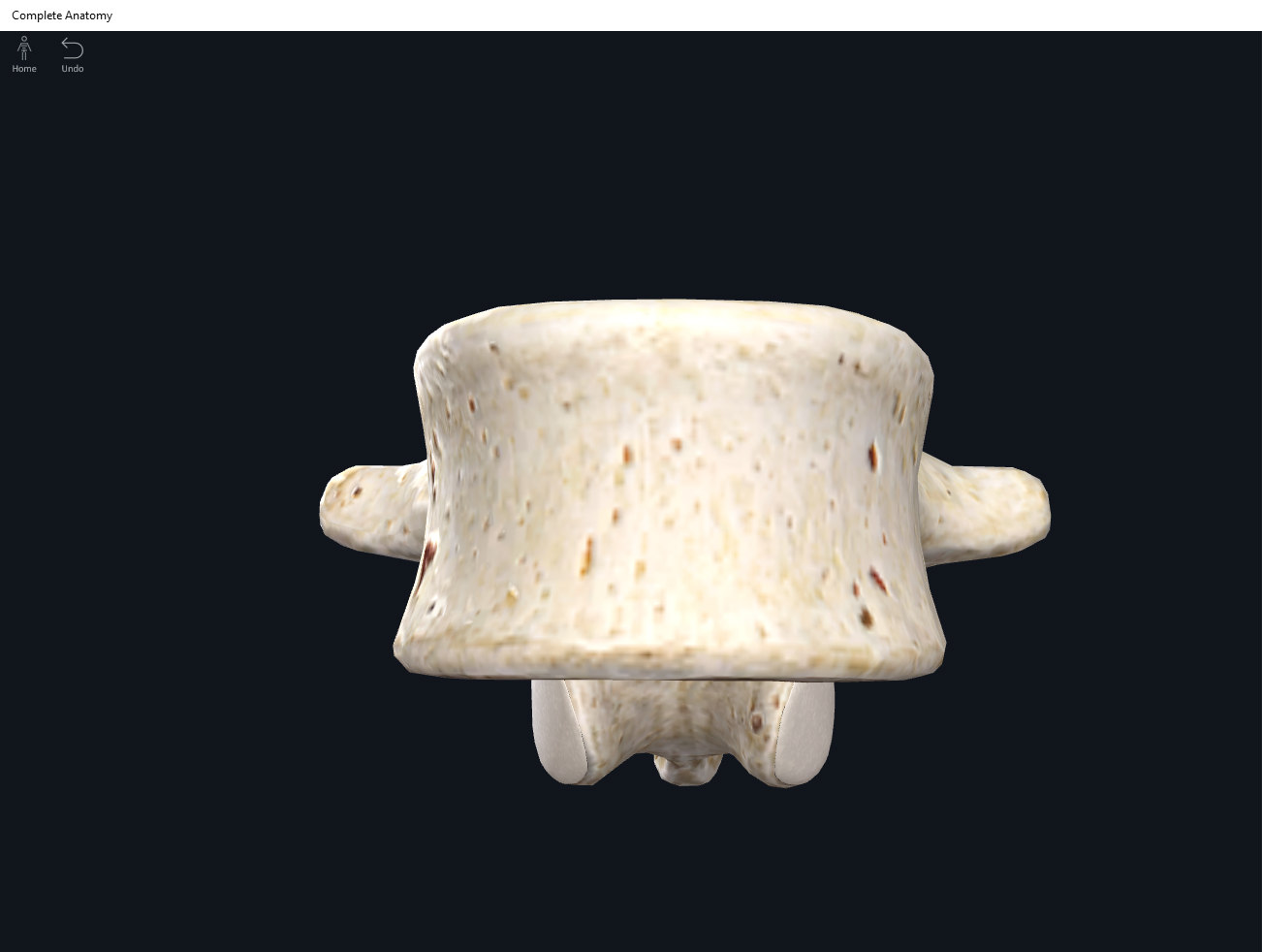
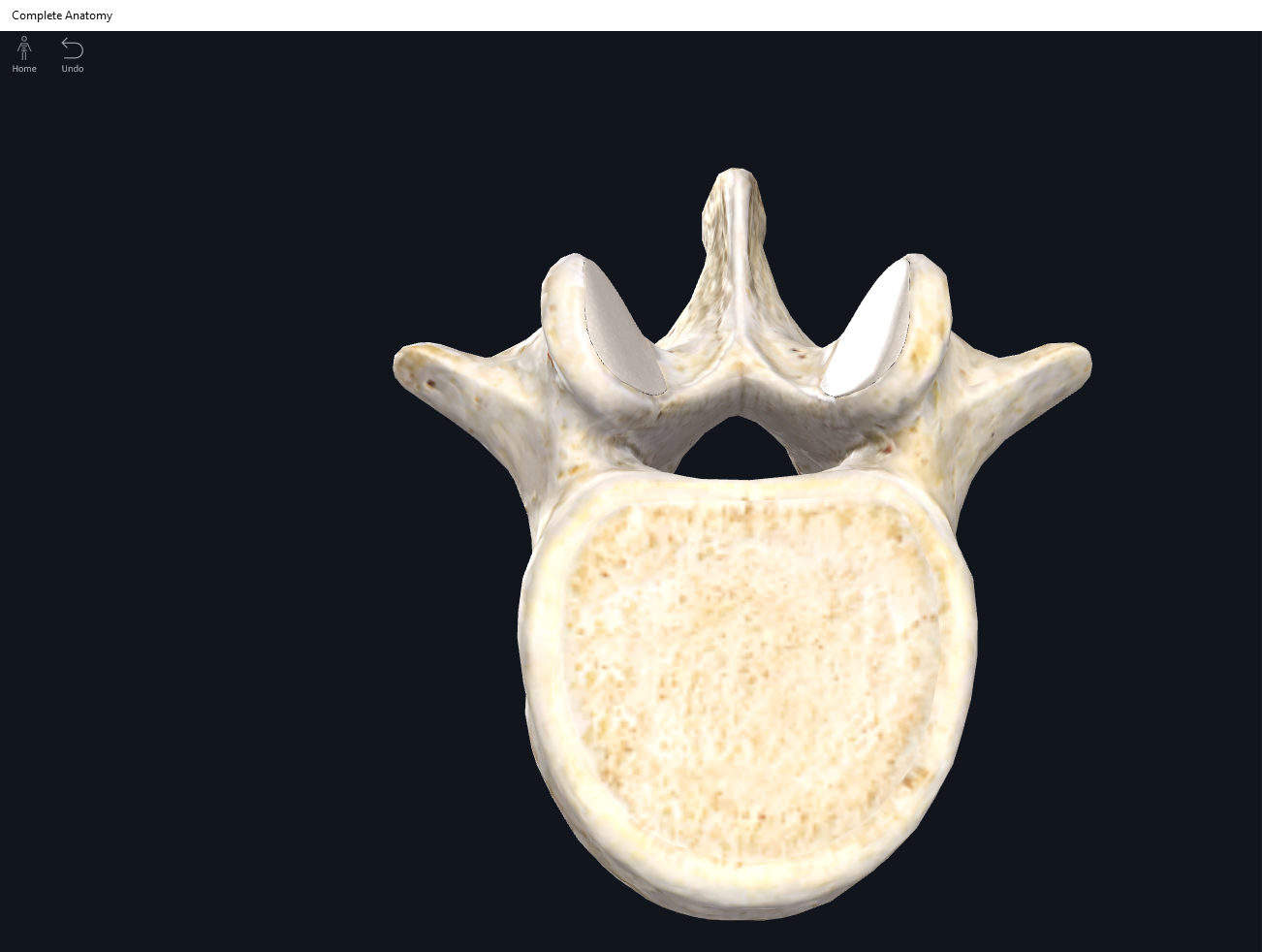
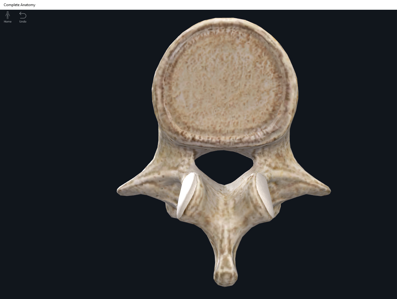
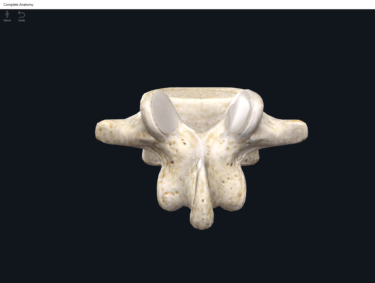
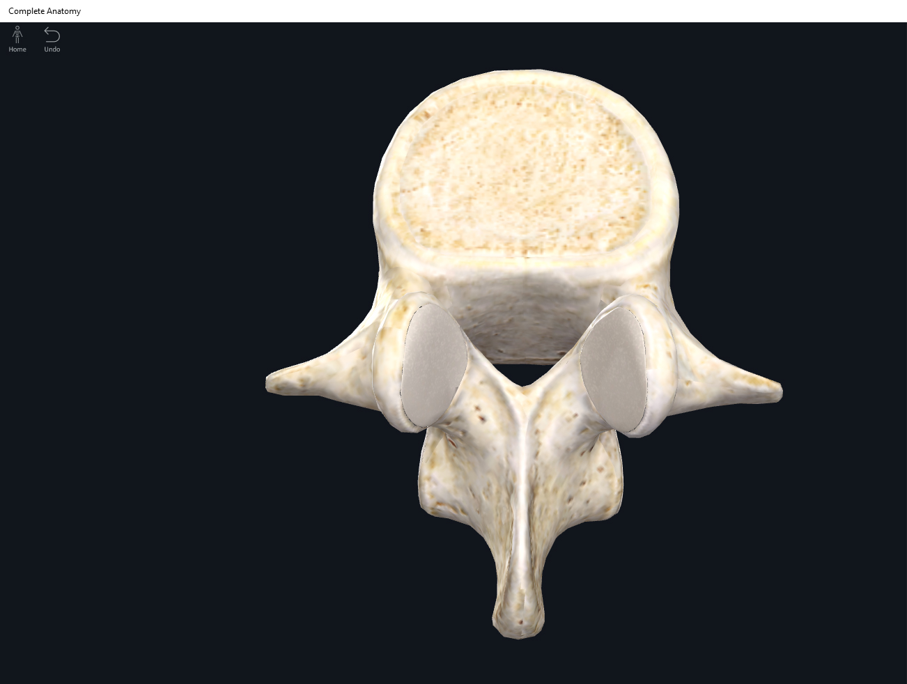
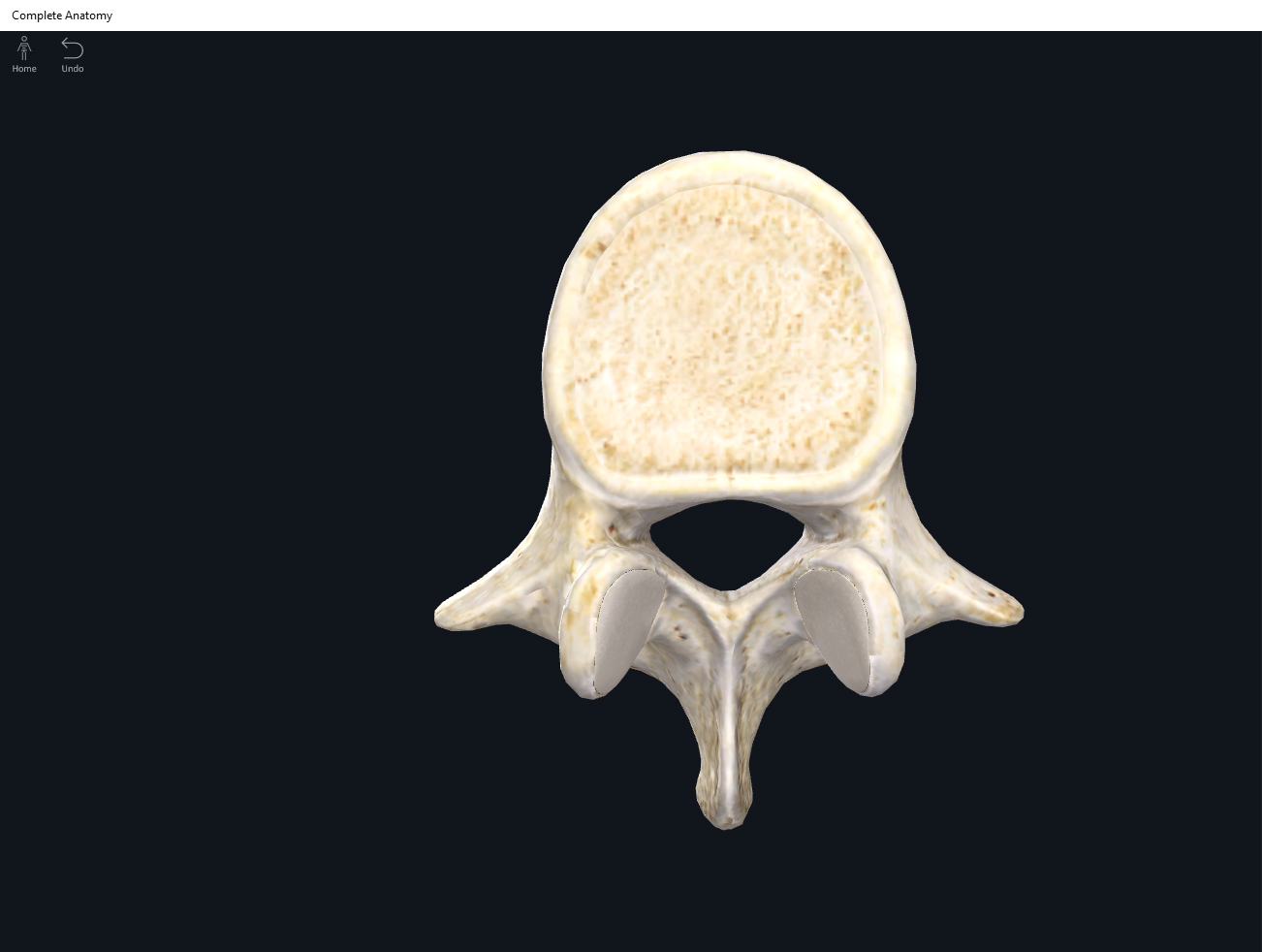
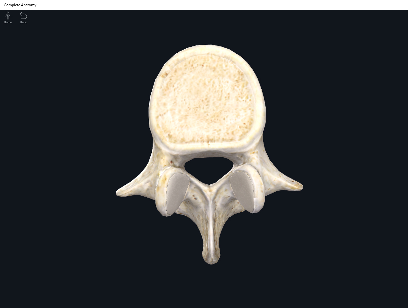
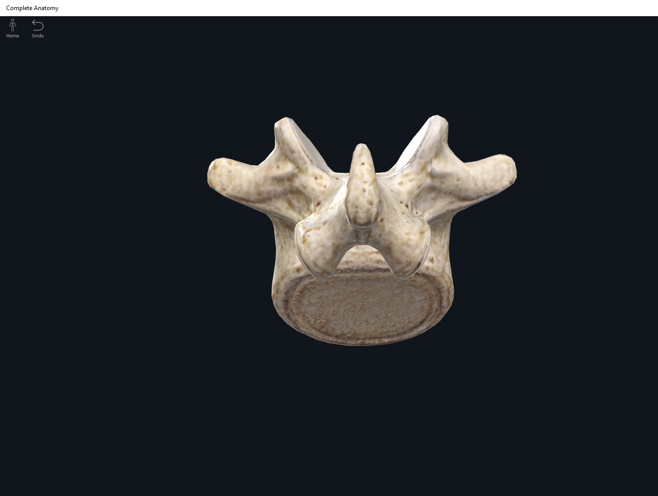
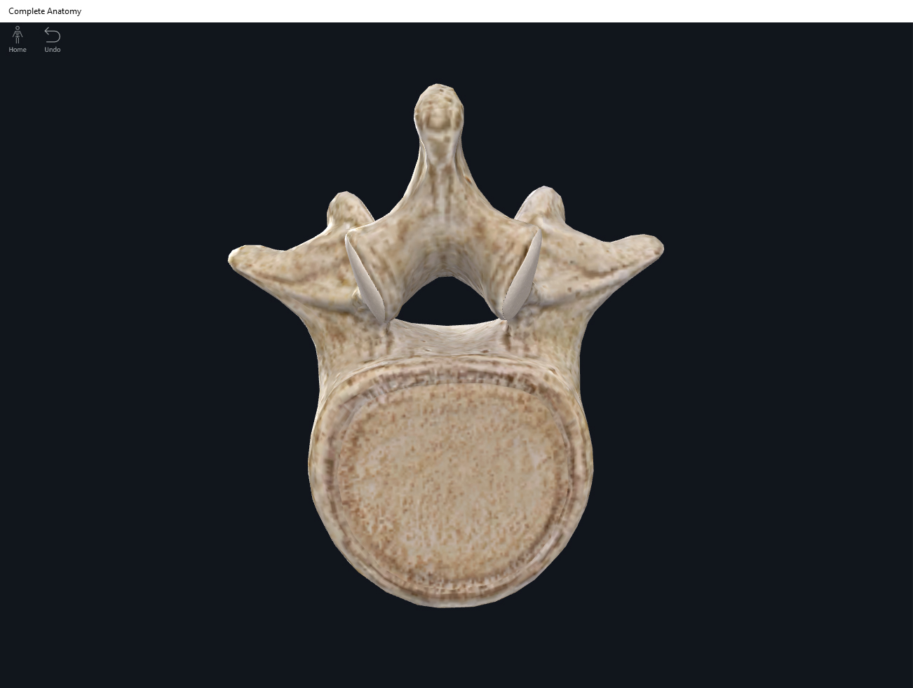
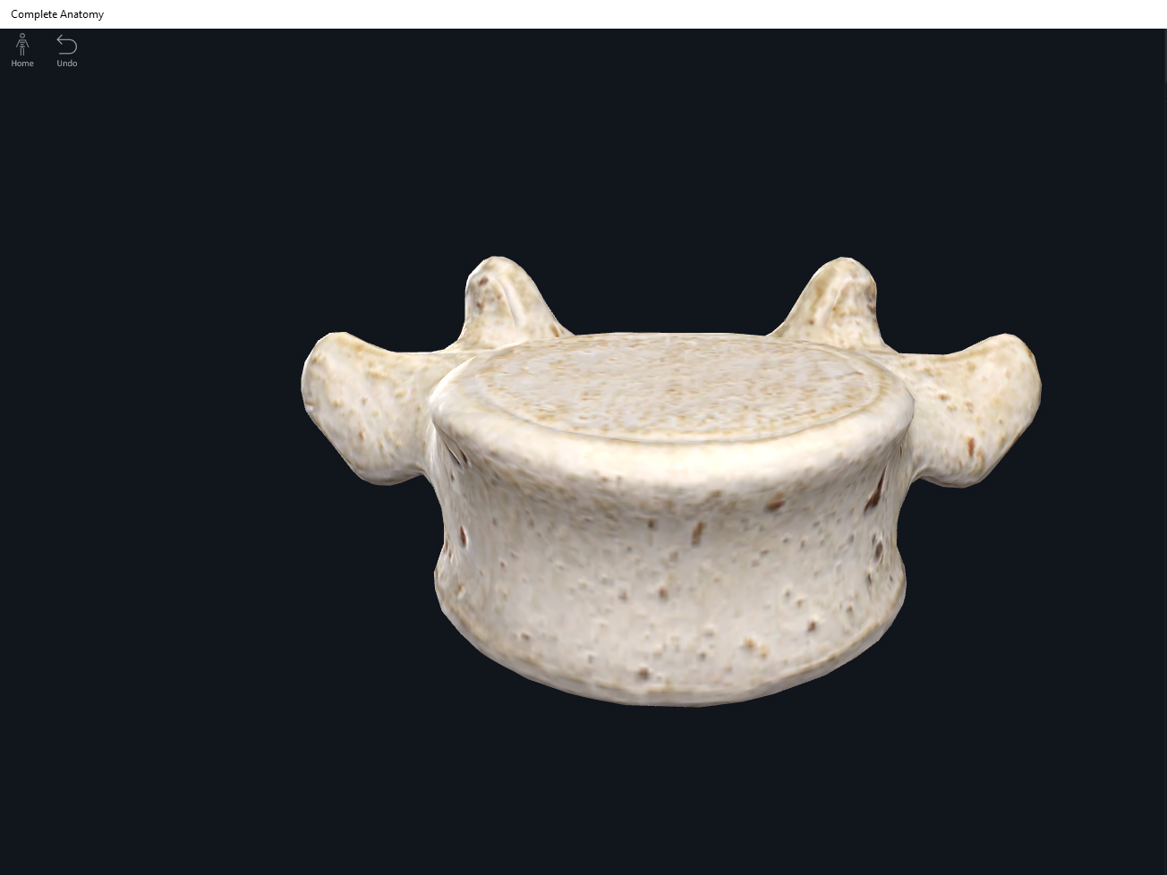
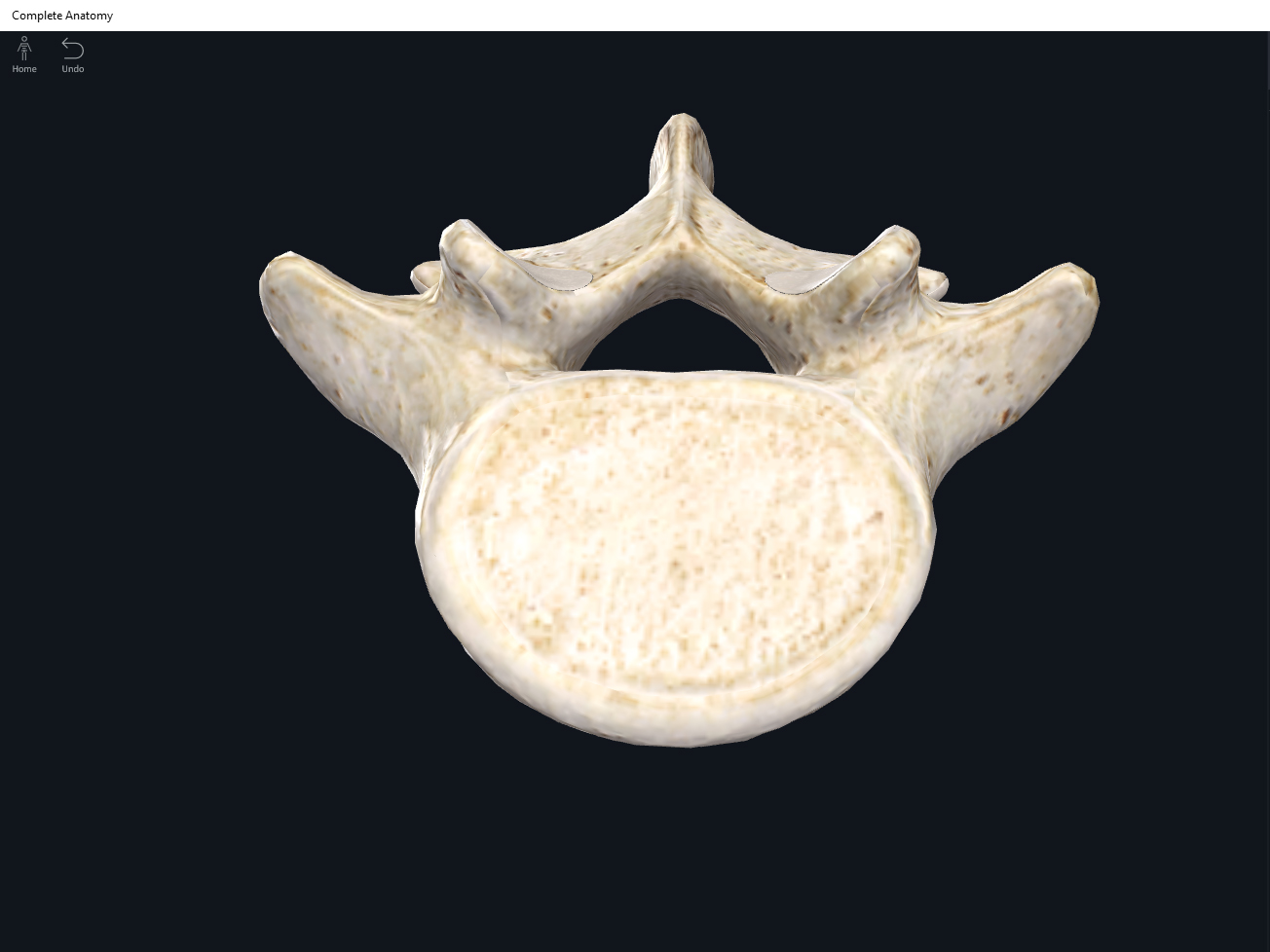
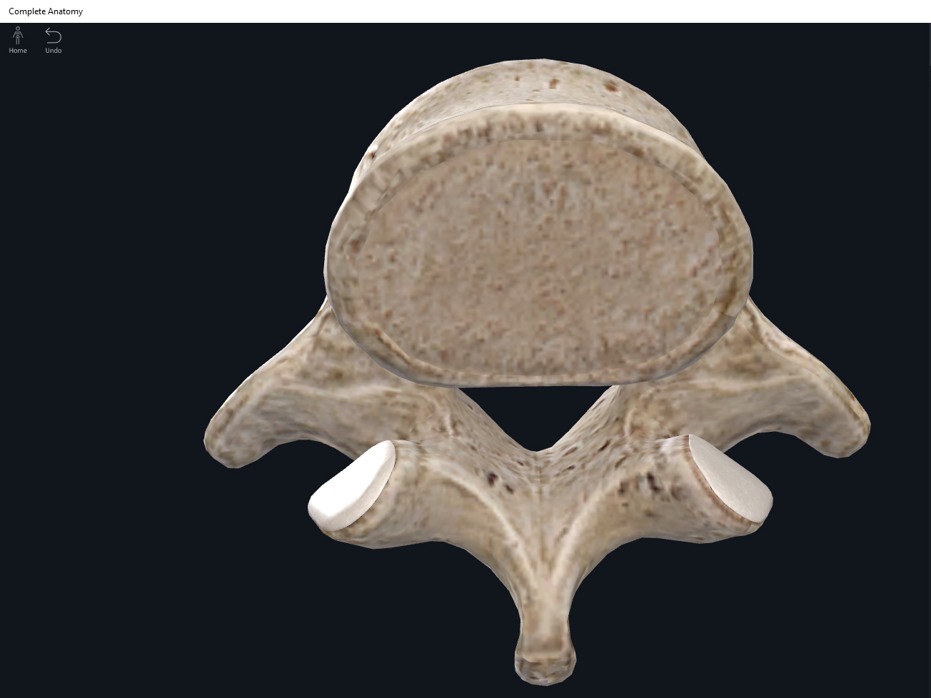
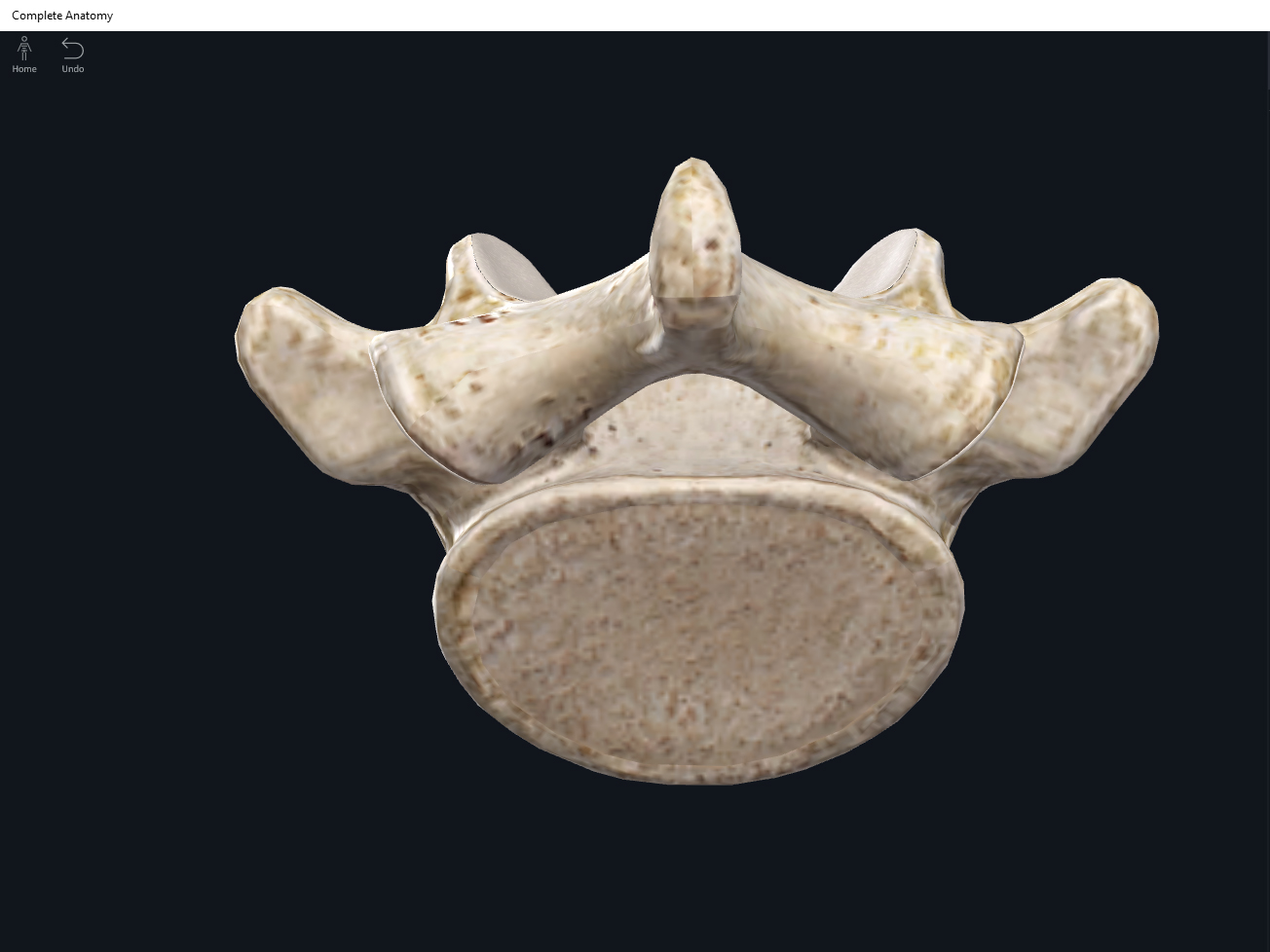
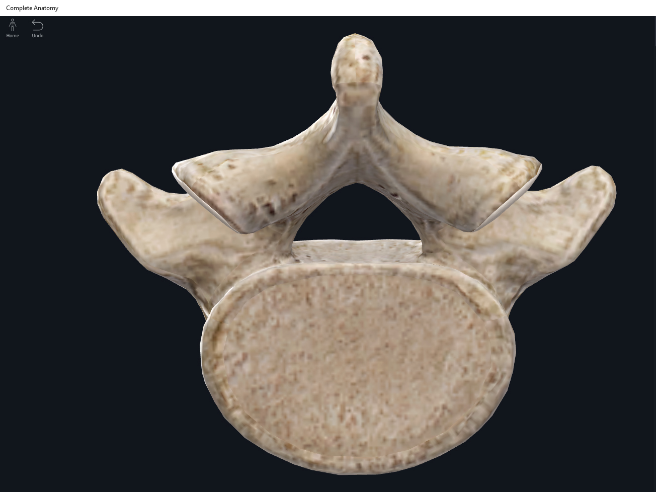
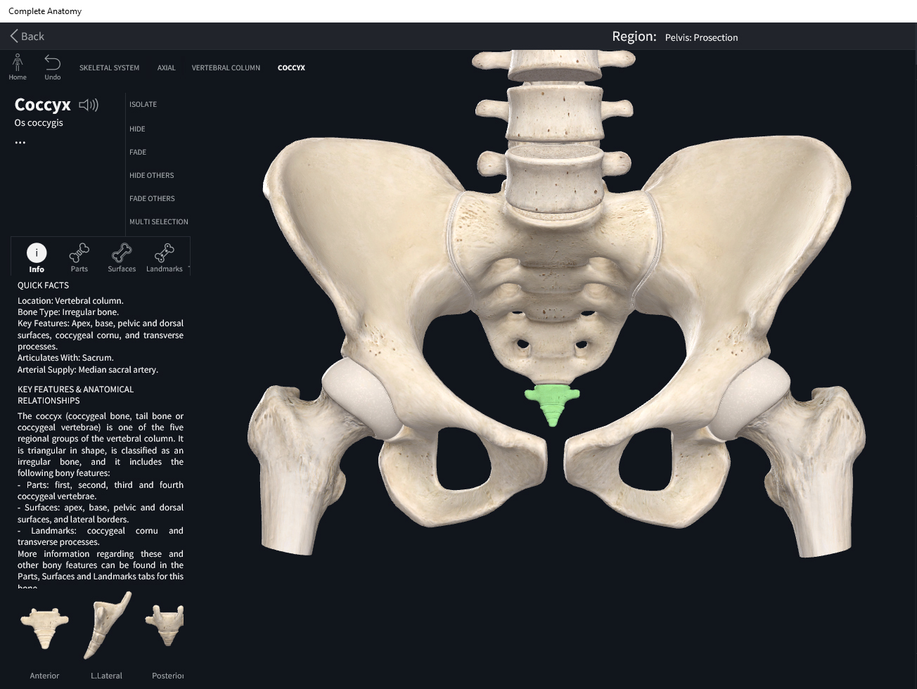
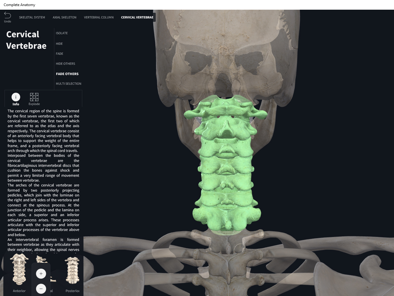
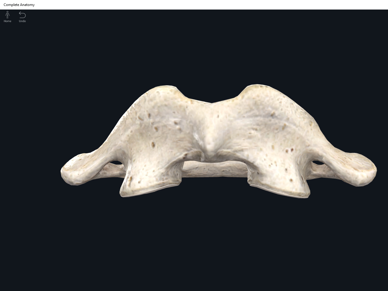
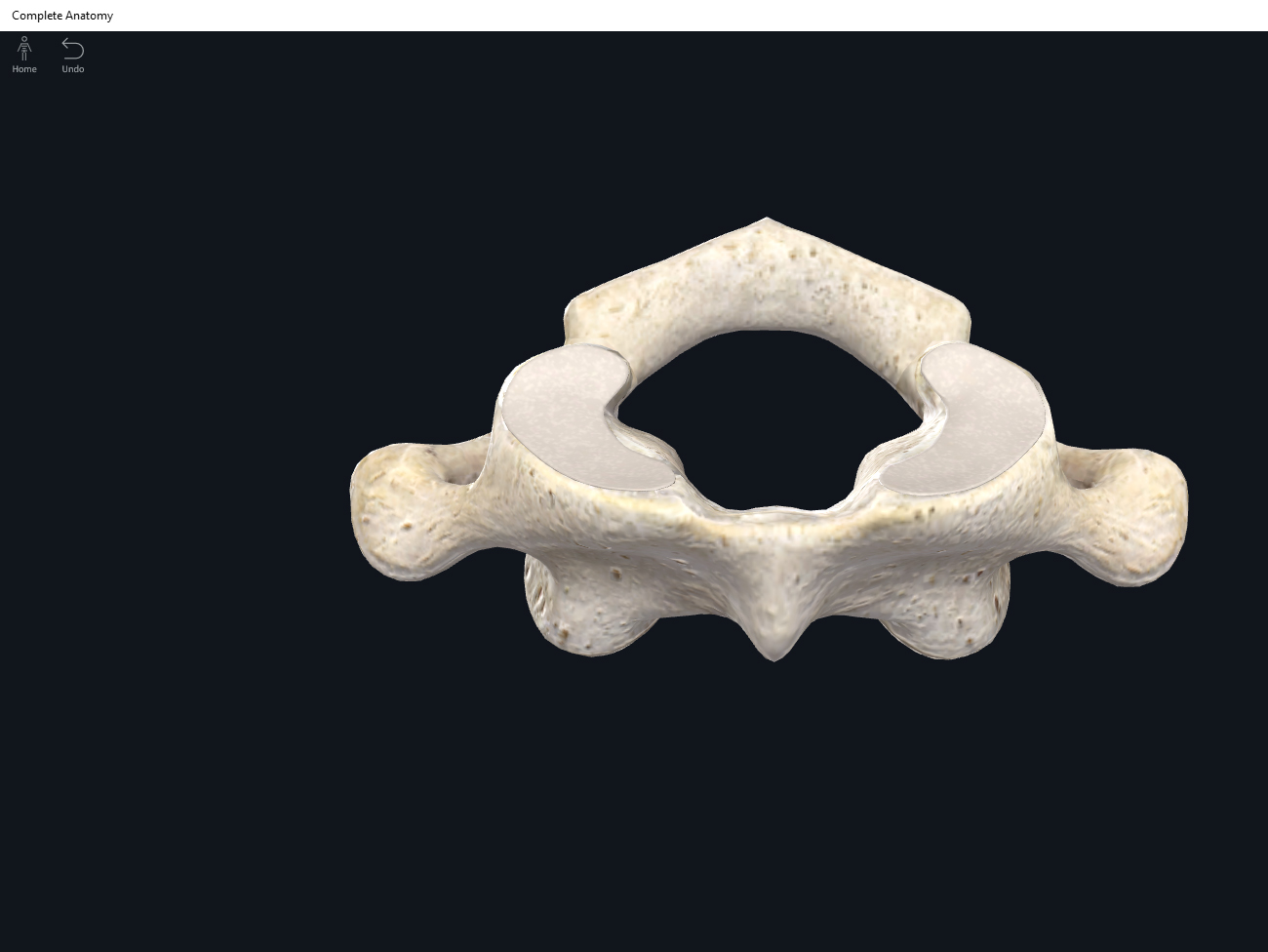
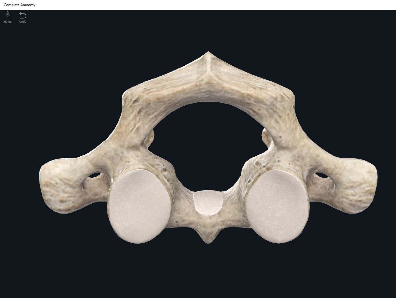
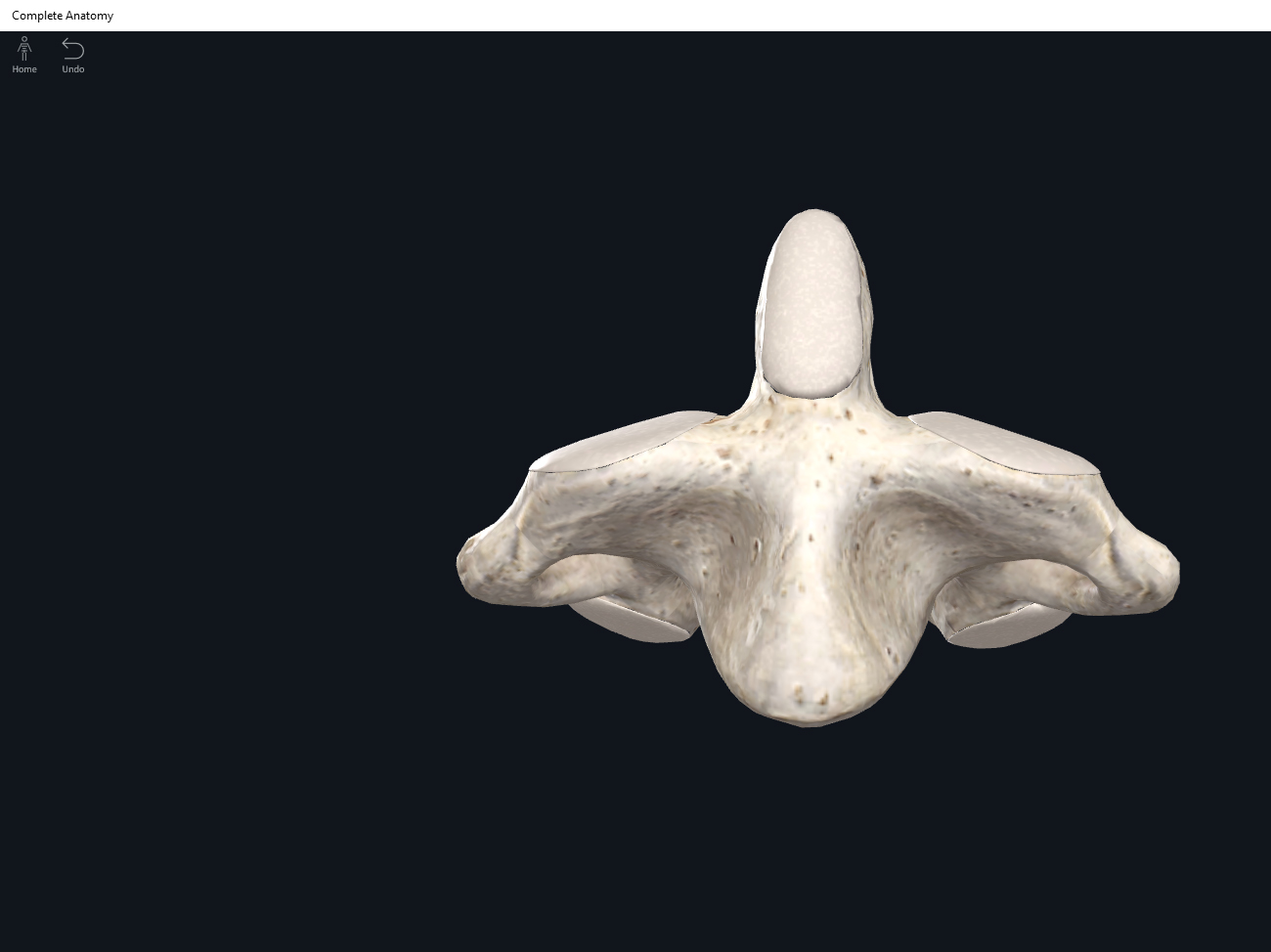
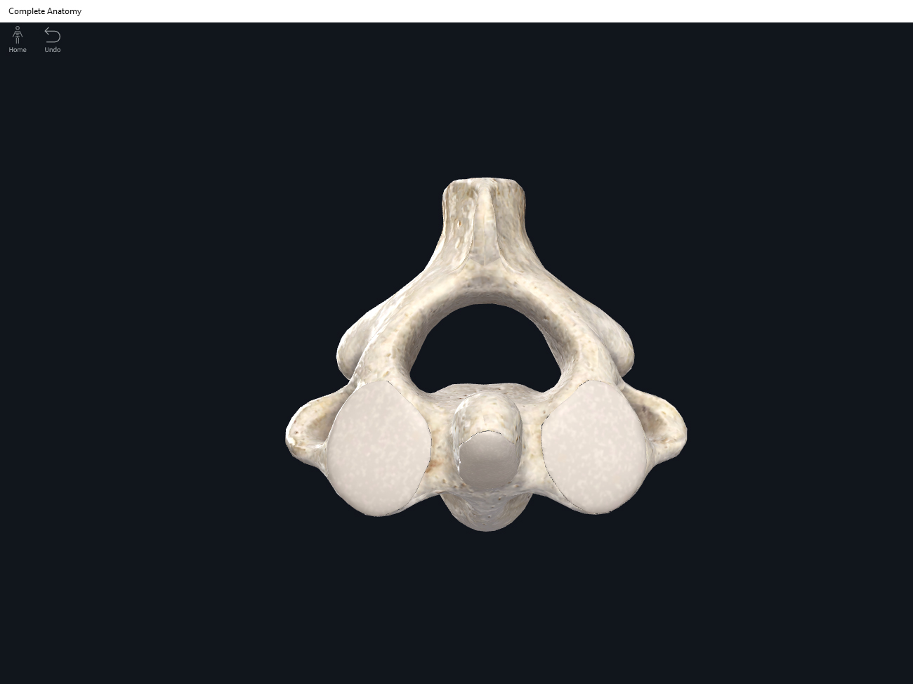
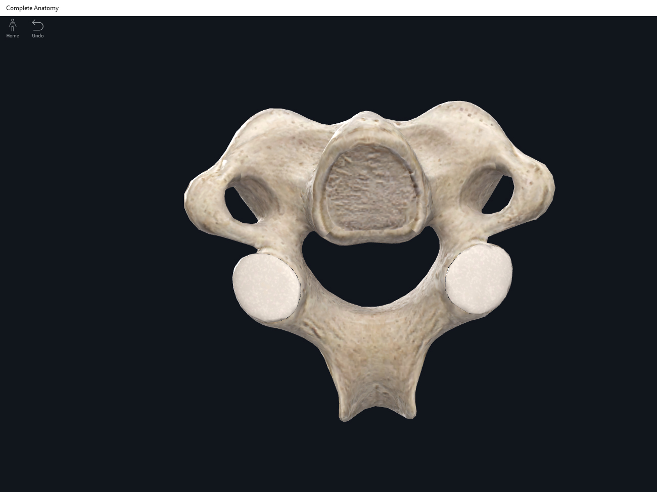
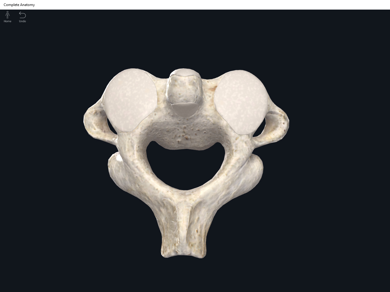
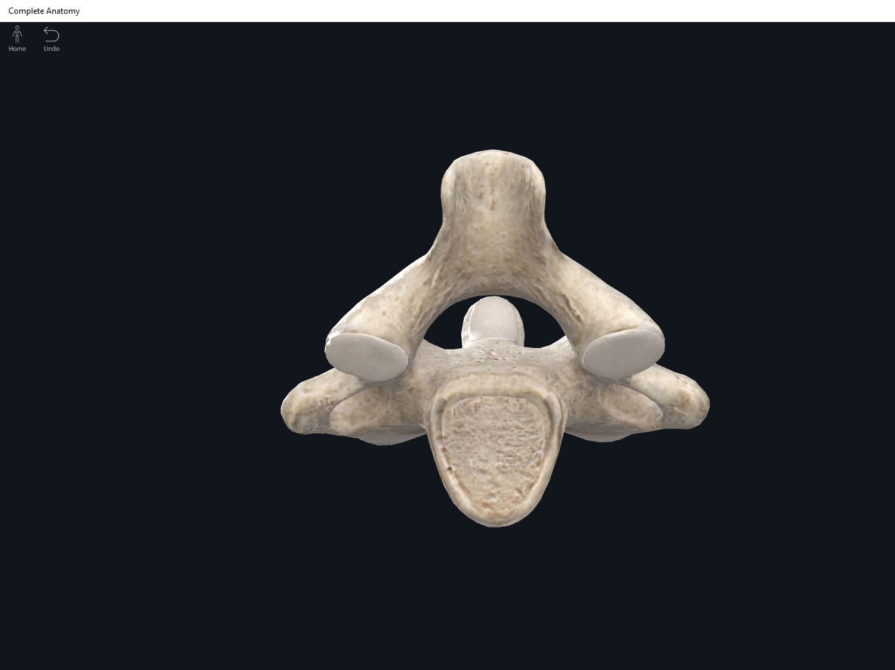
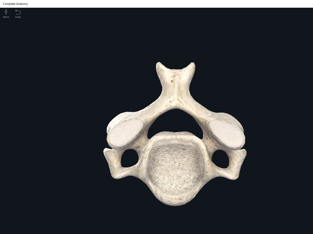
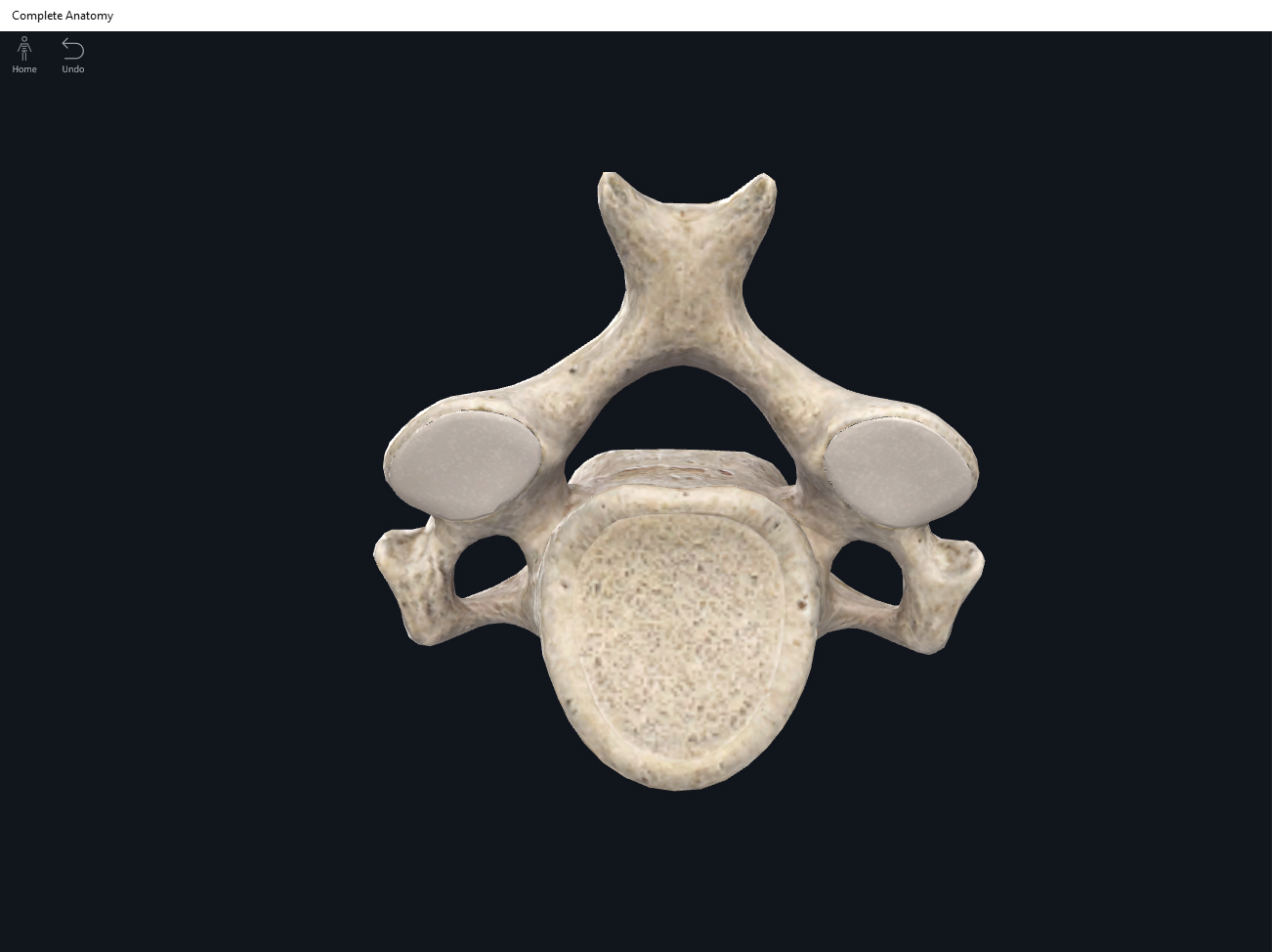
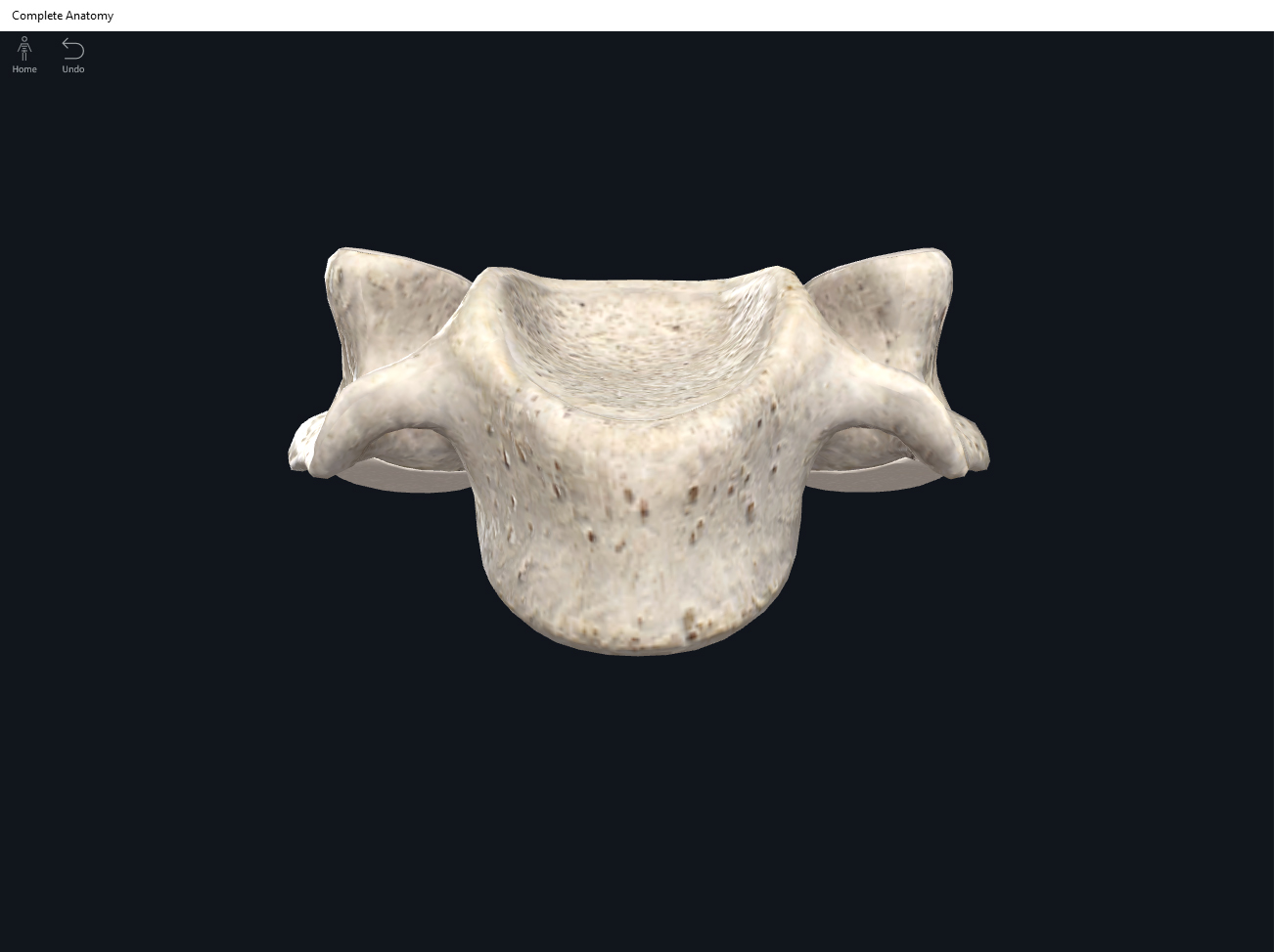
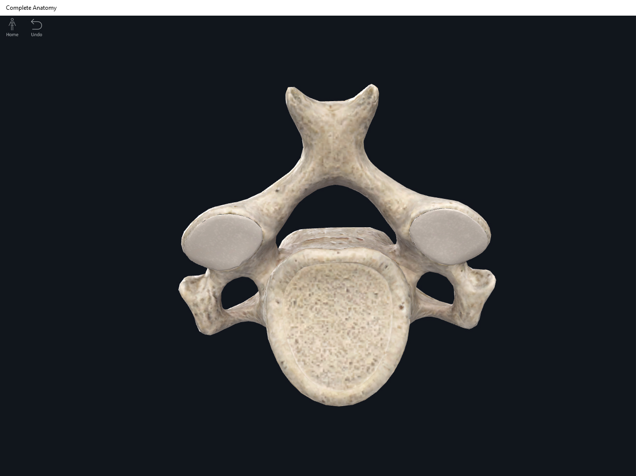
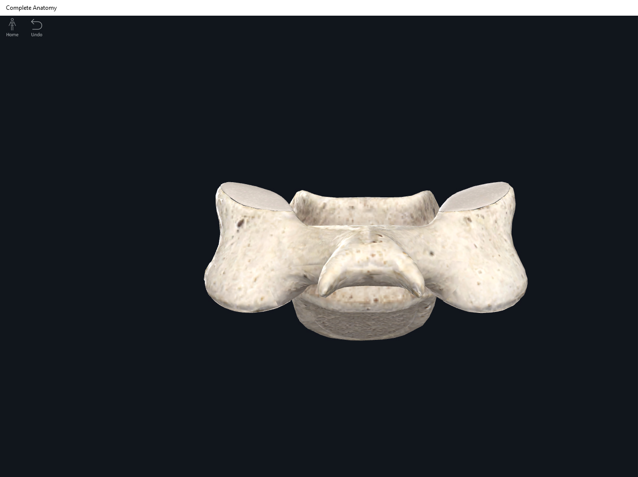
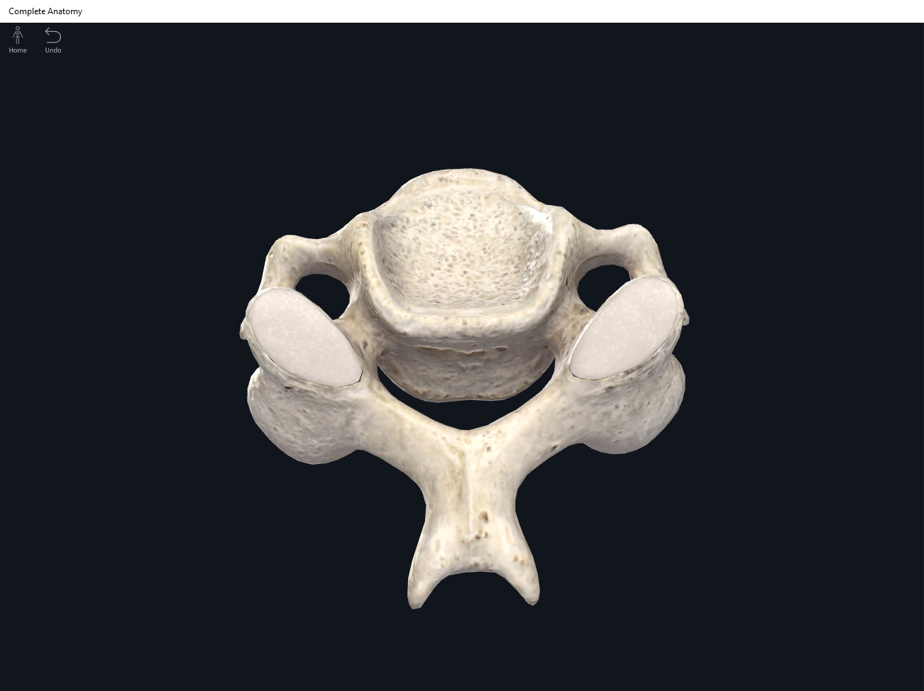
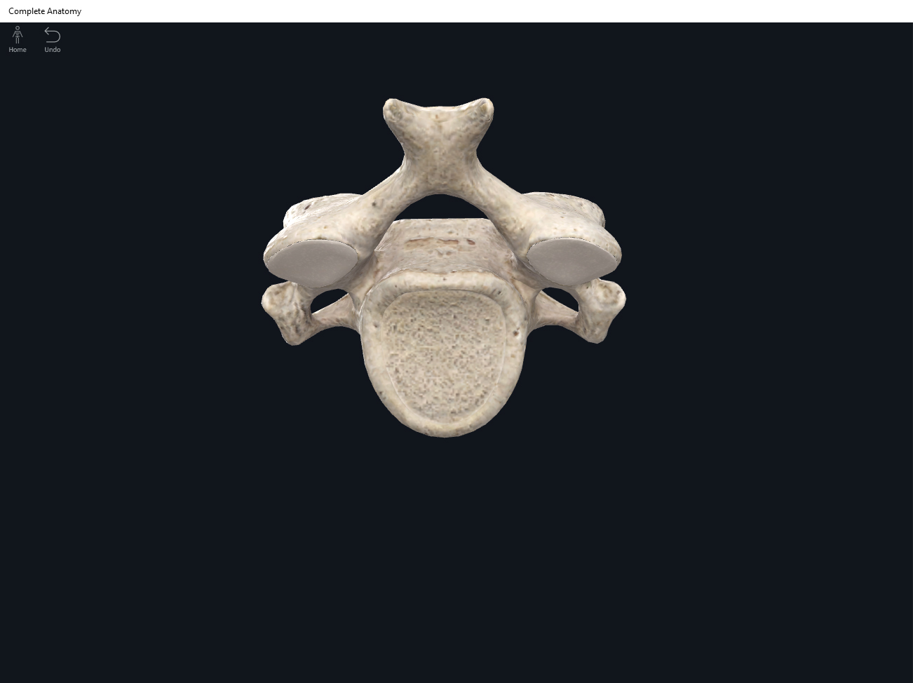
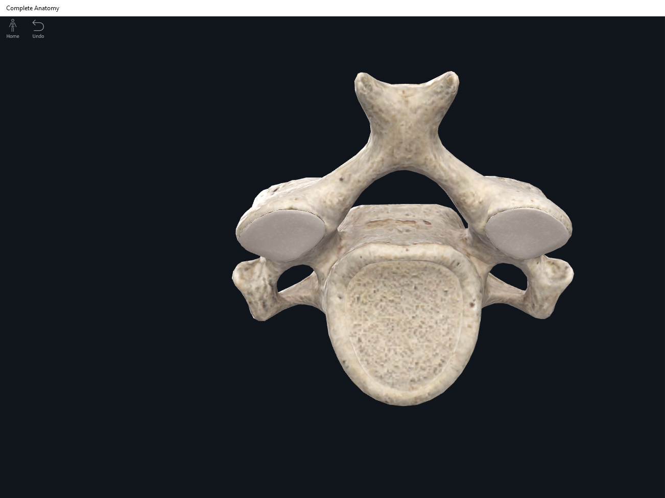
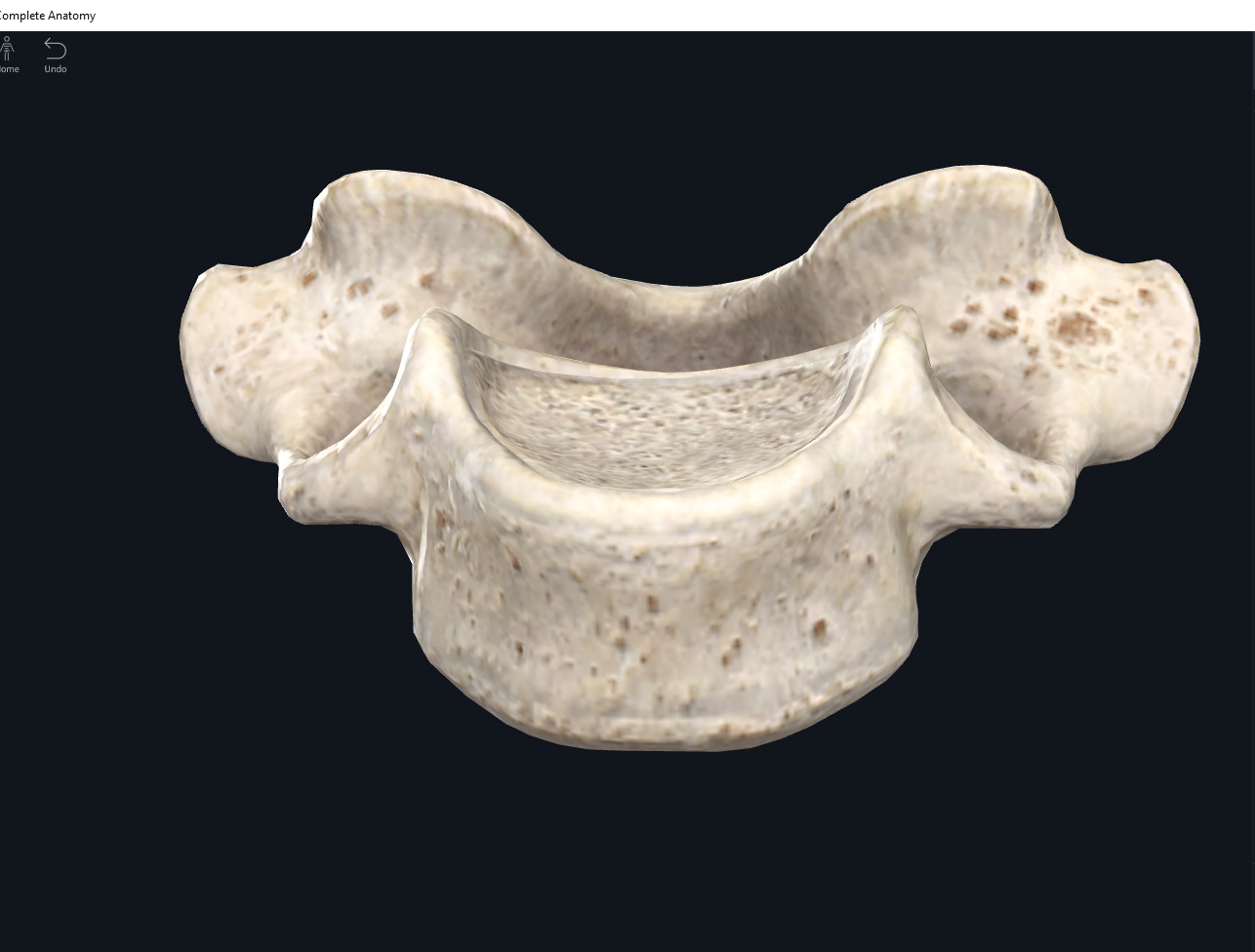
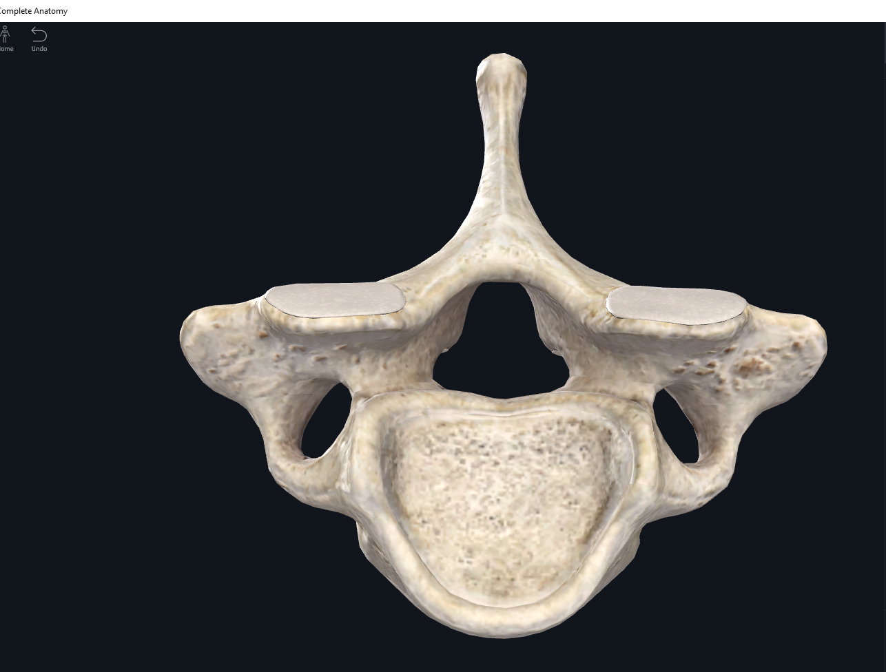
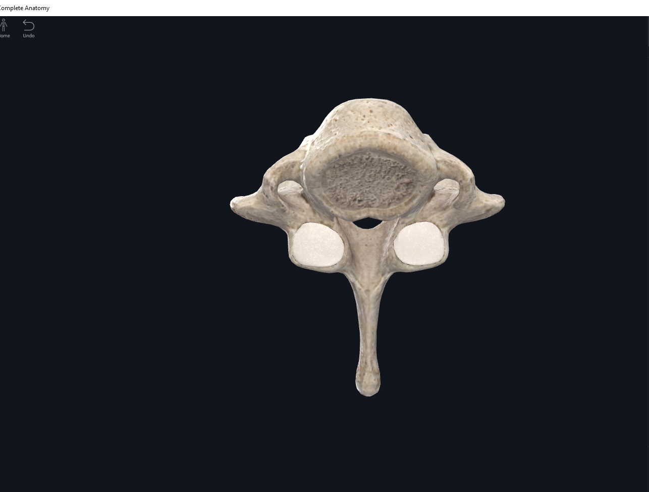
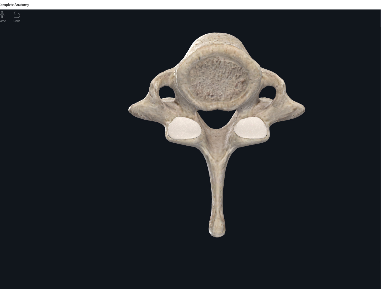
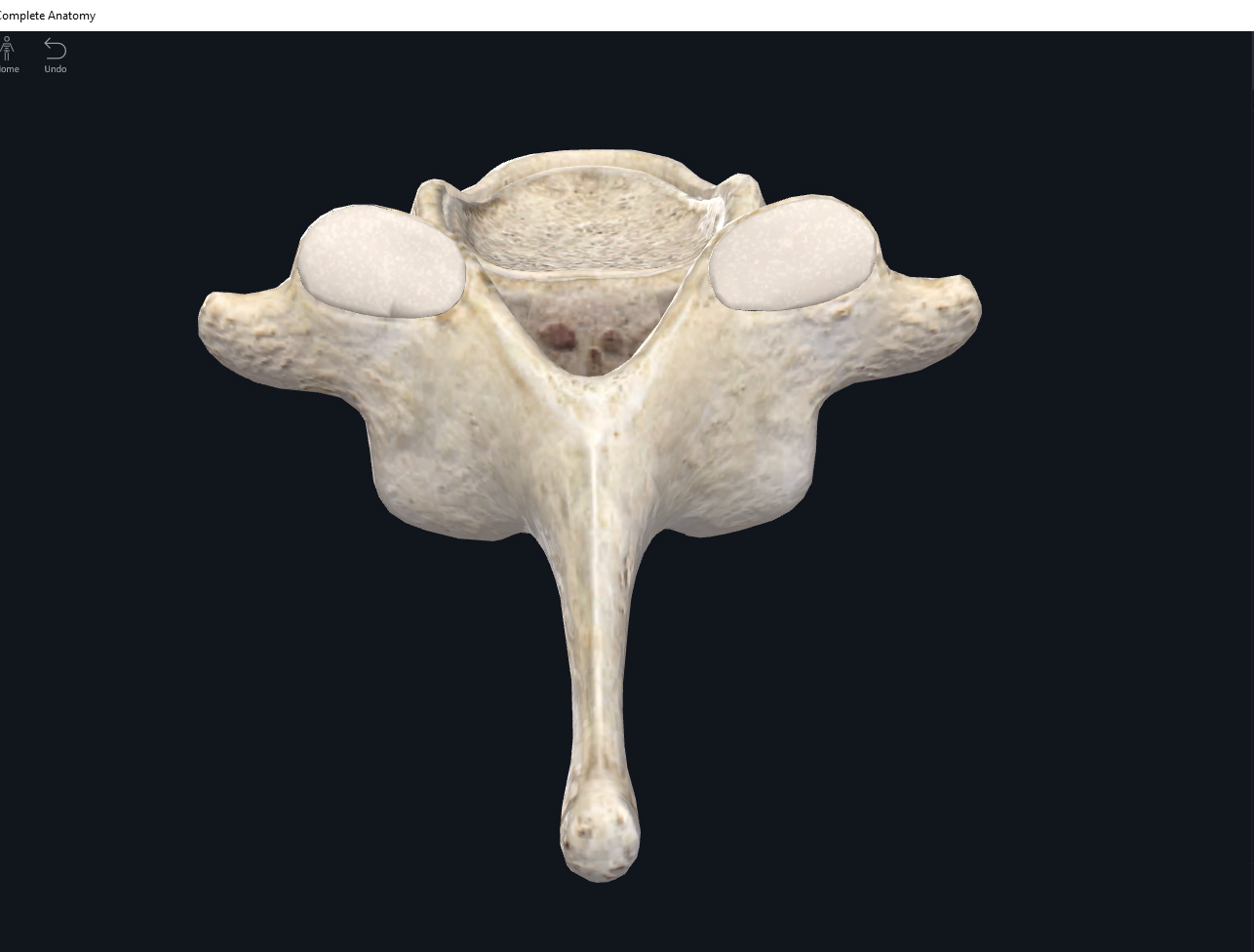
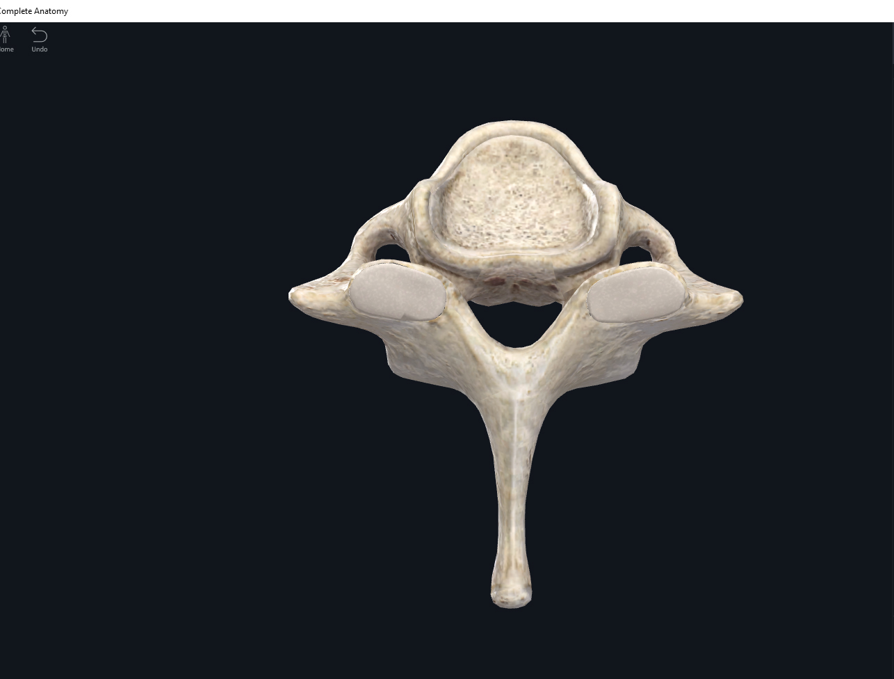
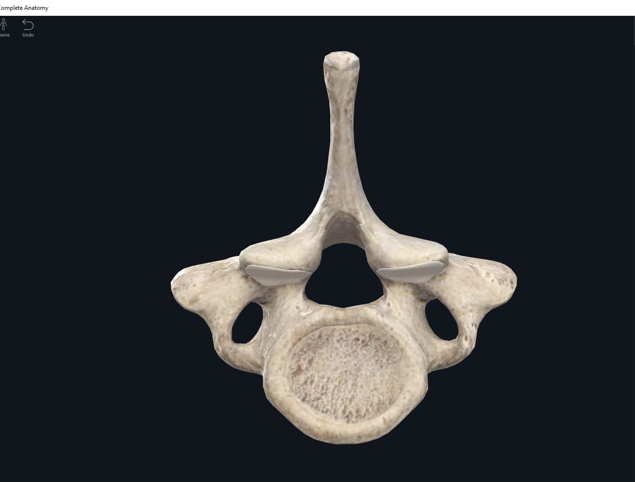
      • Symphasis: ends of bones covered by hyaline cartilage with a broad flattish fibrocartilage connecting the bones. All symphsis occur in the body’s midline. E.g. pubic symphasis; sternal angle between the manubrium and sternal body; intervertebral joints between vertebral bodies.
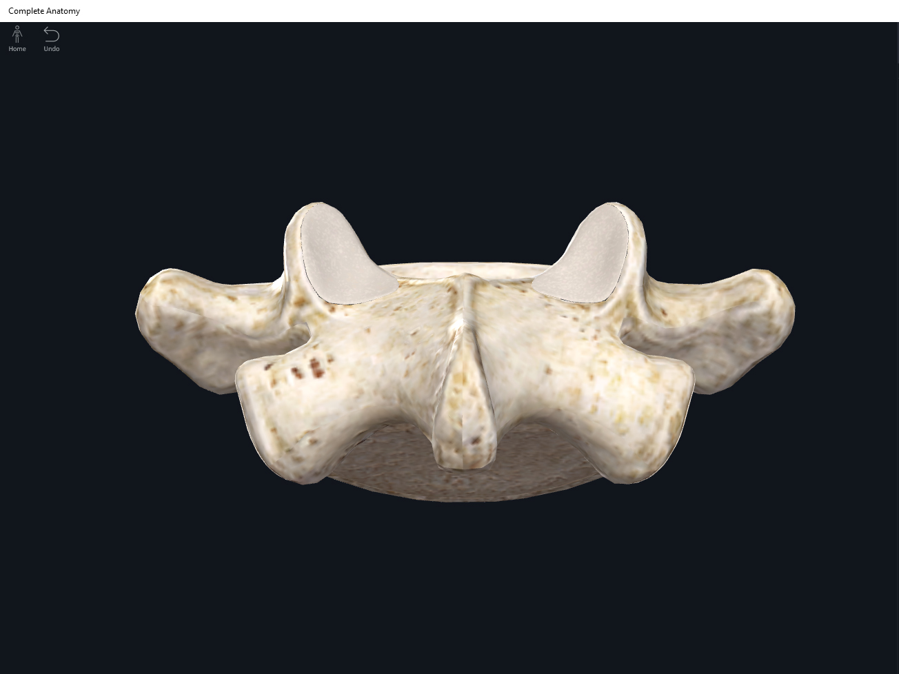
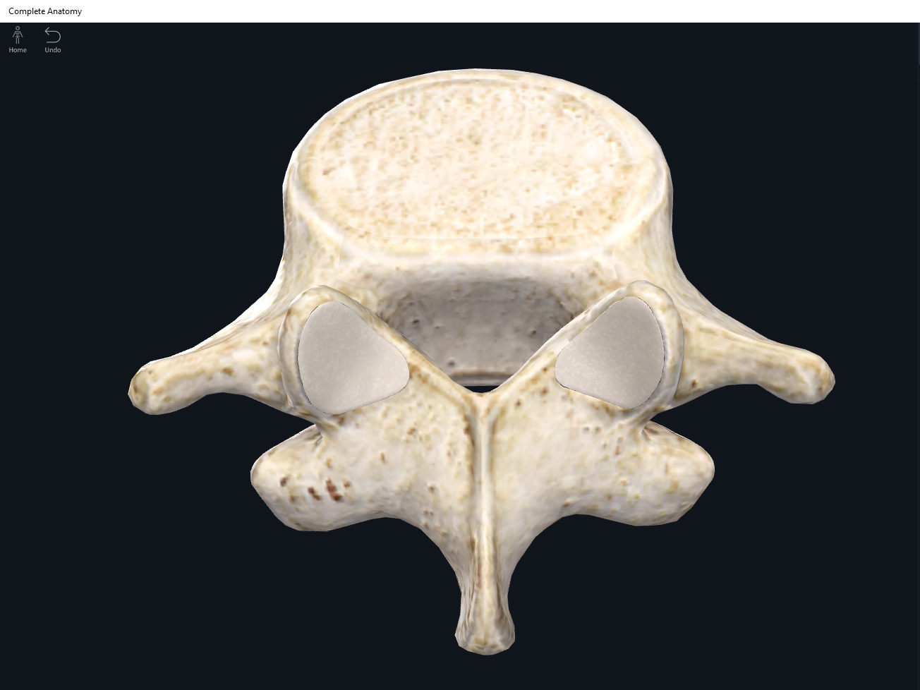
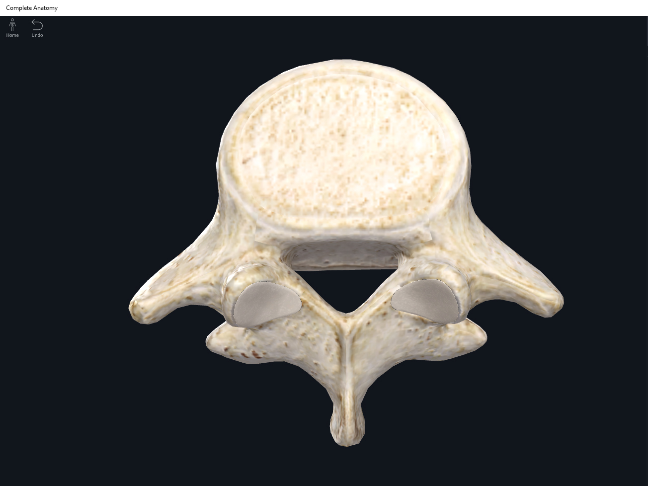
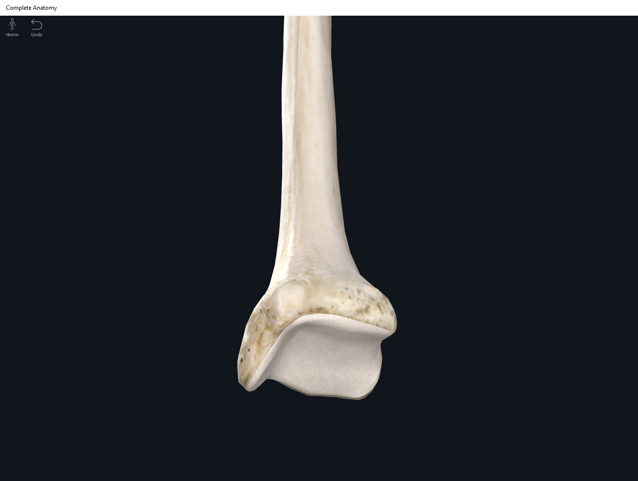
    • Synovial joints: presence of synovial cavity; articular capsule with dense connective tissue and often accessory ligaments present. Diarthrotic (freely moving).
      • Bones covered with articular cartilage (a layer of hyaline) to reduce friction.
      • Articular (joint) capsule: sleevelike; encloses the joint cavity where the two bones articulate with each other. Has 2 layers: fibrous membrane (mostly collagen, dense) attaches to periosteum of bones; synovial membrane (inner membrane) of areolar connective tissue.
      • Articular fat pads: fatty pads act as cushioning.
      • Synovial fluid: lubrication and reduce friction. Clear and viscous. Fibroblast-like cells in synovial membrane secrete this hyaluronic acid. Also some fluid from blood plasma. Has phagocytic cells as “clean-up” crew.
      • Accesory ligaments.
      • Articular menisci: pads of fibrocartilage.
    • Functional classification: based on how much movement is allowed in a joint.
      • Synarthrosis: immoveable.
      • Amphiarthrosis: somewhat moveable.
      • Diarthrosis: freely moveable joint.
    • Bursae: fluid-filled sacs between skin-bones, tendons-bones, muscles-bones, or ligaments-bones.
    • Tendon sheaths: reduce friction at joints. Tubelike bursa wrap around tendons.


Function.


Clinical Significance.


References

Biel, A. (2015). Trail guide to the body: A hands-on guide to locating muscles, bones and more.

Cedars-Sinai. (2018). Vertebrae of the spine. Retrieved from https://www.cedars-sinai.org/health-library/diseases-and-conditions/v/vertebrae-of-the-spine.html

Jenkins, G., & Tortora, G. J. (2012). Anatomy and Physiology: From Science to Life, 3rd Edition International Stu. John Wiley & Sons.

Muscolino, J. E. (2017). The muscular system manual: The skeletal muscles of the human body.


Posted on

Bones: Wrist and Hand.

Download these notes.

Anatomy & Physiology: Bones—Wrist and Hand.

Structure.

  • Wrist (carpus) bones: organized into two rows distal and proximal to the forearm.
    • Proximal row (anterior) starting from medial (pinky side) to lateral: pisiform (mostly viewable from the anterior, sort of lies on top of triquetrum), triquetrum, lunate, scaphoid.
    • Distal row (anterior) starting from medial (pinky side) to lateral:, hamate, capitate, trapezoid, trapezium.
  • Metacarpals: bones between the carpals and phalanges. Start numbering from the thumb (e.g. the first metacarpal or I) to pinky finger (fifth metacarpal or V).
  • Phalanges: Except for the thumb, there are 3 rows of phalanges (proximal, middle, and distal). They are numbered starting from the thumb (e.g. first proximal phalanx) to the pinky (e.g. fifth proximal phalanx).

Function.

Clinical Significance.

References

Biel, A. (2015). Trail guide to the body: A hands-on guide to locating muscles, bones and more.

Cedars-Sinai. (2018). Vertebrae of the spine. Retrieved from https://www.cedars-sinai.org/health-library/diseases-and-conditions/v/vertebrae-of-the-spine.html

Jenkins, G., & Tortora, G. J. (2012). Anatomy and Physiology: From Science to Life, 3rd Edition International Stu. John Wiley & Sons.

Muscolino, J. E. (2017). The muscular system manual: The skeletal muscles of the human body.

Posted on

Bones: Vertebral Column, Thoracic Region.

Download these notes.

Anatomy & Physiology: Bones—Vertebral Column, Thoracic Region.


Structure.

  • The thoracic vertebrae are larger and stronger than the cervical vertebrae.
  • One foramina: vertebral.


  • The spinous processes are long and pointing posterior and inferiorly (downward and back). As compared to the spinous processes of the lumbar region, the thoracic spinous processes resemble the head of a giraffe.
  • The spinous processes of T11 and T12 are “transitional” (into the lumbar vertebrae) in that they are shorter and directed less inferiorly (pointing less downward).
  • Thoracic vertebrae have longer transverse processes as compared to cervical vertebrae.
  • The thoracic vertebrae articulate with the ribs.
  • The body of the thoracic vertebrae have facets/demifacets that articulate with the heads of the ribs.
  • The transverse processes have facets to articulate with the tubercles of ribs.
  • Superior facets: posterolateral.
  • Inferior facets: anteromedial.


Function.


Clinical Significance.


References

Biel, A. (2015). Trail guide to the body: A hands-on guide to locating muscles, bones and more.

Cedars-Sinai. (2018). Vertebrae of the spine. Retrieved from https://www.cedars-sinai.org/health-library/diseases-and-conditions/v/vertebrae-of-the-spine.html

Jenkins, G., & Tortora, G. J. (2012). Anatomy and Physiology: From Science to Life, 3rd Edition International Stu. John Wiley & Sons.

Muscolino, J. E. (2017). The muscular system manual: The skeletal muscles of the human body.

Posted on

Bones: Vertebral Column, Sacrum.

Download these notes.

Anatomy & Physiology: Bones—Vertebral Column, Sacrum.


Structure.

  • Five sacral bones fuse (between ages 16-18) to form the sacrum.
  • Triangular shaped.
  • The sacrum is a keystone structure for the lumbo-pelvic-hip complex.
  • Female sacrum (to accomodate childbirth): shorter, wider, and more curved than male sacrum.
  • The smooth concave portion is directed anteriorly. 4 lines mark the fusion. Bilateral to these lines are 4 sacral foramina from anterior to posterior. Bilaterally like the sacral ala or “wings”.
  • Median sacral crest: fused spinous processes.
  • Lateral sacral crest: fused transverse processes.
  • Sacral hiatus: the gap created when the laminae of the 4th and 5th sacral vertebra fail to meet or fail to align neatly (can happen sometimes).
  • Sacral promontory: the anterior projecting border of the superior sacram. This is used as a landmark for measuring the pelvis.
  • Auricular surface: “ear”; bilaterally. Articulates with the ilium of the hip bone forming the sacroiliac joint (SI joint).
  • Superior articular processes articulate with the inferior articular processes of L5.


Function.


Clinical Significance.


References

Biel, A. (2015). Trail guide to the body: A hands-on guide to locating muscles, bones and more.

Cedars-Sinai. (2018). Vertebrae of the spine. Retrieved from https://www.cedars-sinai.org/health-library/diseases-and-conditions/v/vertebrae-of-the-spine.html

Jenkins, G., & Tortora, G. J. (2012). Anatomy and Physiology: From Science to Life, 3rd Edition International Stu. John Wiley & Sons.

Muscolino, J. E. (2017). The muscular system manual: The skeletal muscles of the human body.

Posted on

Bones: Vertebral Column, Lumbar Region.

Download these notes.

Anatomy & Physiology: Bones—Vertebral Column, Lumbar Region.


Structure.

  • The largest and strongest of the vertebrae as compared to the cervical and thoracic vertebrae.
  • Supports and transmits a low of bodyweight/forces.
  • One foramina: vertebral.
  • Projections are short and thick.
  • The body is thick and substantial.
  • Spinous processes are thick and broad, and resemble mooseheads.
  • Superior articular processes are pointing more medially instead of superiorly.
  • The inferior articular processes are pointing more laterally instead of inferiorly.
  • Superior facets: medial.
  • Inferior facets: lateral.


Function.


Clinical Significance.


References

Biel, A. (2015). Trail guide to the body: A hands-on guide to locating muscles, bones and more.

Cedars-Sinai. (2018). Vertebrae of the spine. Retrieved from https://www.cedars-sinai.org/health-library/diseases-and-conditions/v/vertebrae-of-the-spine.html

Jenkins, G., & Tortora, G. J. (2012). Anatomy and Physiology: From Science to Life, 3rd Edition International Stu. John Wiley & Sons.

Muscolino, J. E. (2017). The muscular system manual: The skeletal muscles of the human body.

Posted on

Bones: Vertebral Column, Coccyx.

Download these notes.

Anatomy & Physiology: Bones—Vertebral Column, Coccyx.


Structure.

  • The adult coccyx is formed from the fusion of 4 coccygeal vertebrae (Co1-Co4) between the ages of 20-30 years old.
  • Triangular shape.
  • Superior articular processes of Co1 articulates superiorly with the sacrum.
  • Female: coccyx is pointing inferiorly to allow passage for birth.
  • Male: coccyx points anteriorly.


Function.


Clinical Significance.


References

Biel, A. (2015). Trail guide to the body: A hands-on guide to locating muscles, bones and more.

Cedars-Sinai. (2018). Vertebrae of the spine. Retrieved from https://www.cedars-sinai.org/health-library/diseases-and-conditions/v/vertebrae-of-the-spine.html

Jenkins, G., & Tortora, G. J. (2012). Anatomy and Physiology: From Science to Life, 3rd Edition International Stu. John Wiley & Sons.

Muscolino, J. E. (2017). The muscular system manual: The skeletal muscles of the human body.

Posted on

Bones: Vertebral Column, Cervical Region.

Download these notes.

Anatomy & Physiology: Bones—Vertebral Column, Cervical Region.


Structure.

  • Cervical vertebrae: C1 (atlas), C2 (axis), C3-C7. (Remember breakfast at 7). C7 known as the vertebral prominens as it distinctly protrudes at the base of the neck at the transition to the thoracic vertebrae.
  • Except for the sacrum, the cervical vertebrae are smaller than the thoracic and lumbar vertebrae.
  • All cervical vertebrae have 3 foramina: vertebral (x1) and transverse (x2).
  • The vertebral foramen is the largest (due to the enlargement of the spinal cord at top) as compared to the thoracic and lumbar vertebrae.
  • The vertebral artery passes through the transverse foramina.
  • Spinous processes of C2-C6 are often bifid (split into two small processes at the terminal end) and slender.
  • Small transverse processes.
  • Superior facets: posterosuperior.
  • Inferior facets: anteroinferior.
  • C1 (atlas): supports head. Has no body and no spinous process. Ring-shaped with anterior and posterior arches. The superior portion have two superior articular facets which articulate with the occipital condyles (allows for the head to nod “yes”). The inferior articular facets articulate with C2. The anterior portion articulates with the dens (tooth-like projection) of C2 and this complex allows for the turning of the head (like turning the head to say “no”).
  • C2 (axis): the superior articular facets articulate with C1; the inferior articular facets articulate with C3. The dens is a peg/tooth-like projection that articulates with the anterior portion of C1 and allows the head to turn (as if to say “no”).
  • C7 (vertebra prominens): has a very prominent spinous process (non-bifid) that may be felt/palpated. The spinous process C7 makes sense as it is a transition from the cervical to the thoracic vertebrae.

Function.


Clinical Significance.


References

Biel, A. (2015). Trail guide to the body: A hands-on guide to locating muscles, bones and more.

Cedars-Sinai. (2018). Vertebrae of the spine. Retrieved from https://www.cedars-sinai.org/health-library/diseases-and-conditions/v/vertebrae-of-the-spine.html

Jenkins, G., & Tortora, G. J. (2012). Anatomy and Physiology: From Science to Life, 3rd Edition International Stu. John Wiley & Sons.

Muscolino, J. E. (2017). The muscular system manual: The skeletal muscles of the human body.

Posted on

Bones: Vertebral Column.

Download these notes.

Anatomy & Physiology: Bones—Vertebral Column.


Structure.

  • Cervical vertebrae: C1 (atlas), C2 (axis), C3-C7. (Remember breakfast at 7). C7 known as the vertebral prominens as it distinctly protrudes at the base of the neck at the transition to the thoracic vertebrae.
  • Thorax vertebrae: T1-T12. (Remember lunch at noon, 12).
  • Lumbar vertebrae: L1-L5. (Remember dinner at 5).
  • Sacrum: S1-S5, 5 fused vertebrae.
  • Coccyx: 4 fused coccygeal vertebrae. “Coccyx” the cuckoo bird.
  • Concave “caving in”. Convex “bulging outward”.
  • Intervertebral discs: between the vertebrae.
    • Annulus fibrosus: outer fibrous ring made of fibrocartilage.
    • Nucleus pulposus: inner part of disk that is soft, pulpy, and elastic.
  • Common structures of the vertebrae are: vertebral body, vertebral arch, and processes.
  • Vertebral arch: when looking top-down (bird’s eye view) onto a “generic” vertebra, the rounded mass anteriorly is the “body” and “everything else” is the “arch”.
    • The pedicles (2 per vertebrate) are the short, stumpy “feet” that connect the rest of the arch to the body. You can imagine them as trunks.
    • The vertebral foramen is the hole between the arch and body for the spinal cord to pass through. It’s sometimes called the spinal canal in the vernacular. The vertebral foramen contains: spinal cord, adipose, areolar connective tissue, and blood vessels.
    • Intervertebral foramen is the space or “hole” that is formed when the vertebrae are stacked together. Each intervertebral foramen allows for a single spinal nerve to pass through.
    • The spinous process is the most significant posterior projection.
    • Lamina: located on either side of the spinous process is like a “roof” supporting and protecting the spinal cord.


  • Processes: 7 projections called processes originate from the vertebral arch.
    • Transverse process (x2): it is the projection between the pedicle and lamina. Attachment points for muscles.
    • Spinous process (x1): the most significant posterior projection. Forms the highest point in the arch. Attachment point for muscles.
    • Superior articular processes (x2): between the pedicle and transverse process, lies a slight projection with a concave “seat”, the facet. The facet of the superior process articulates with the inferior process of the vertebrate above. The facet are the articulating surfaces and are covered in hyaline cartalige.
    • Inferior articular processes (x2): located on the underside of the vertebra articulate with the superior articular processes on the vertebra below.


Function.


Clinical Significance.


References

Biel, A. (2015). Trail guide to the body: A hands-on guide to locating muscles, bones and more.

Cedars-Sinai. (2018). Vertebrae of the spine. Retrieved from https://www.cedars-sinai.org/health-library/diseases-and-conditions/v/vertebrae-of-the-spine.html

Jenkins, G., & Tortora, G. J. (2012). Anatomy and Physiology: From Science to Life, 3rd Edition International Stu. John Wiley & Sons.

Muscolino, J. E. (2017). The muscular system manual: The skeletal muscles of the human body.

Posted on

Bones: Ulna.

Download these notes.

Anatomy & Physiology: Bones—Ulna.


Structure.

  • The forearm bone on the “pinky” little finger side.
  • Is larger, longer, and more massive than the radius.
  • Olecranon: located proximally forms the elbow, and is easily palpateable. When the elbow is extended the olecranon tucks into the olecranon fossa of the humerus.
  • Coronoid process: a projection on the anterior proximal end of the ulna. This process tucks into the coronoid fossa of the humerus.
  • Trochlear notch: articulates with the trochlea of the humerus. It lies between the olecranon and coronoid process.
  • Radial notch: located lateral and inferior to the coronoid process, this notch articulates with the radial head.
  • Head: located on the distal end of the ulna.
  • Styloid process: distal and posterior end of the ulna.
  • Ulnar tuberosity.


Function.


Clinical Significance.


References

Biel, A. (2015). Trail guide to the body: A hands-on guide to locating muscles, bones and more.

Cedars-Sinai. (2018). Vertebrae of the spine. Retrieved from https://www.cedars-sinai.org/health-library/diseases-and-conditions/v/vertebrae-of-the-spine.html

Jenkins, G., & Tortora, G. J. (2012). Anatomy and Physiology: From Science to Life, 3rd Edition International Stu. John Wiley & Sons.

Muscolino, J. E. (2017). The muscular system manual: The skeletal muscles of the human body.

Posted on

Bones: Tibia.

Download these notes.

Anatomy & Physiology: Bones—Tibia.


Structure.

  • The shin bone.
  • Articulates with: femur and fibula (proximallly); fibula and talus (distally).
  • Lateral condyle: inferior surface of latereal condyle articulates with the fibula.
  • Medial condyle.
  • Intercondylar eminence: a ridge on the condylar surface.
  • Tibial tuberosity: anterior superior surface; easily palpateable.
  • Anterior border: the shin.
  • Medial malleolus: distil end that articulates with the talus.
  • Fibular notch: distil end; articulates with fibula.


Function.


Clinical Significance.


References

Biel, A. (2015). Trail guide to the body: A hands-on guide to locating muscles, bones and more.

Cedars-Sinai. (2018). Vertebrae of the spine. Retrieved from https://www.cedars-sinai.org/health-library/diseases-and-conditions/v/vertebrae-of-the-spine.html

Jenkins, G., & Tortora, G. J. (2012). Anatomy and Physiology: From Science to Life, 3rd Edition International Stu. John Wiley & Sons.

Muscolino, J. E. (2017). The muscular system manual: The skeletal muscles of the human body.