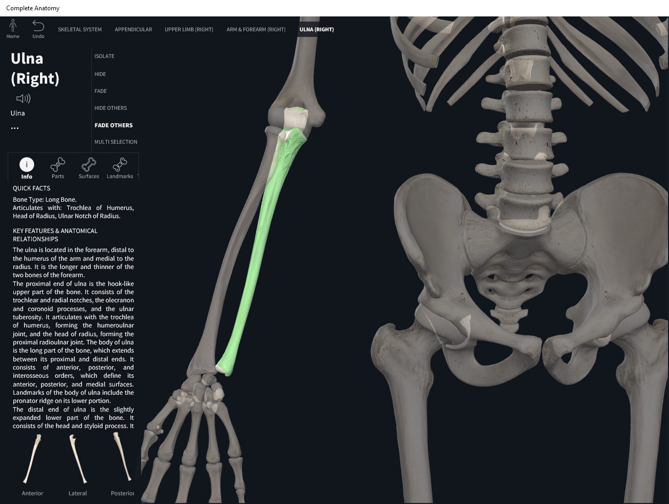
Anatomy & Physiology: Bones—Wrist and Hand.
Structure.
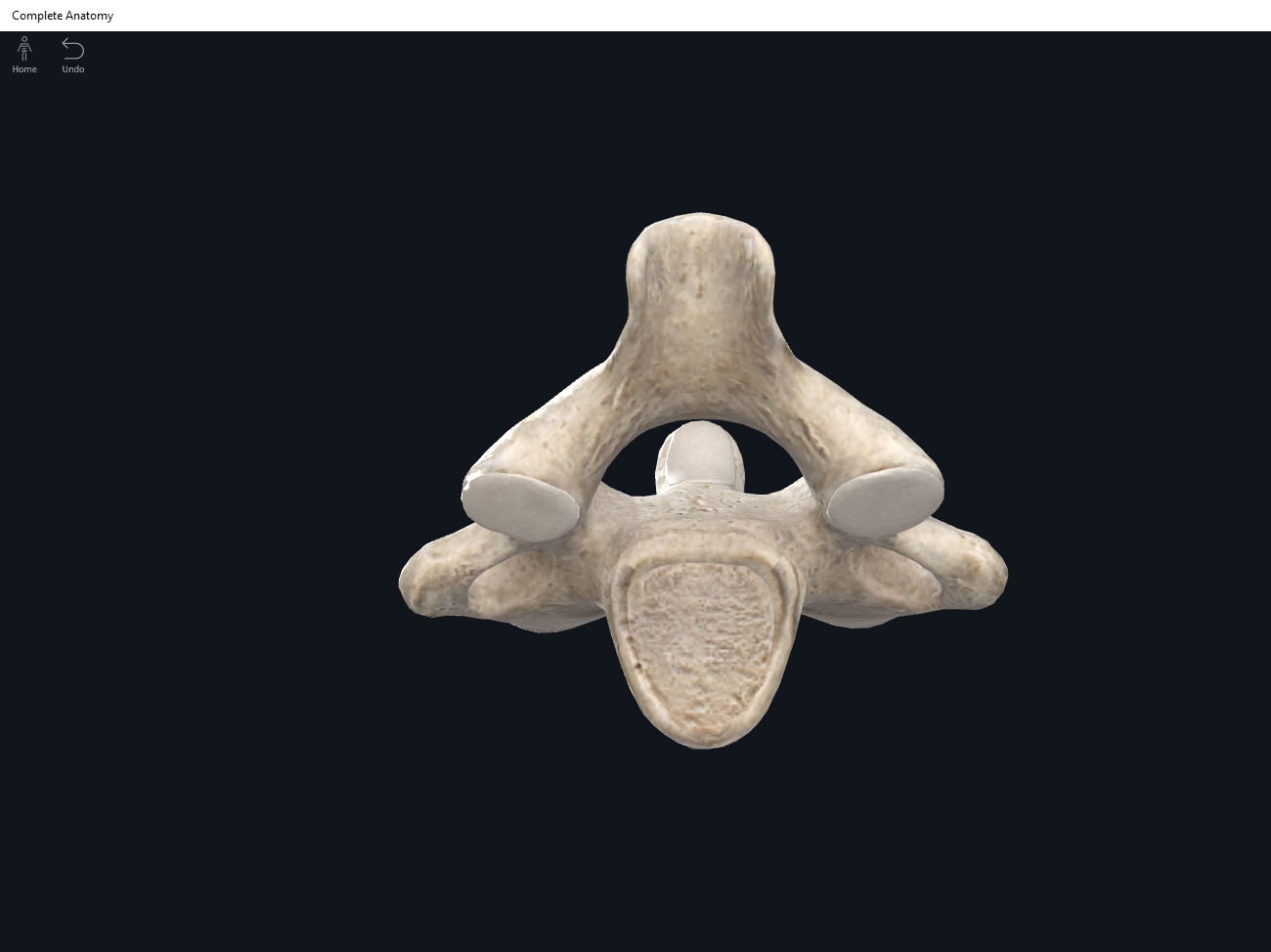
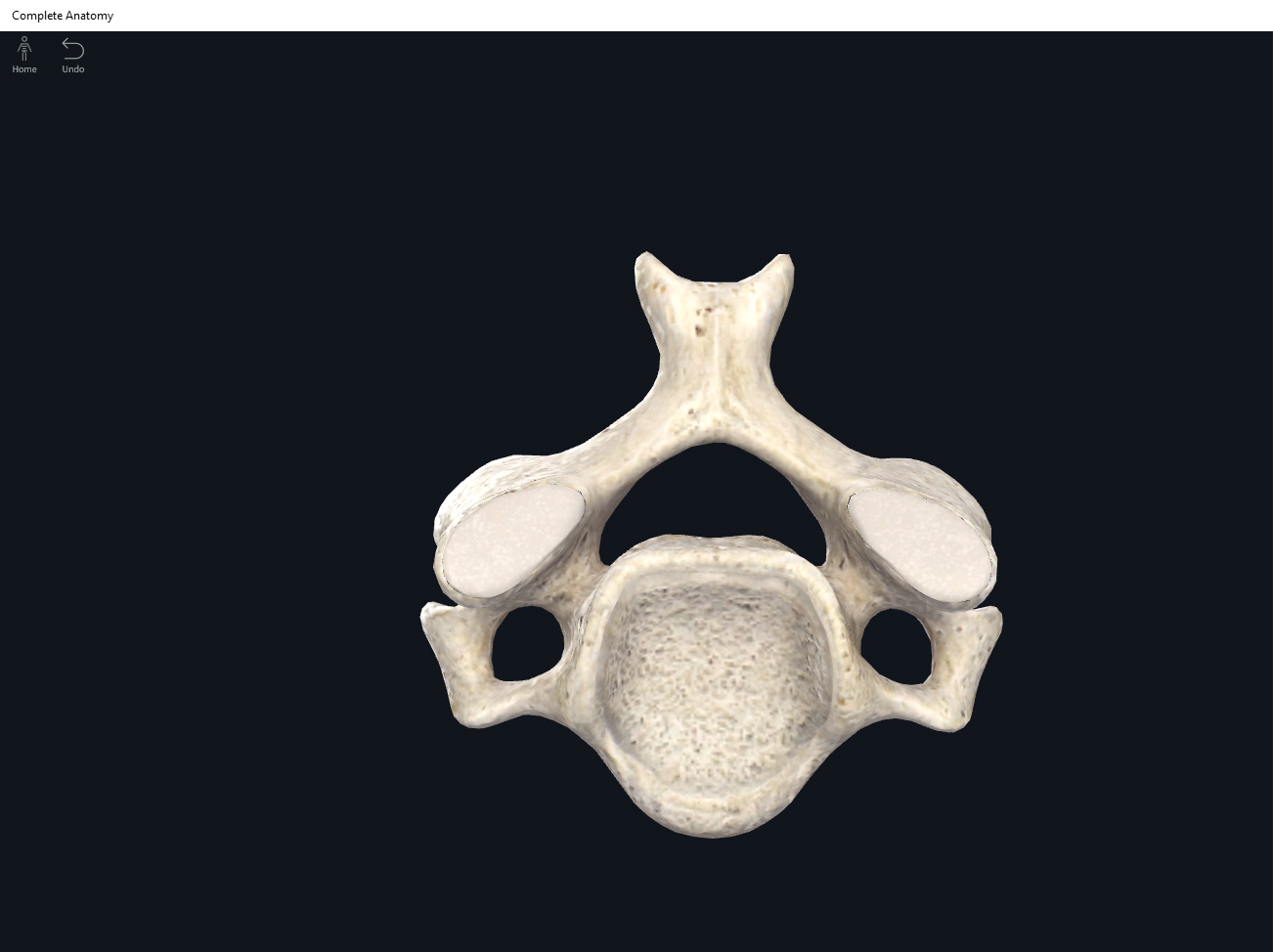
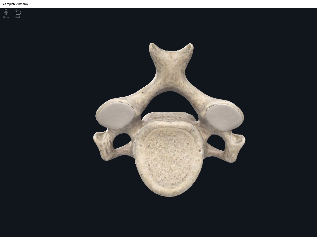
- Wrist (carpus) bones: organized into two rows distal and proximal to the forearm.
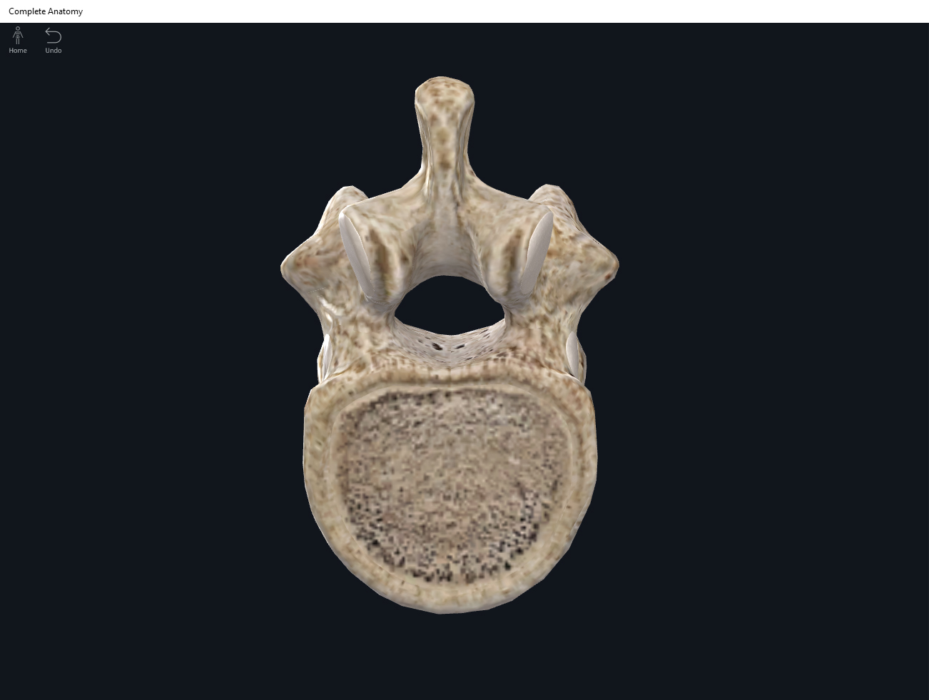
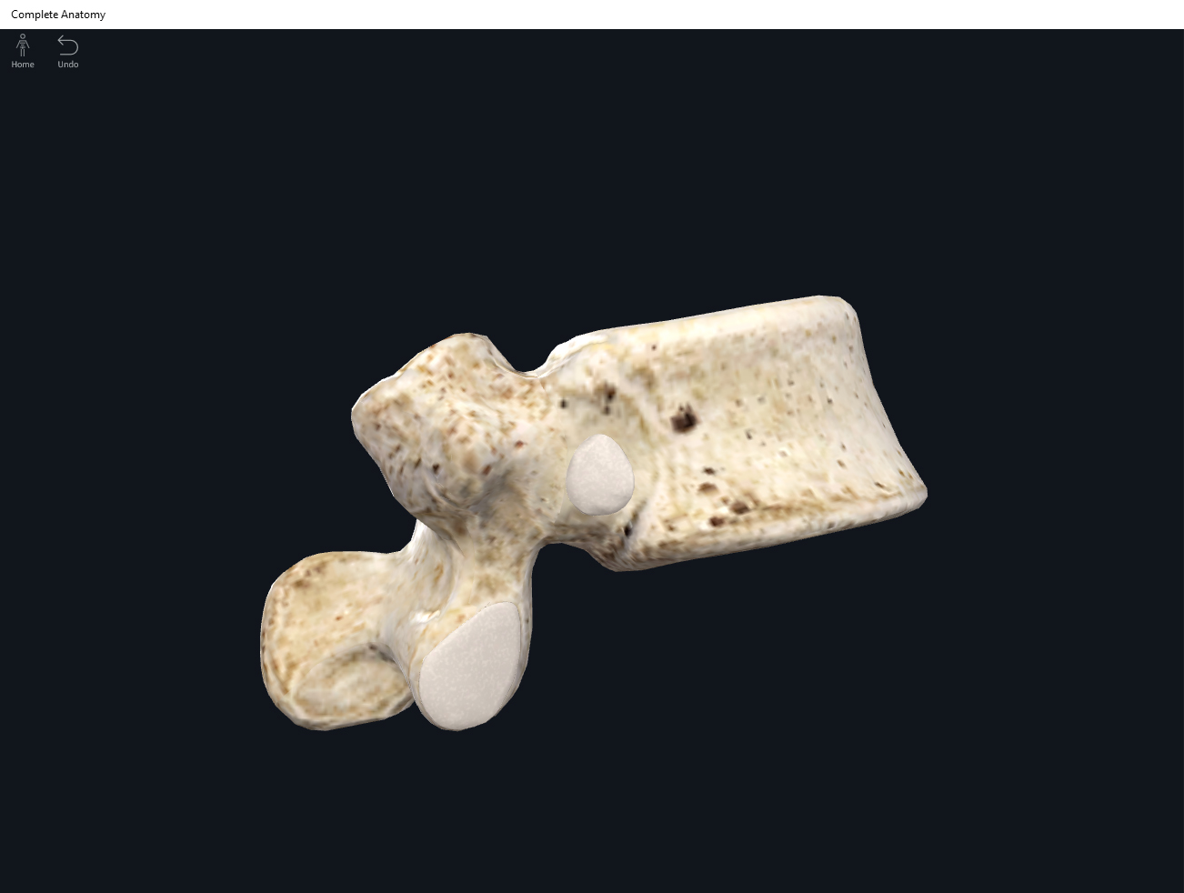
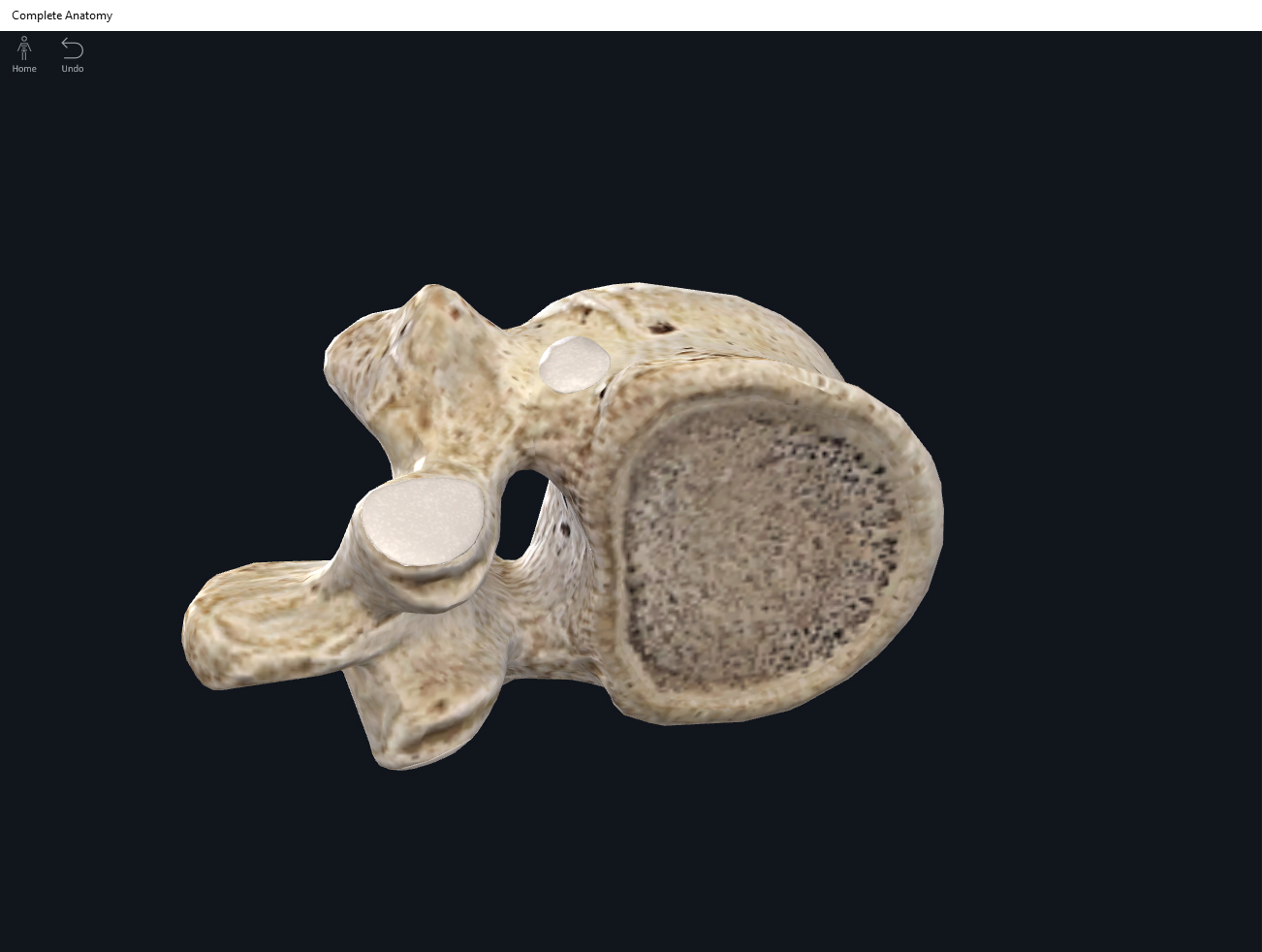
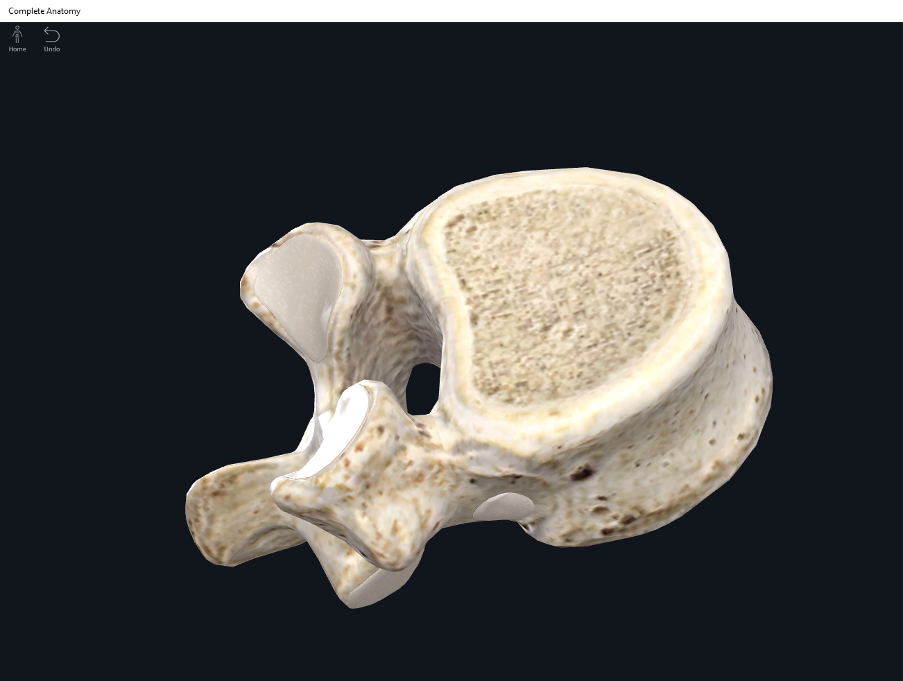
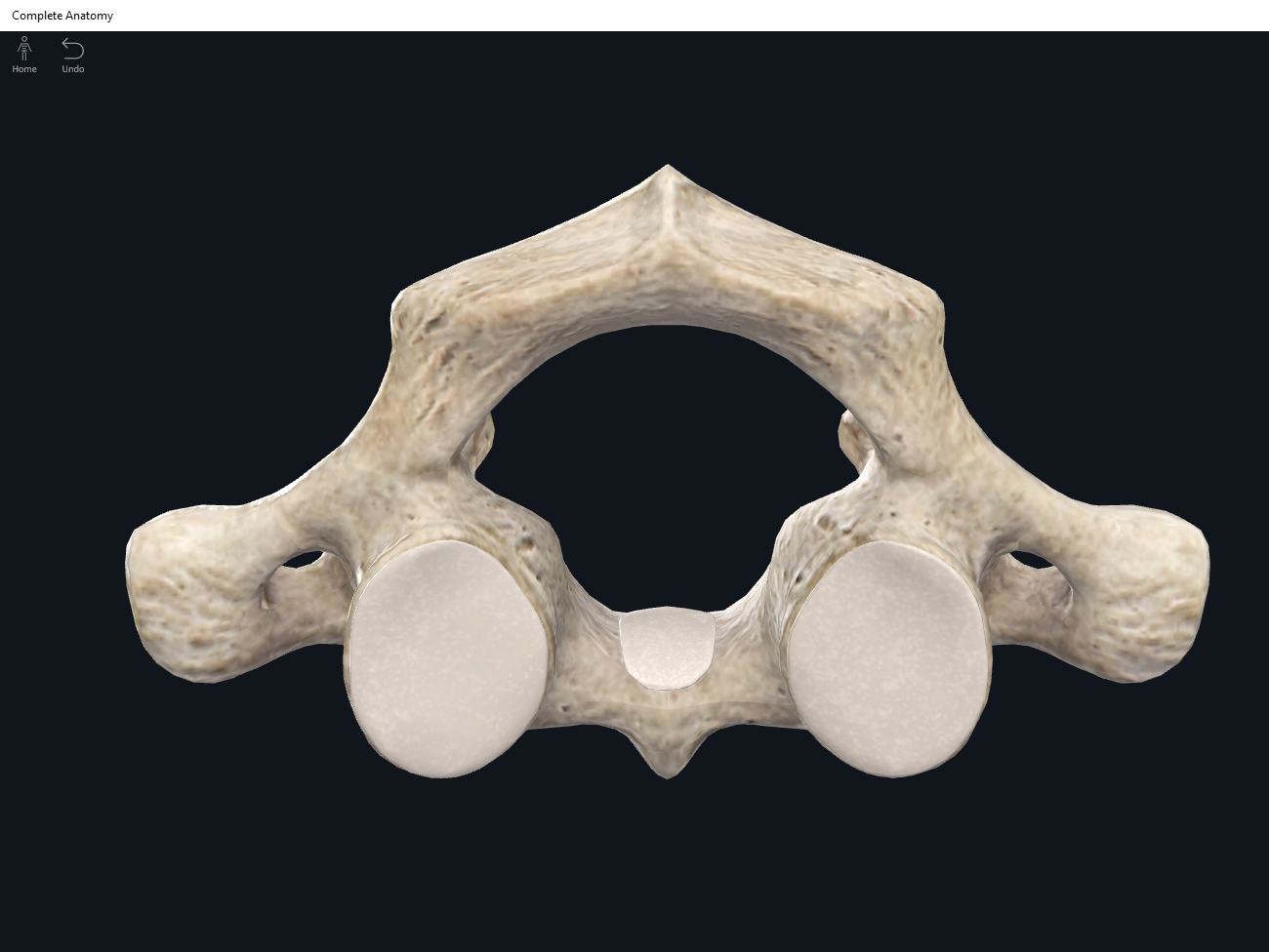
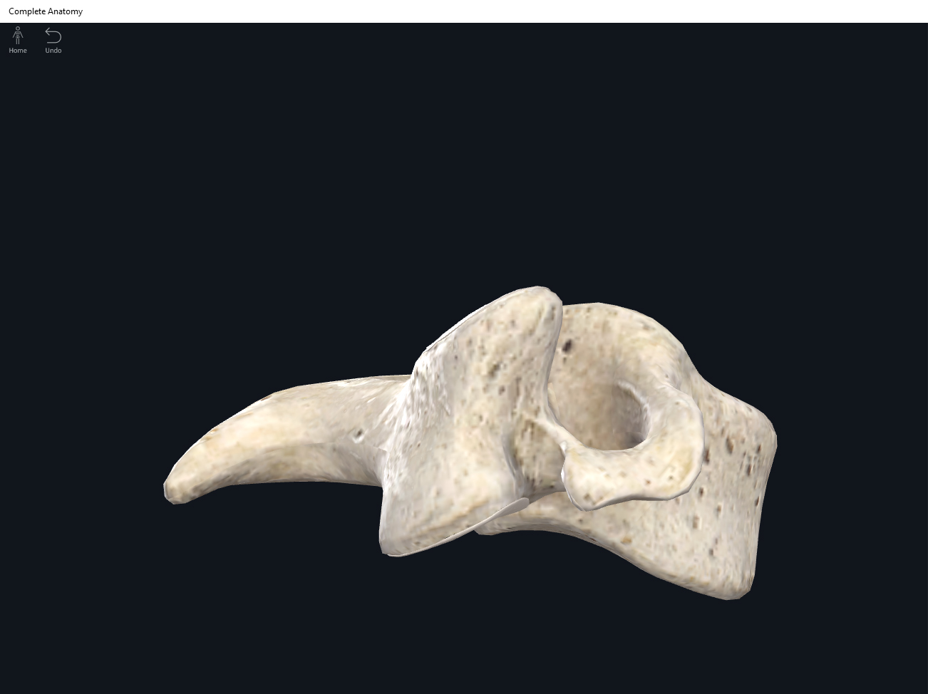
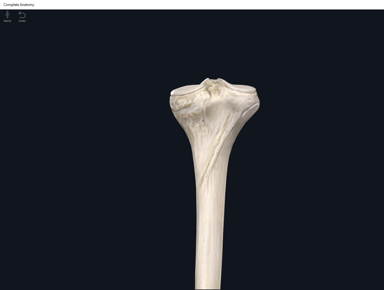
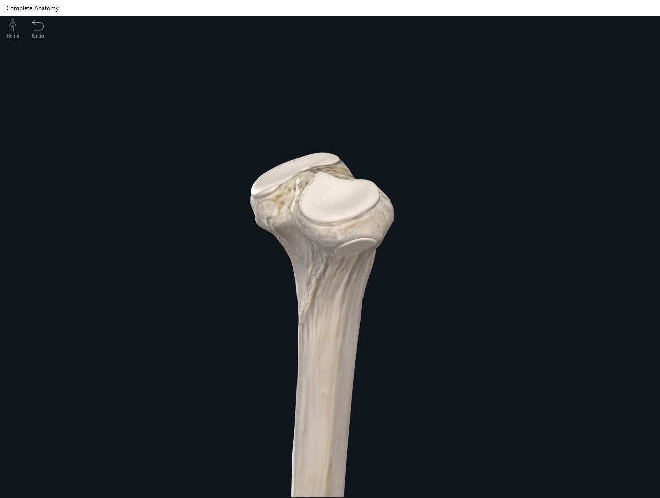
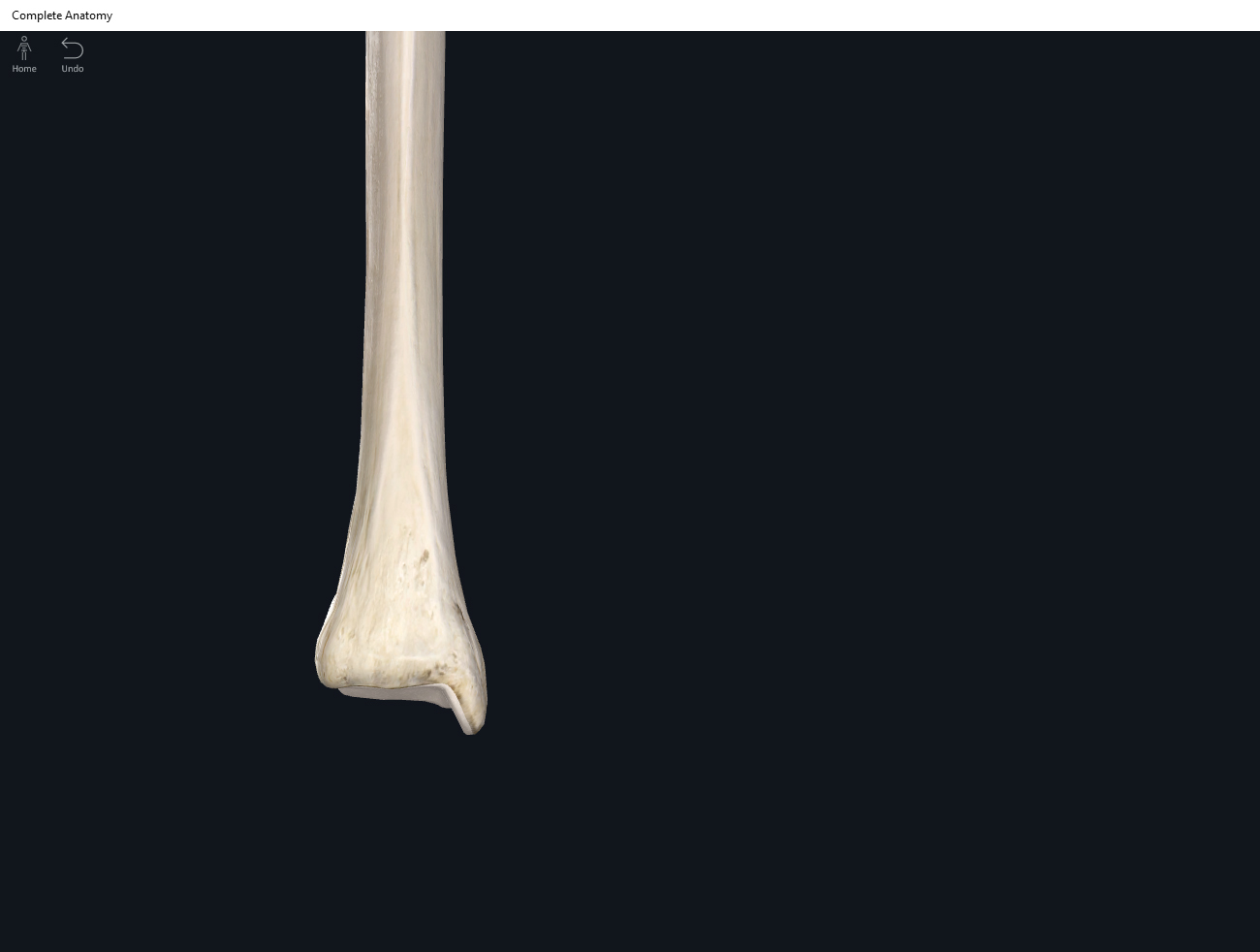
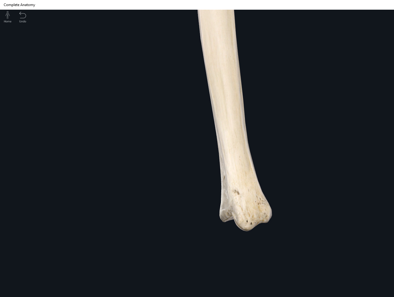
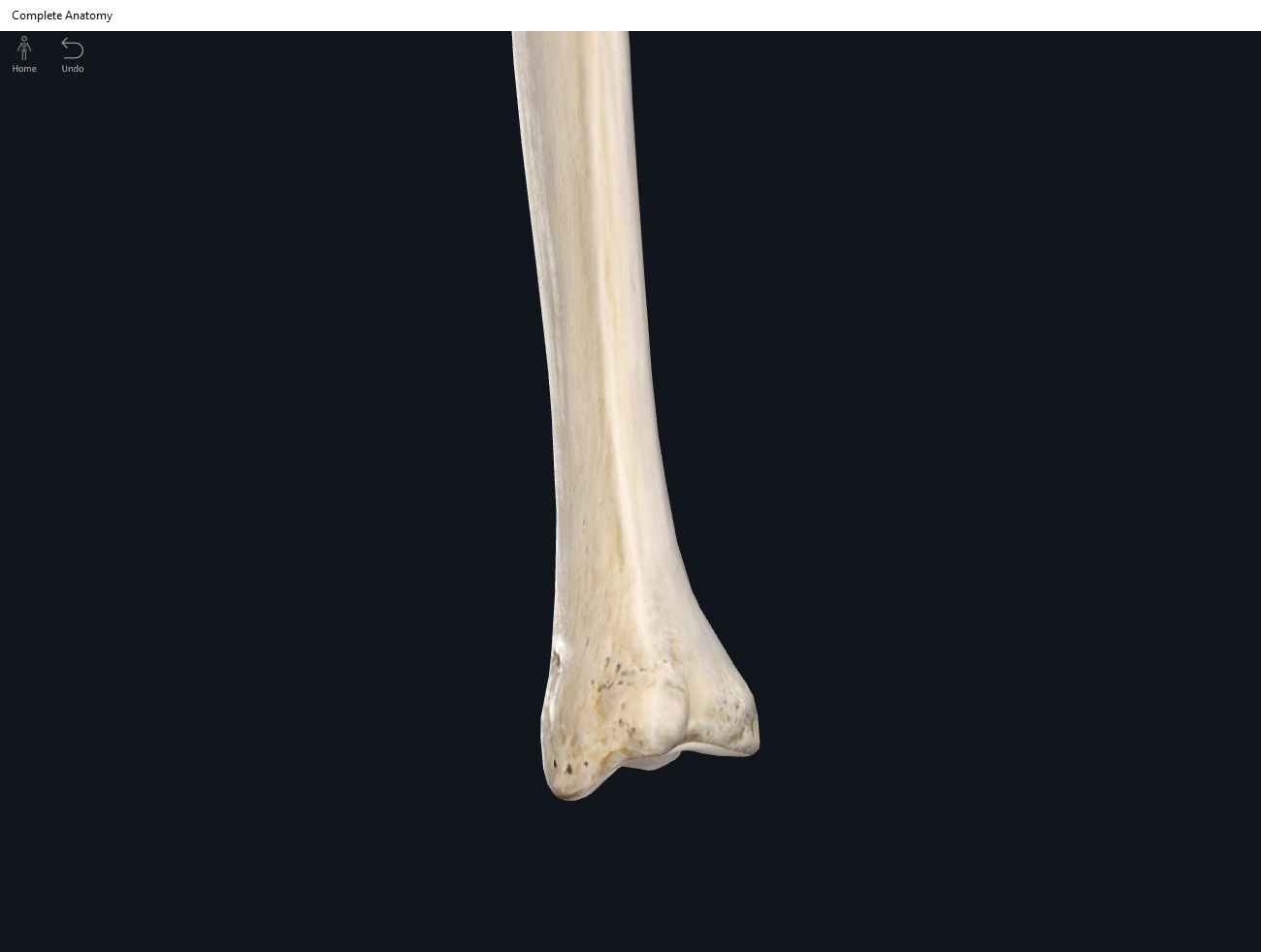
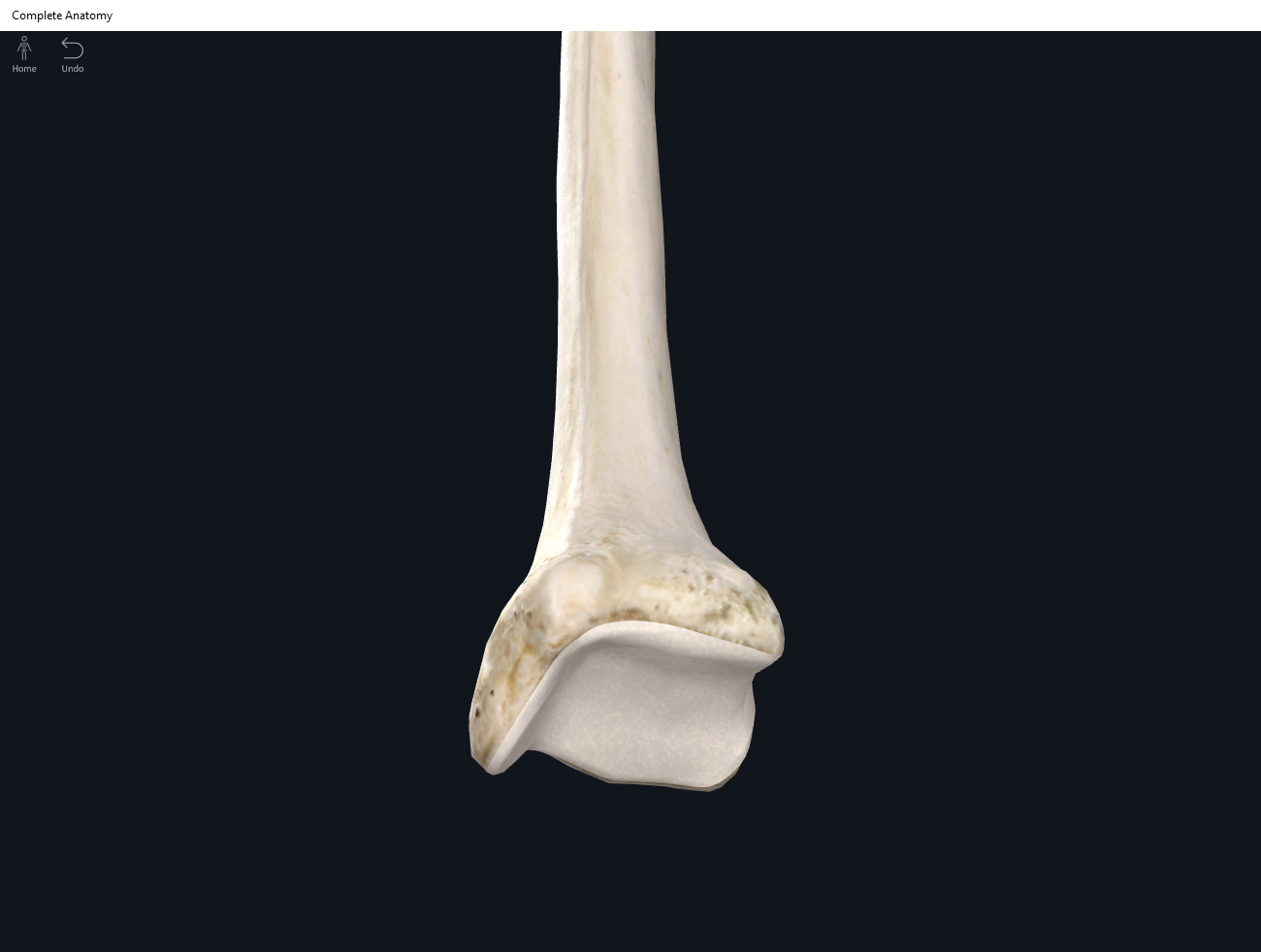
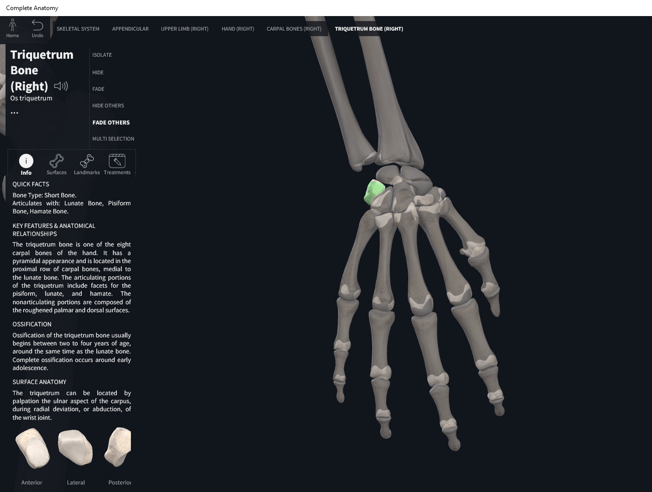
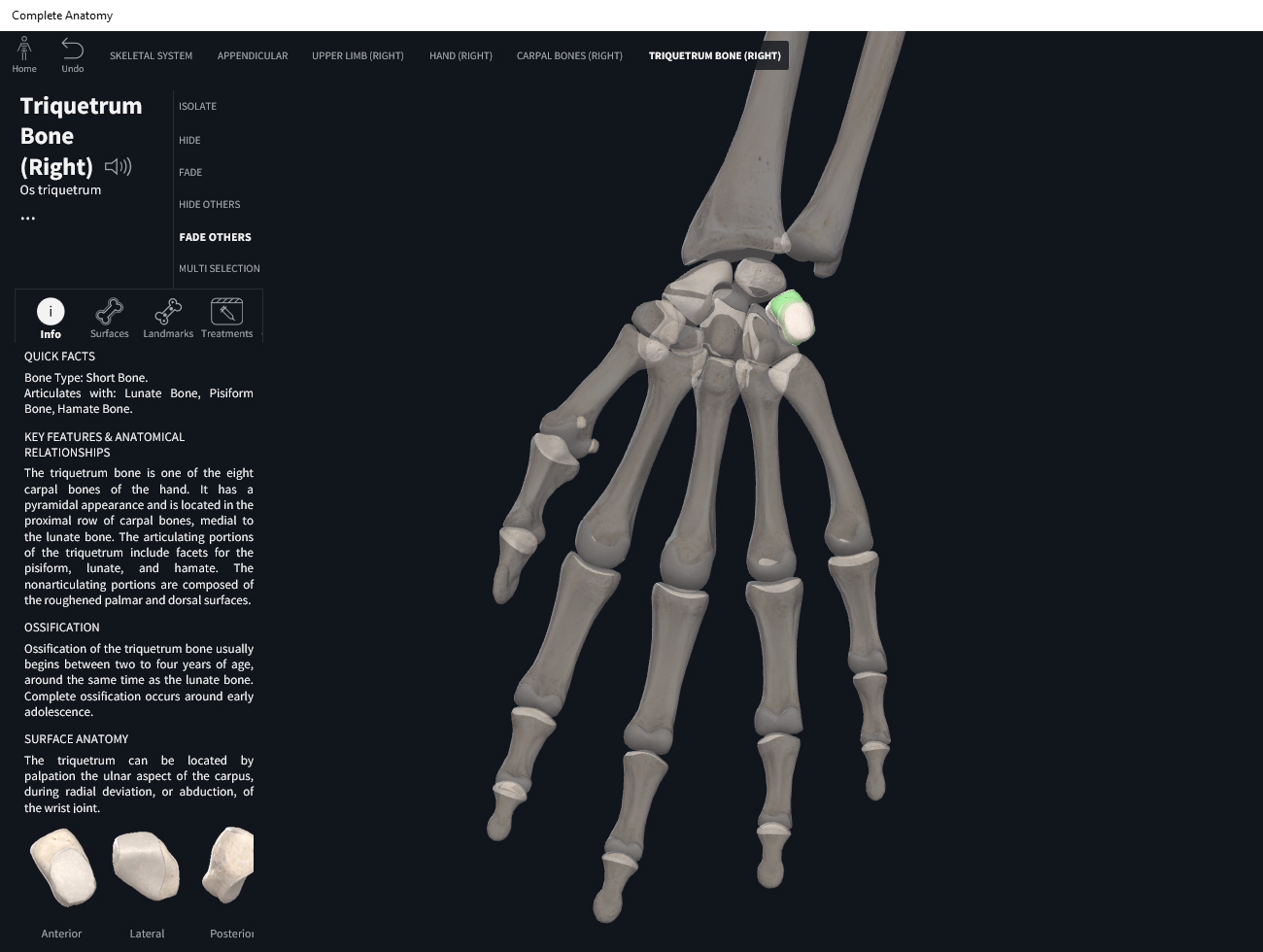
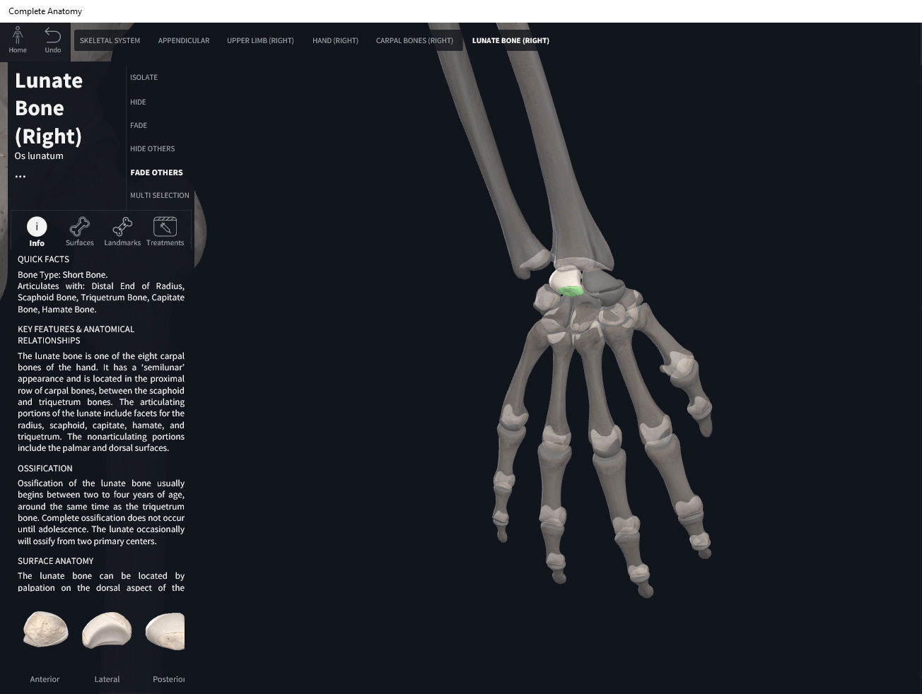
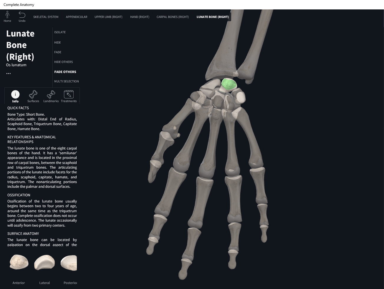
- Proximal row (anterior) starting from medial (pinky side) to lateral: pisiform (mostly viewable from the anterior, sort of lies on top of triquetrum), triquetrum, lunate, scaphoid.
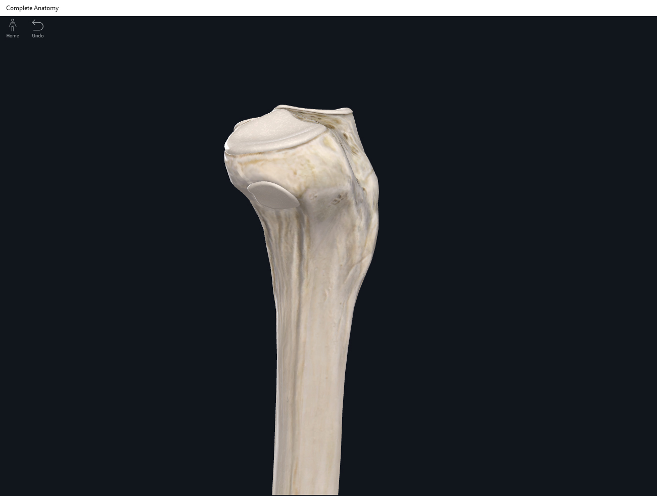
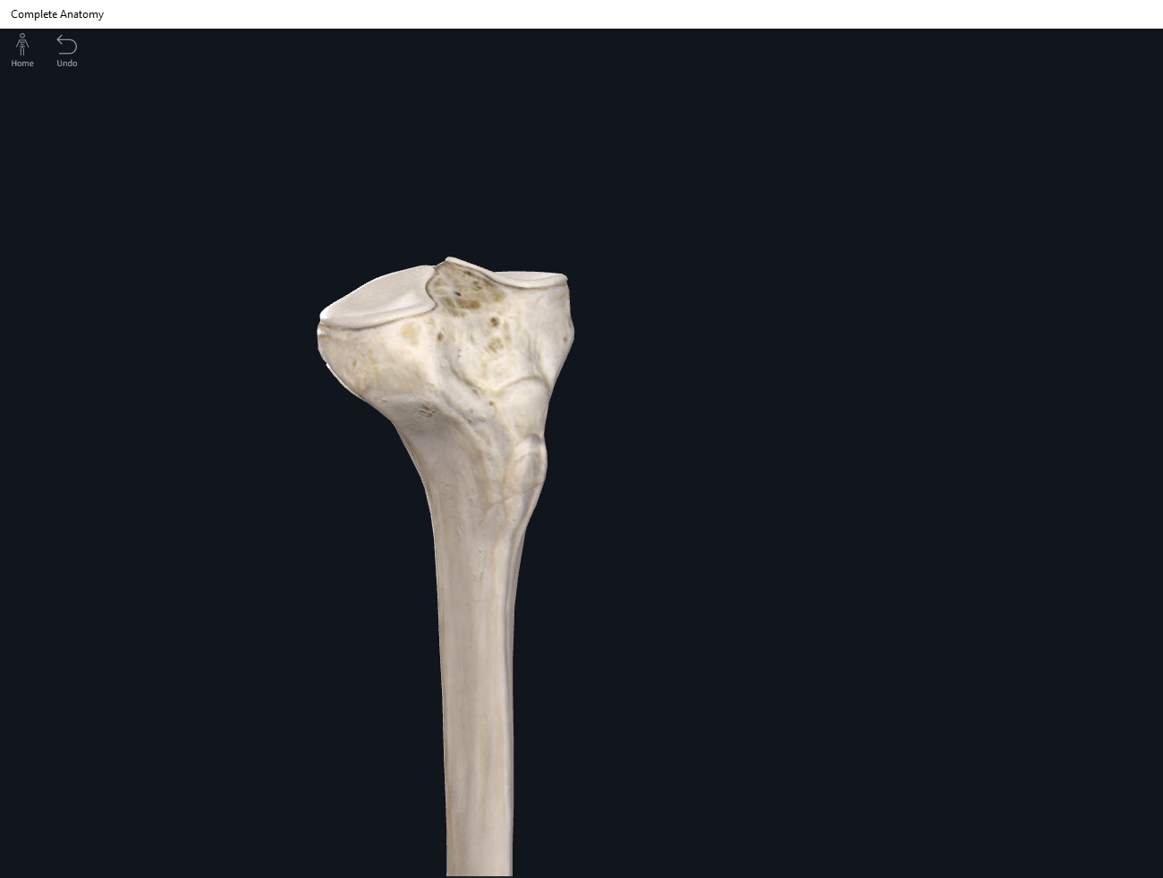
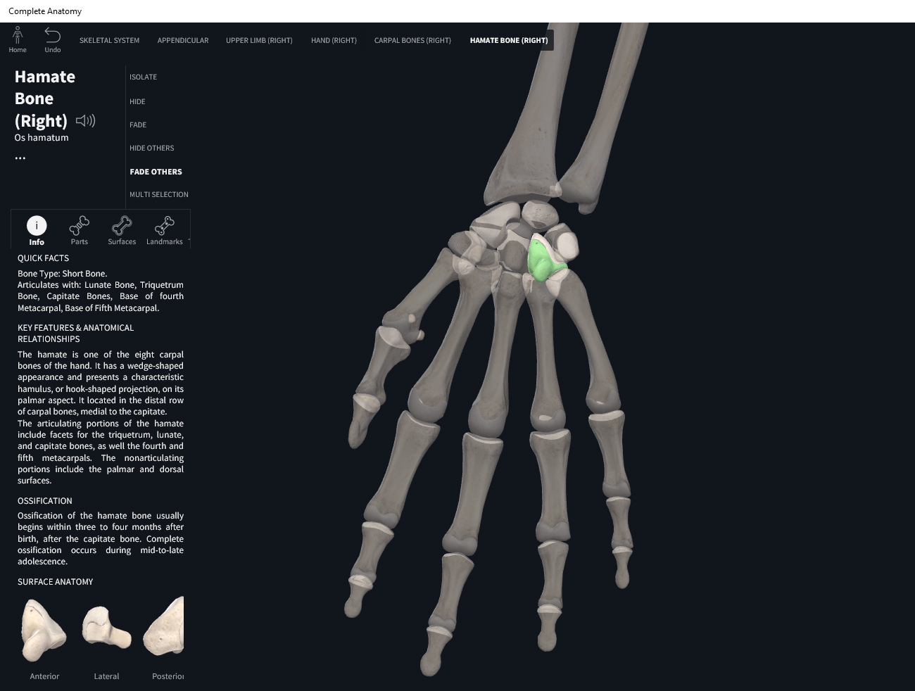
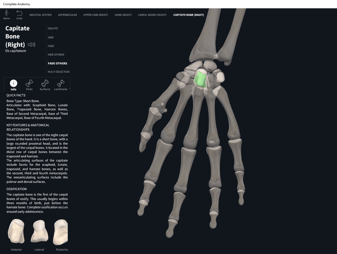
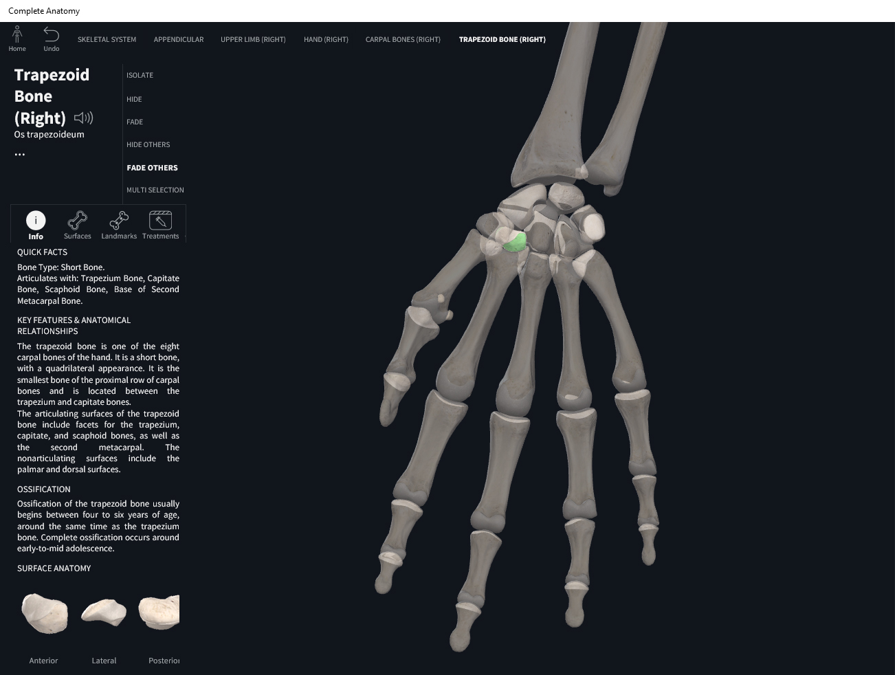
- Distal row (anterior) starting from medial (pinky side) to lateral:, hamate, capitate, trapezoid, trapezium.
- Metacarpals: bones between the carpals and phalanges. Start numbering from the thumb (e.g. the first metacarpal or I) to pinky finger (fifth metacarpal or V).
- Phalanges: Except for the thumb, there are 3 rows of phalanges (proximal, middle, and distal). They are numbered starting from the thumb (e.g. first proximal phalanx) to the pinky (e.g. fifth proximal phalanx).
Function.
Clinical Significance.
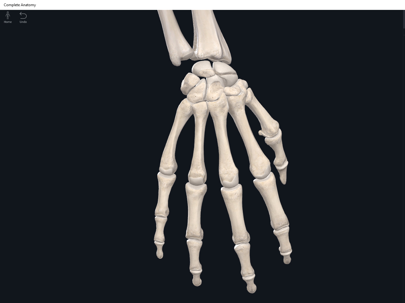
Hand, right. Used with permission by 3D4Medical.

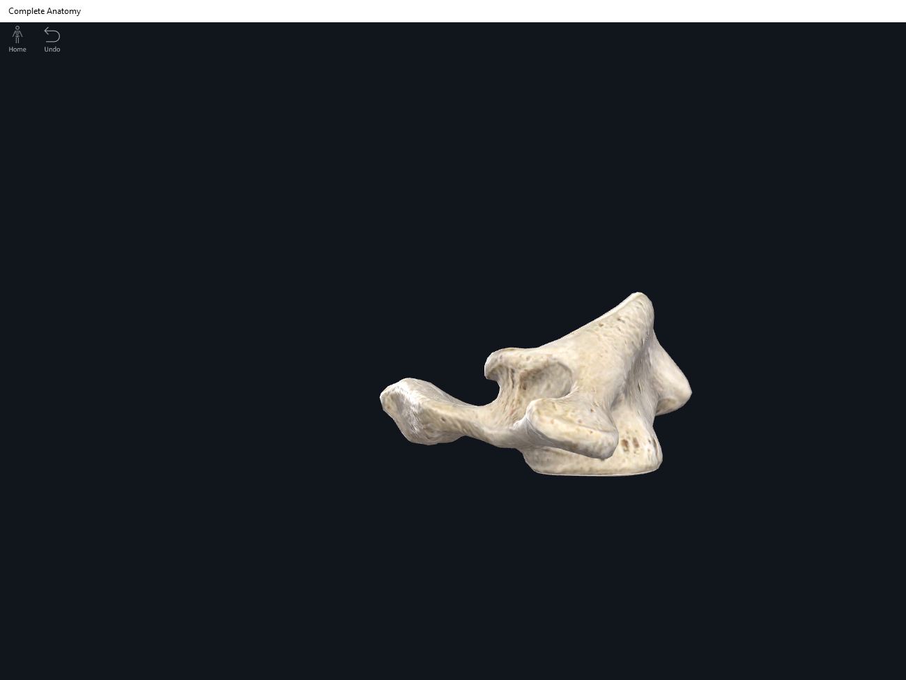
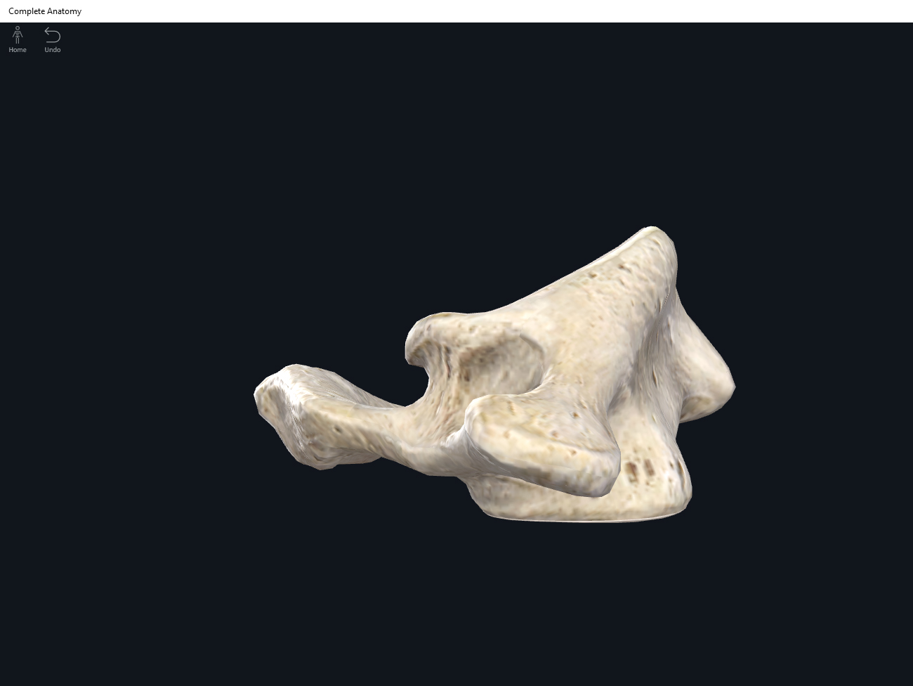
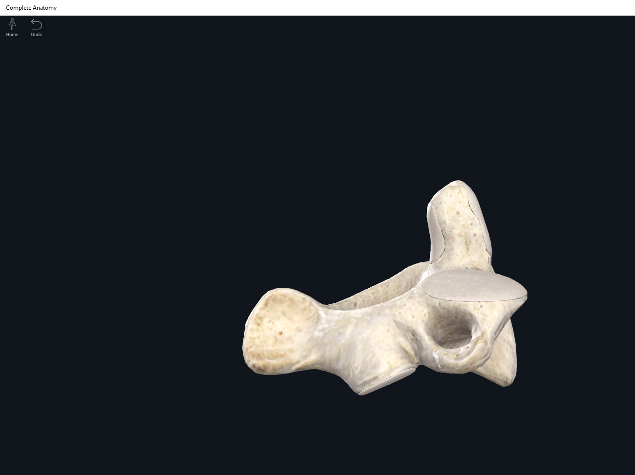
Pisiform. Used with permission by 3D4Medical.

Triquetrum. Used with permission by 3D4Medical.
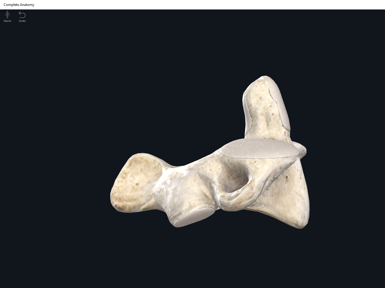
Lunate. Used with permission by 3D4Medical.
Scaphoid. Used with permission by 3D4Medical.

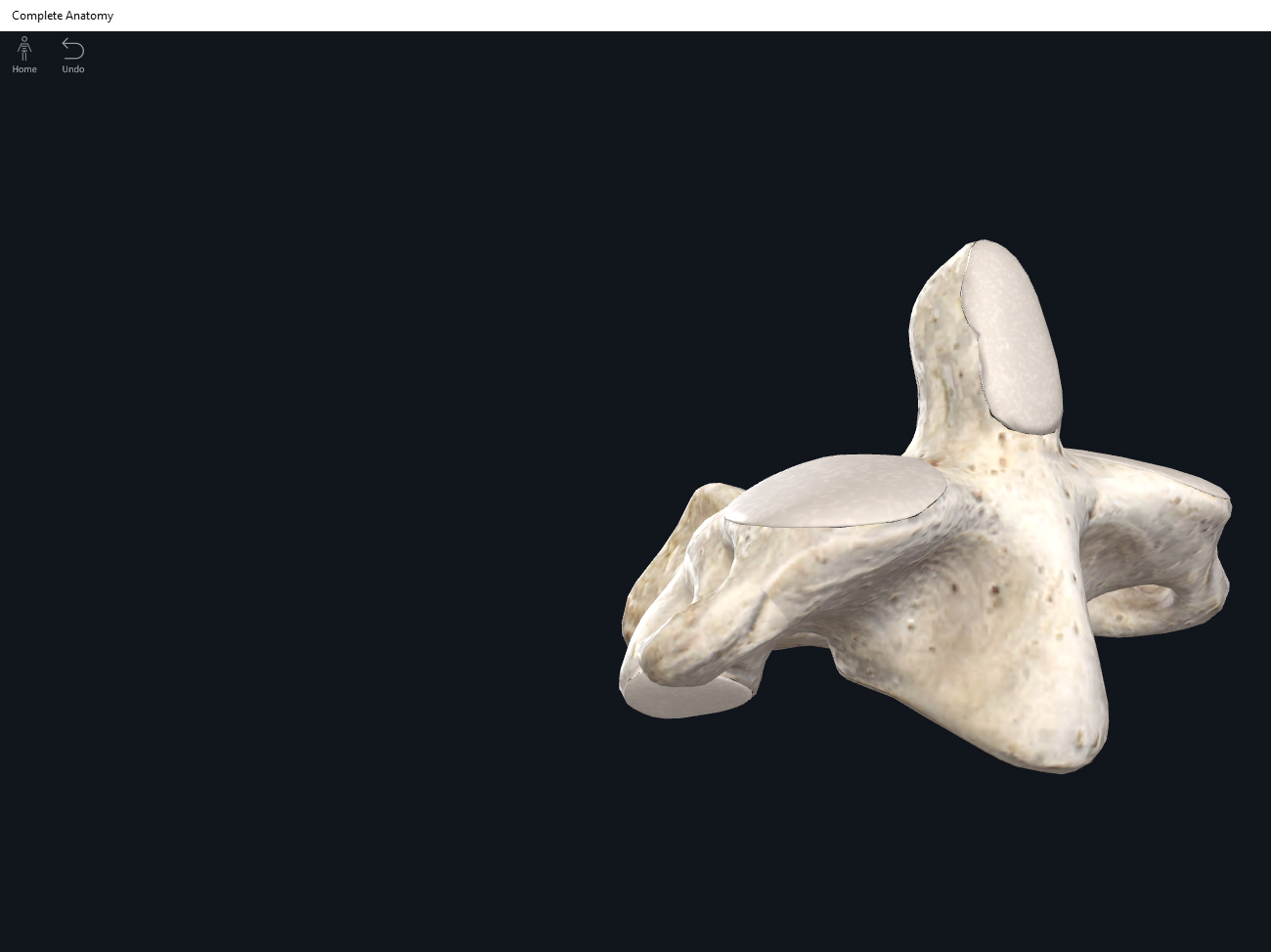
Hamate. Used with permission by 3D4Medical.
Capitate. Used with permission by 3D4Medical.
Trapezoid. Used with permission by 3D4Medical.
Trapezium. Used with permission by 3D4Medical.

References
Biel, A. (2015). Trail guide to the body: A hands-on guide to locating muscles, bones and more.
Cedars-Sinai. (2018). Vertebrae of the spine. Retrieved from https://www.cedars-sinai.org/health-library/diseases-and-conditions/v/vertebrae-of-the-spine.html
Jenkins, G., & Tortora, G. J. (2012). Anatomy and Physiology: From Science to Life, 3rd Edition International Stu. John Wiley & Sons.
Muscolino, J. E. (2017). The muscular system manual: The skeletal muscles of the human body.