Posted on

Bones: Thoracic Cage.

Download these notes.

Anatomy & Physiology: Bones—Thoracic Cage.


Structure.

  • The thoracic cage refers to the space enclosed by the sternum, ribs, costal cartilages, and the thoracic vertebral bodies.
  • The superior end is narrower than the inferior end.


Function.


Clinical Significance.


References

Biel, A. (2015). Trail guide to the body: A hands-on guide to locating muscles, bones and more.

Cedars-Sinai. (2018). Vertebrae of the spine. Retrieved from https://www.cedars-sinai.org/health-library/diseases-and-conditions/v/vertebrae-of-the-spine.html

Jenkins, G., & Tortora, G. J. (2012). Anatomy and Physiology: From Science to Life, 3rd Edition International Stu. John Wiley & Sons.

Muscolino, J. E. (2017). The muscular system manual: The skeletal muscles of the human body.


Posted on

Bones: Sternum.

Download these notes.

Anatomy & Physiology: Bones—Sternum.


Structure.

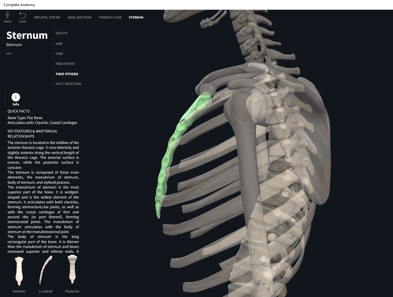
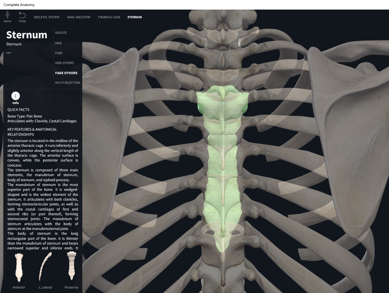
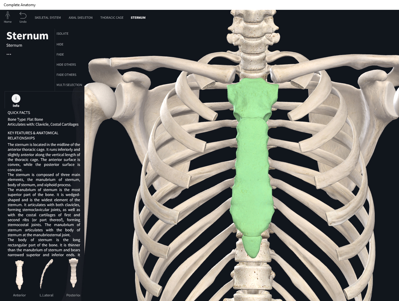
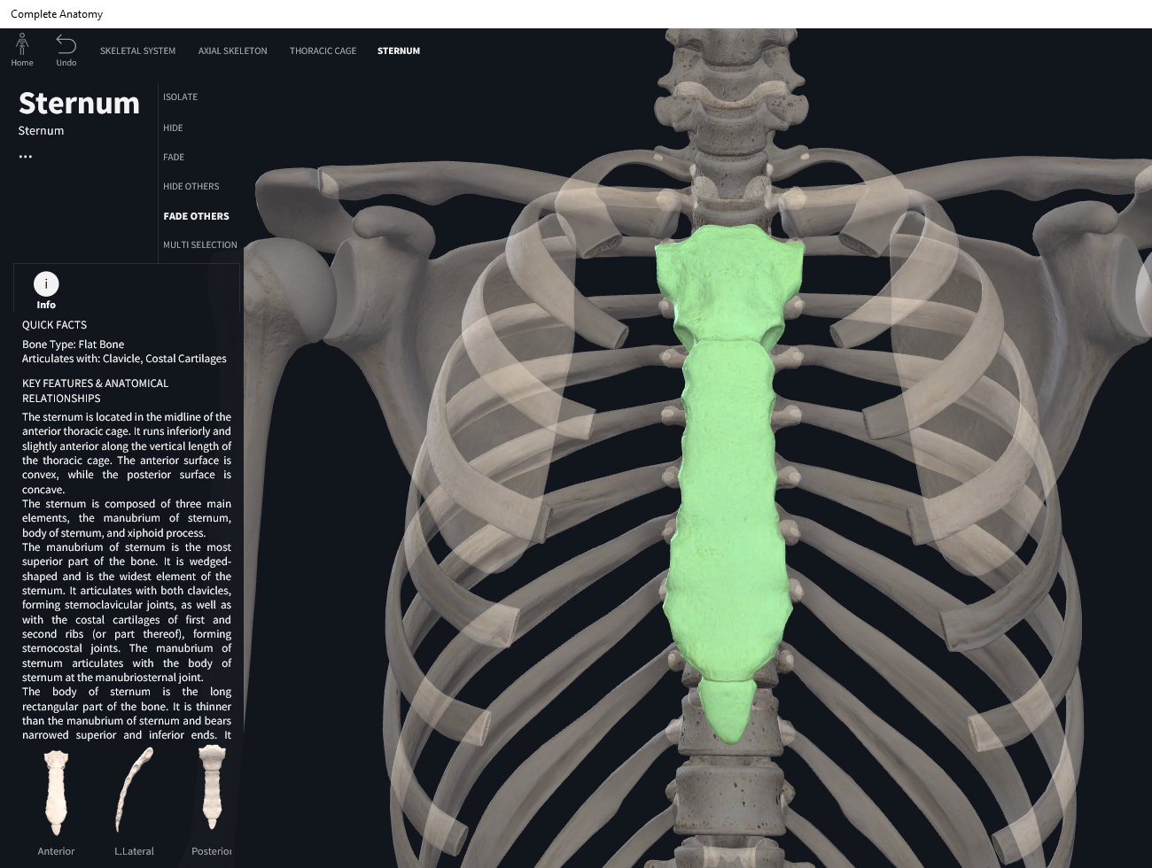
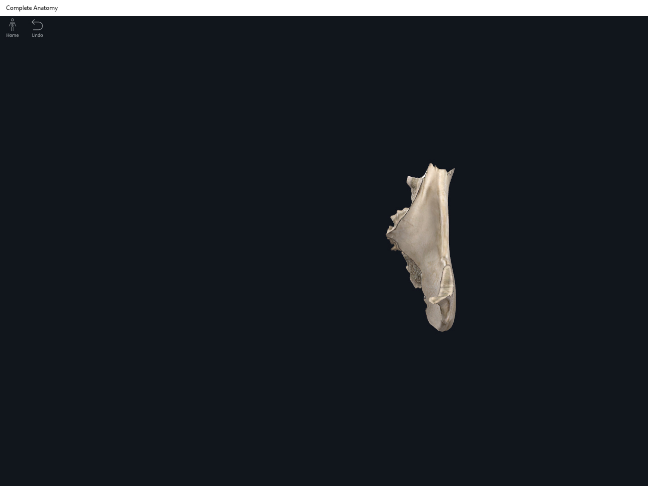
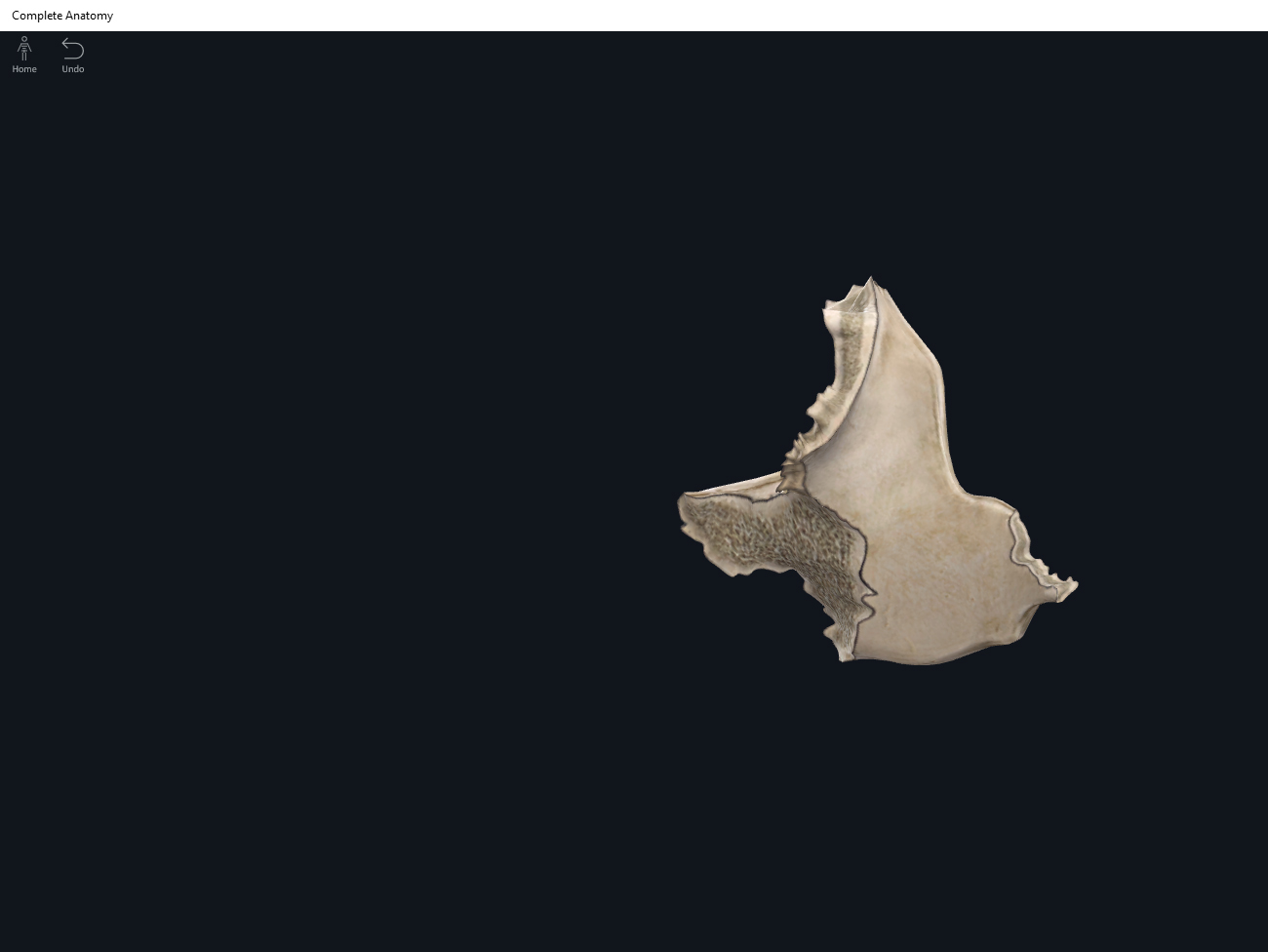
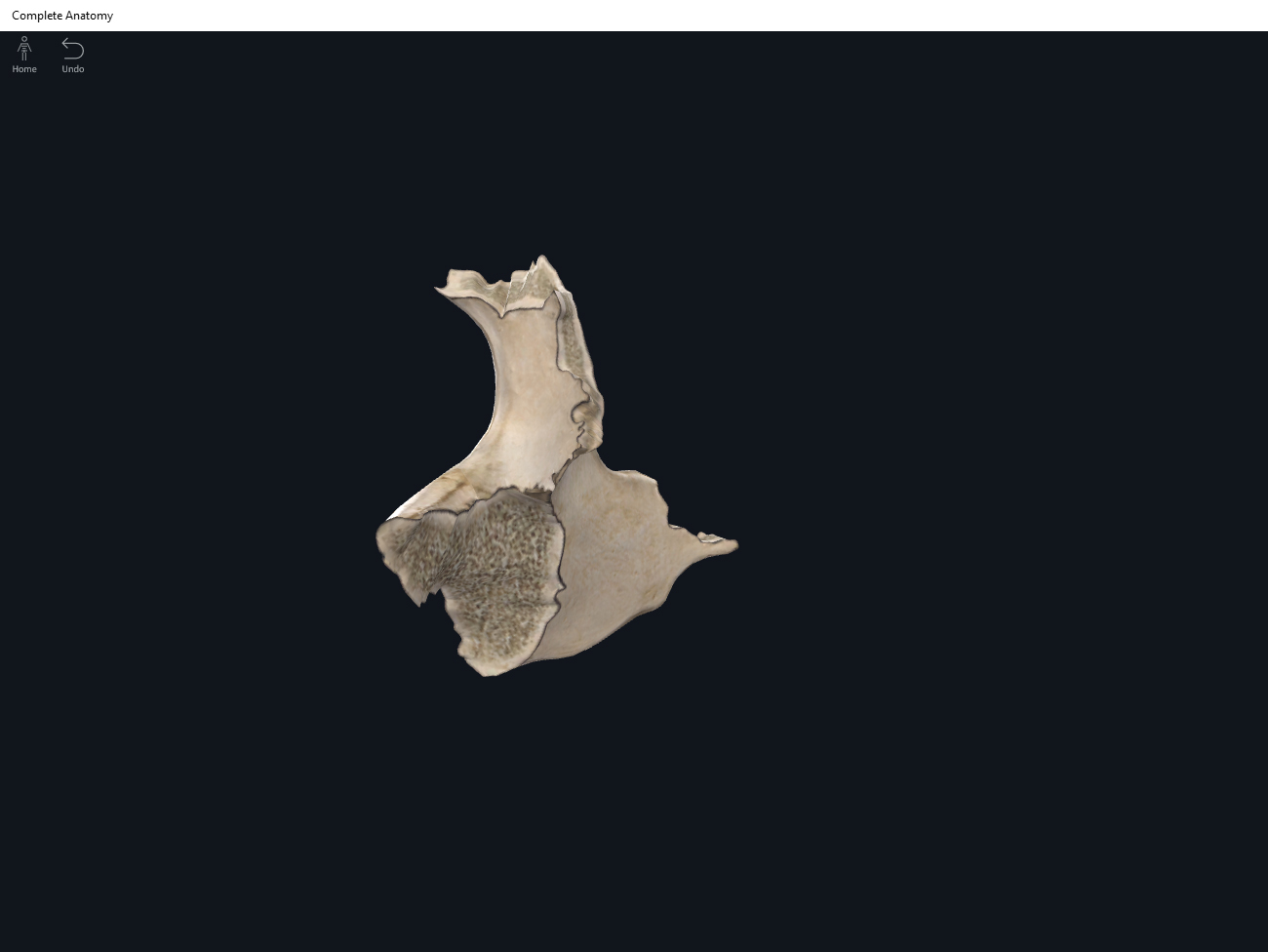
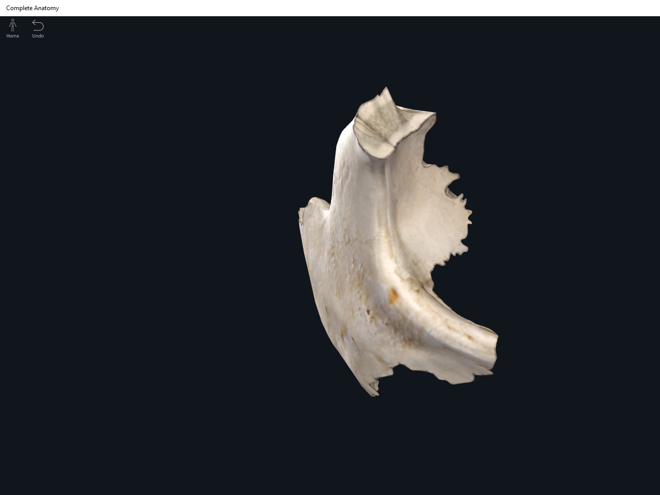
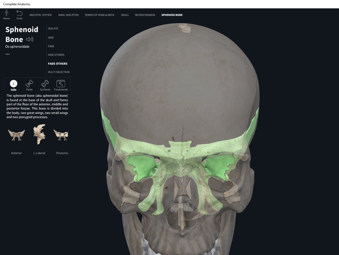
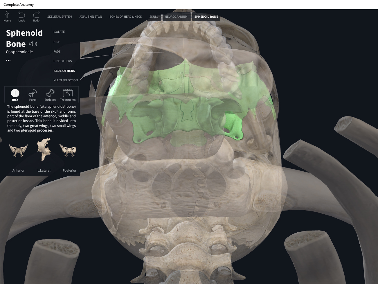
  • The sternum is comprised of 3 parts (descending order): manubrium, body, xiphoid process.
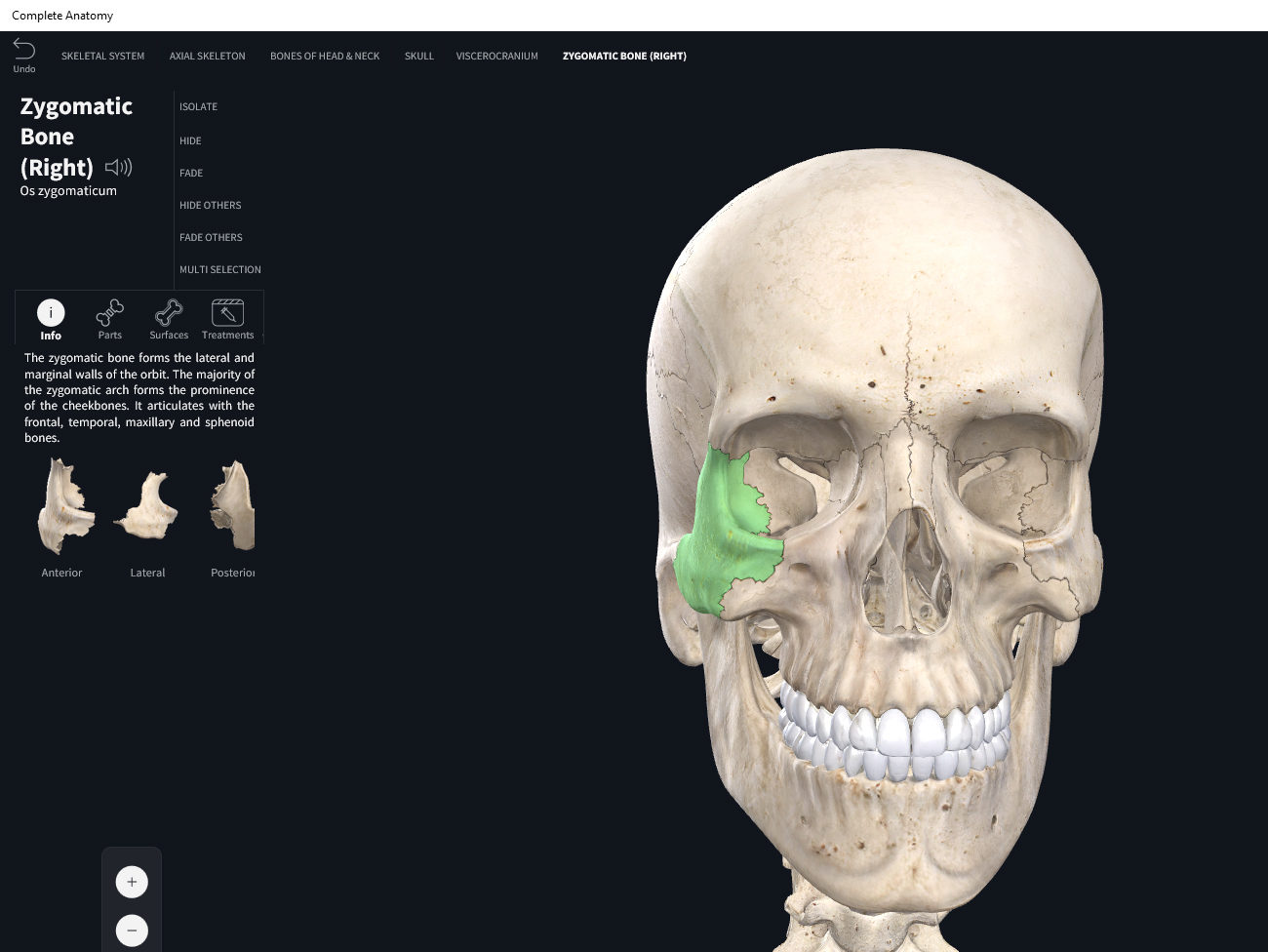
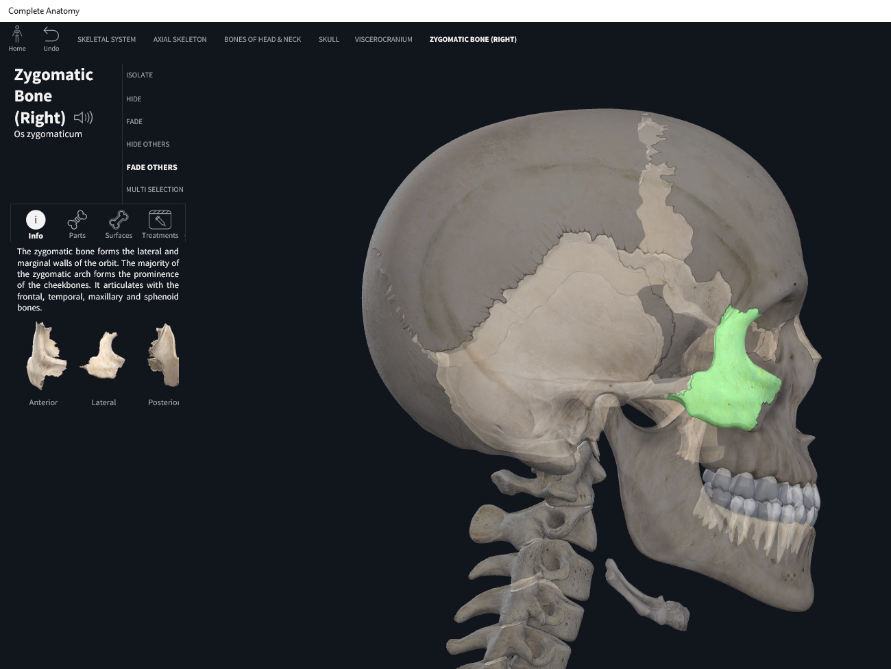
  • Manubrium: most superior of the 3 parts; flat; name means “handle-like” or the vernacular “breastbone”; the suprasternal notch is distinguishable upon inspection and easily palpateable. The manubrium articulates with the medial ends of the clavicles on either side at the clavicular notch.
  • Body: largest piece of the sternum; flattish and elongated. Articulates with the manubrium at the sternal angle. Articulates with the costal cartilages of ribs 2-10.
  • Xiphoid process: the most inferior and smallest portion of the sternum. It is triangle-shaped. It’s made of hyaline and doesn’t ossify until about 40 yrs old. The xiphoid is an attachment site for some abdominal muscles. The xiphoid is palpateable and it has a “give”; it can also be painful to even slightly depress that area.


Function.


Clinical Significance.

  • Sternal angle: clinical landmark for locating rib #2 and counting ribs as reference points to internal organs like the heart.
  • Xiphoid process: incorrect alignment for CPR may cause the xiphoid to fracture off and drive it into (and damage) the internal organs.
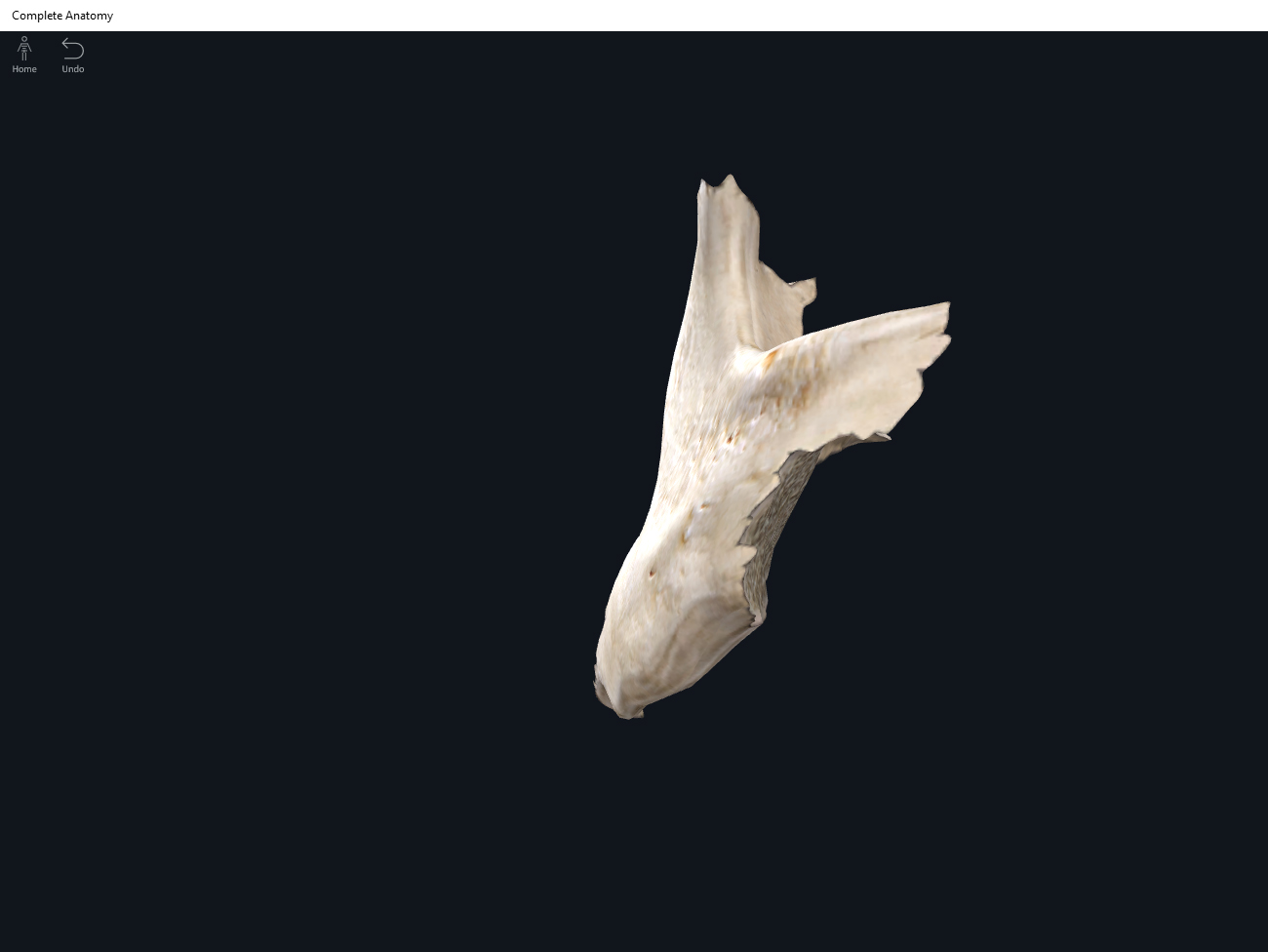
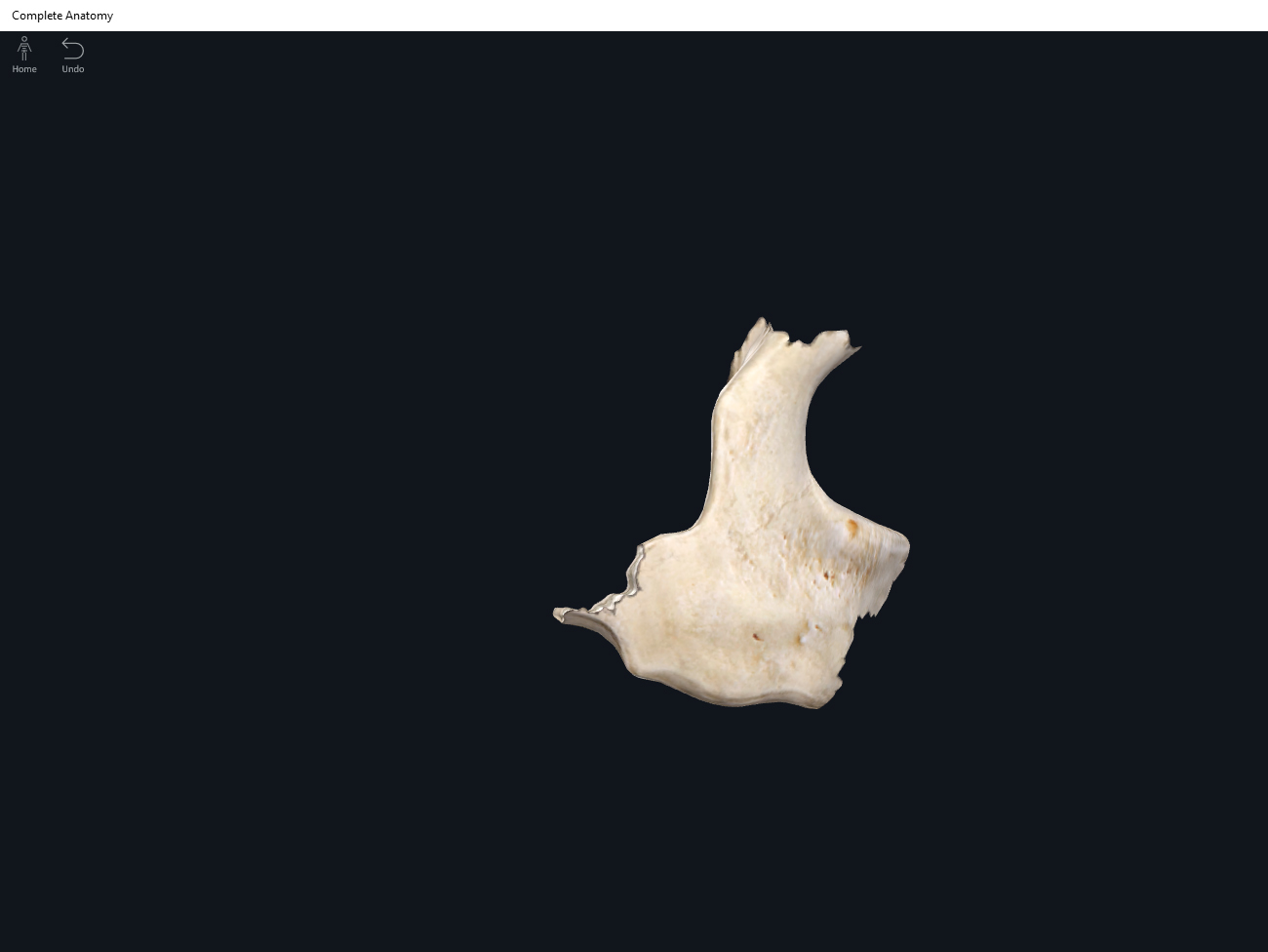
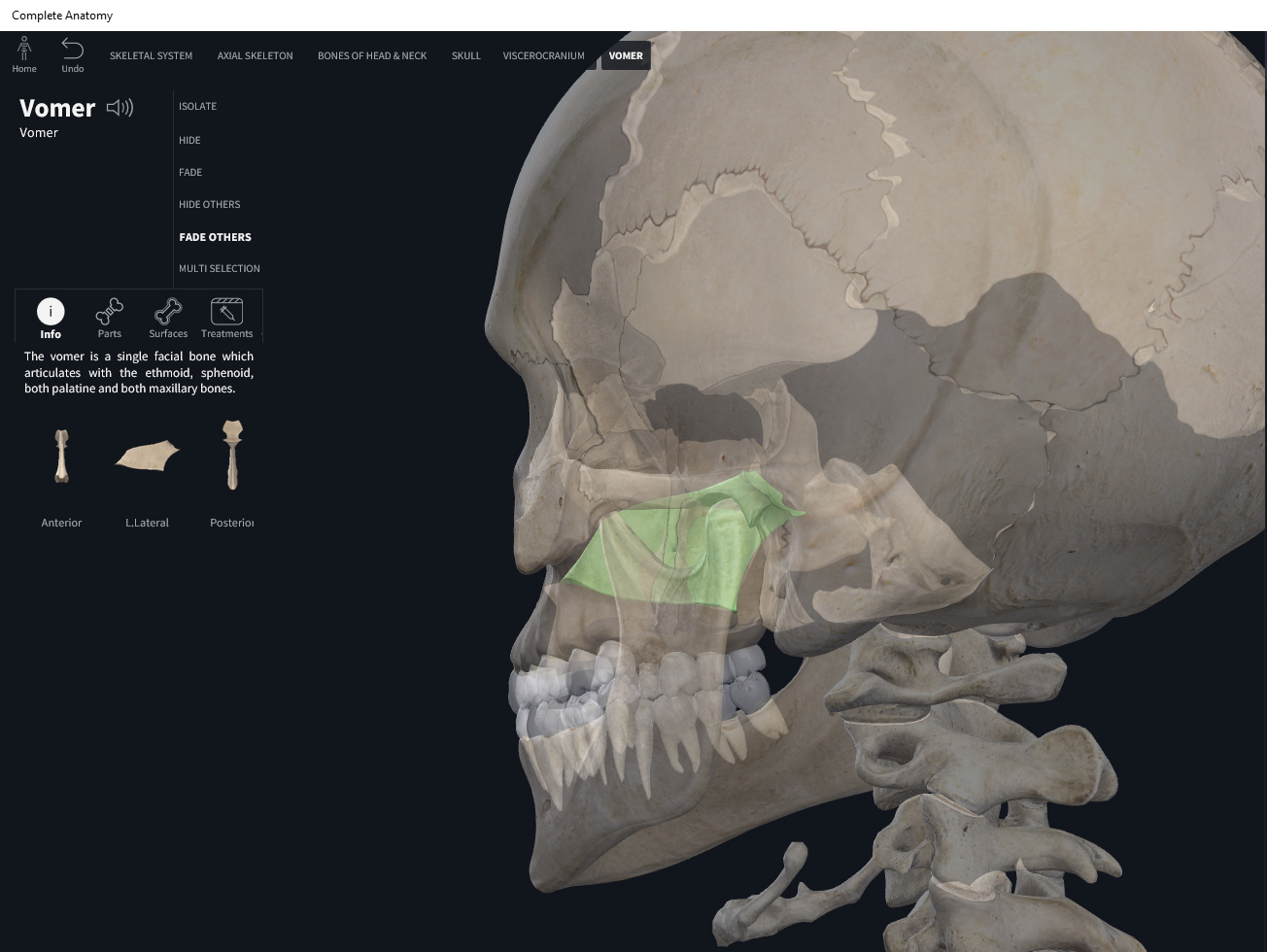
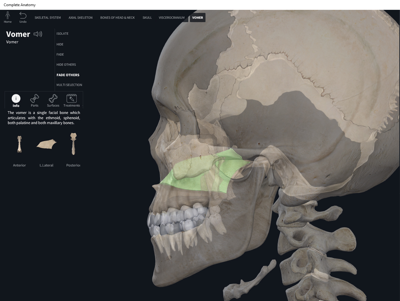
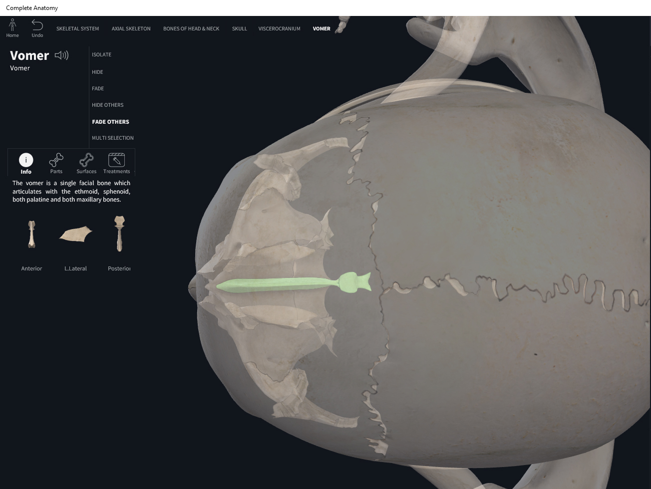
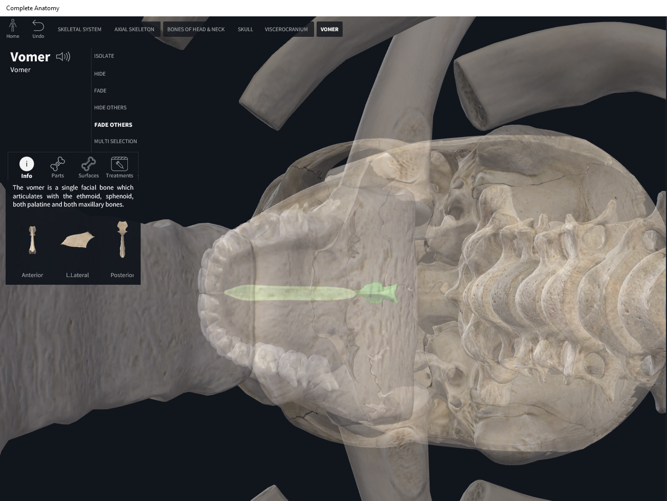
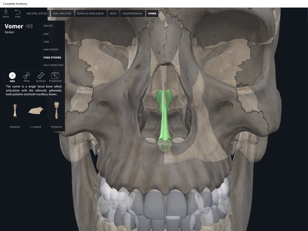
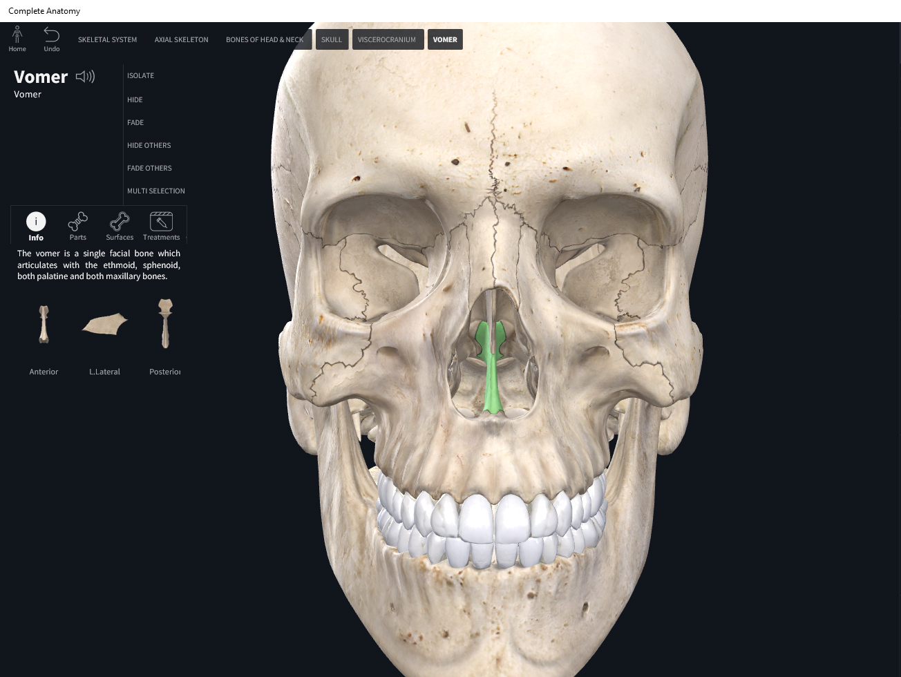
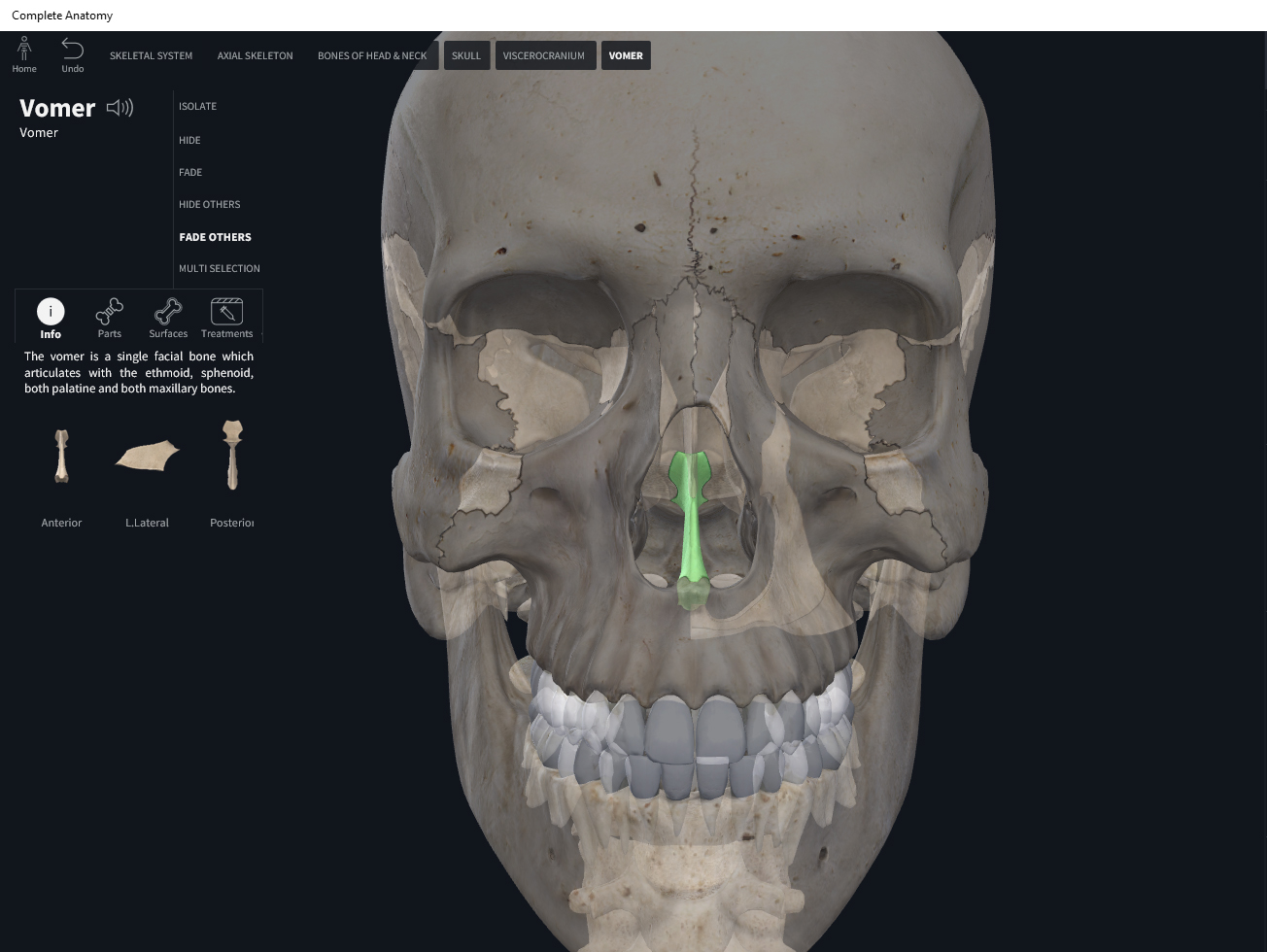
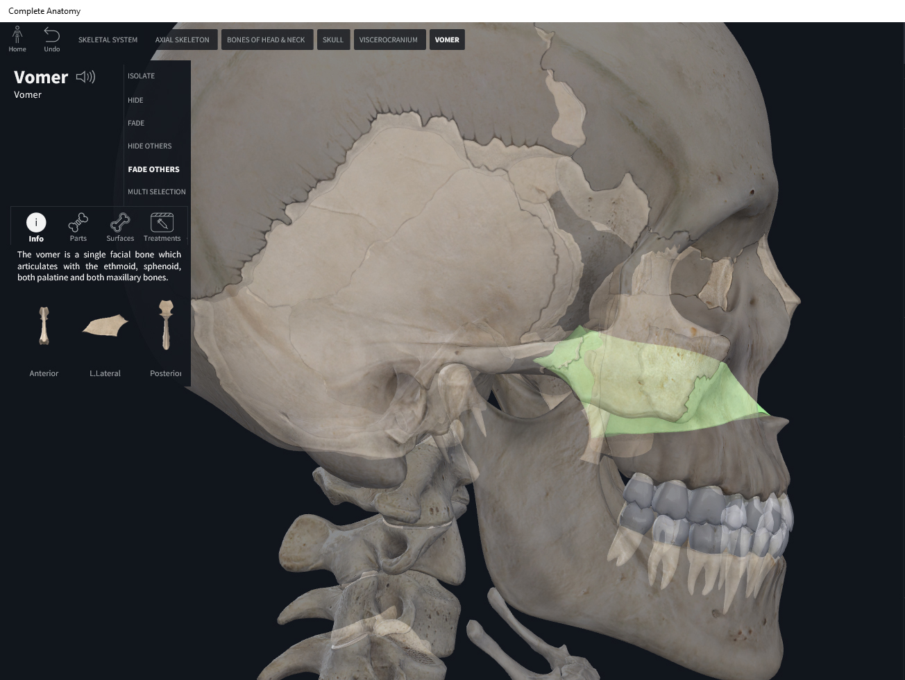
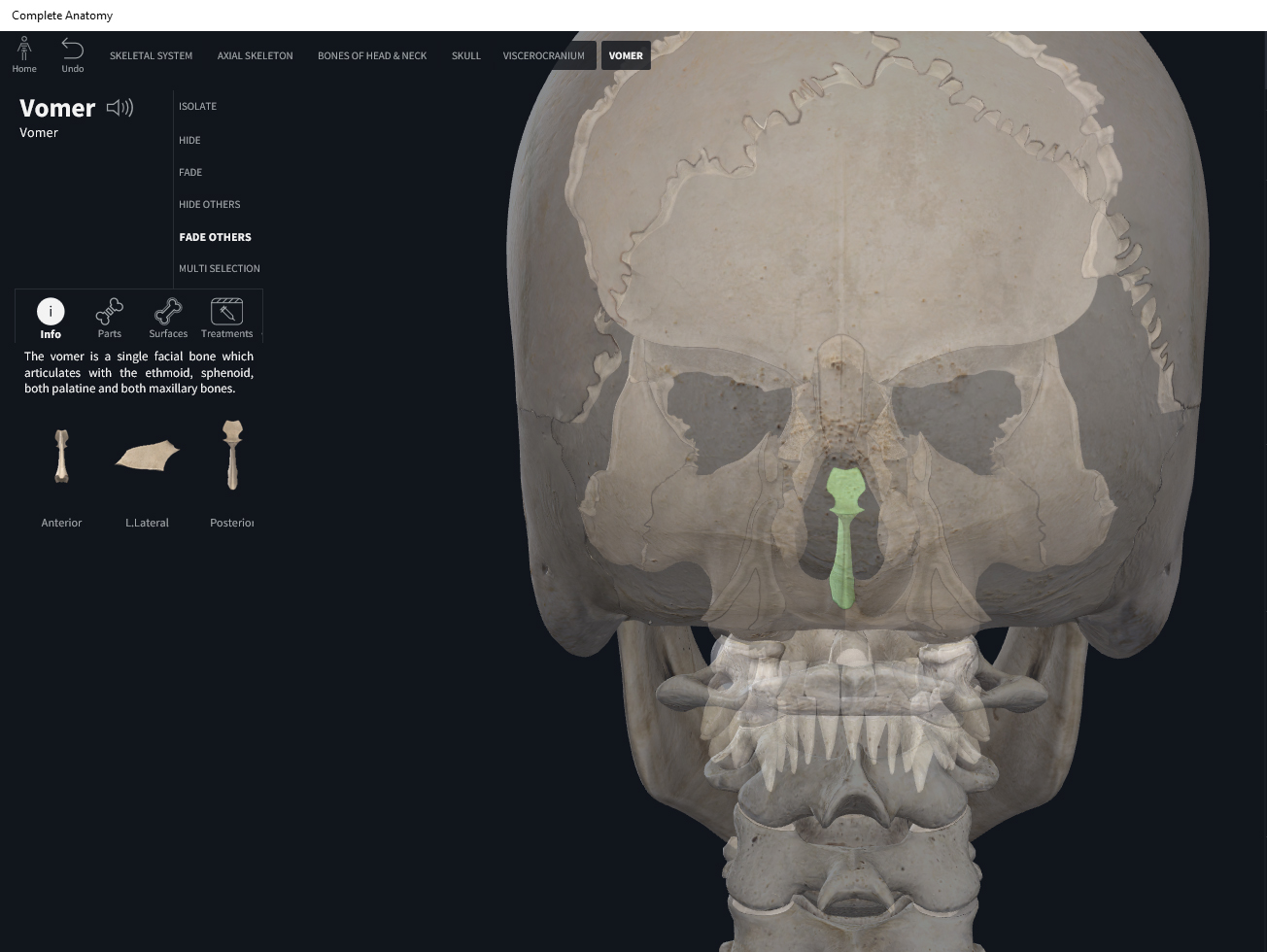
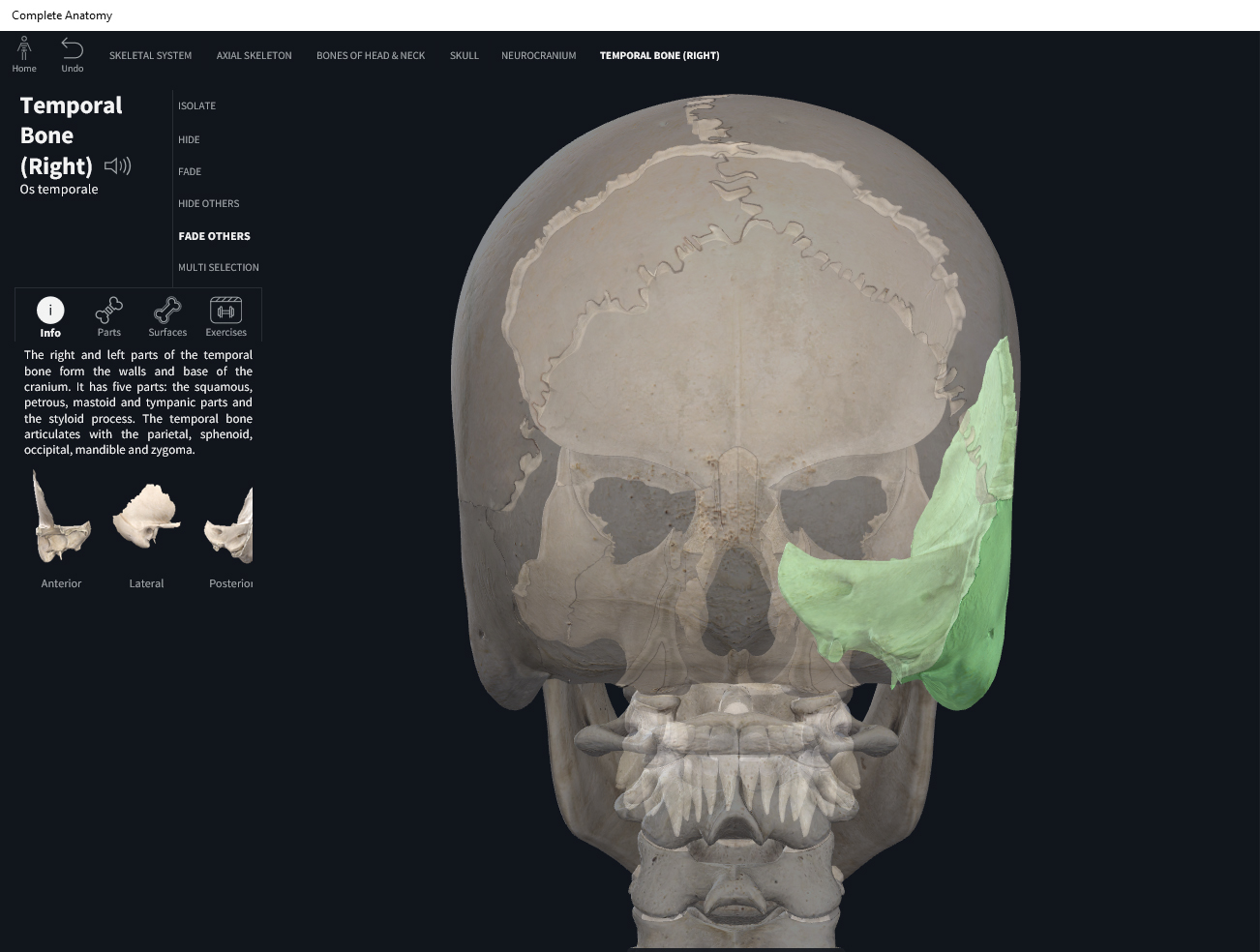
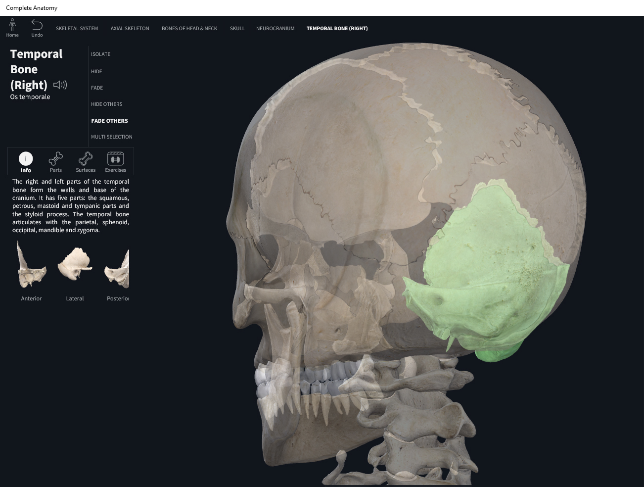
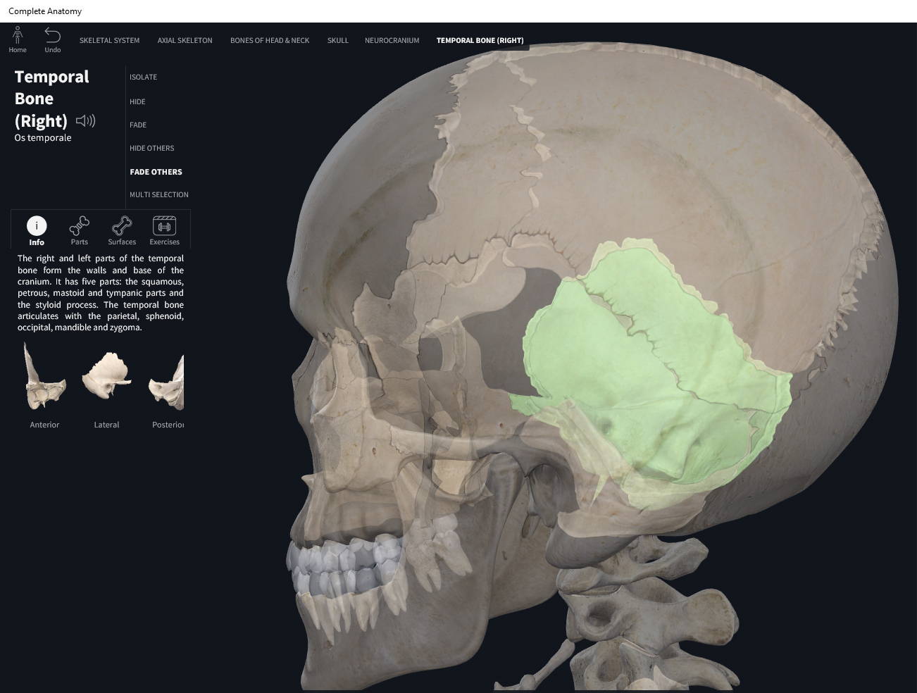
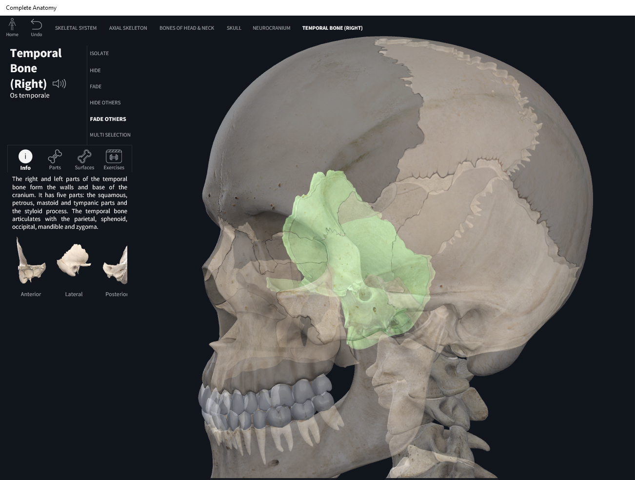
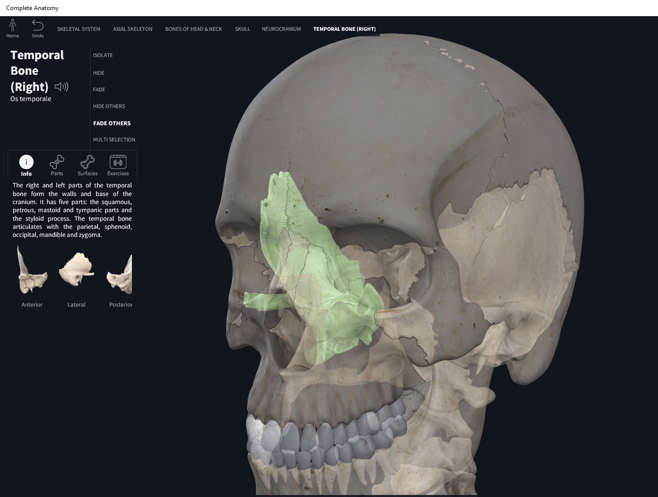
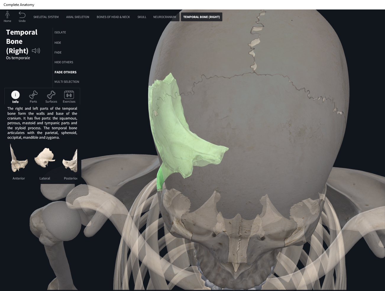
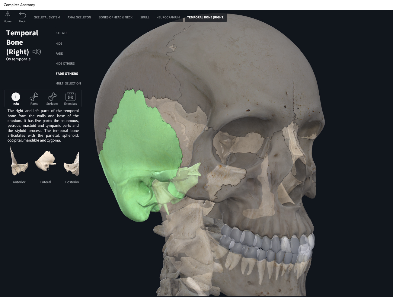
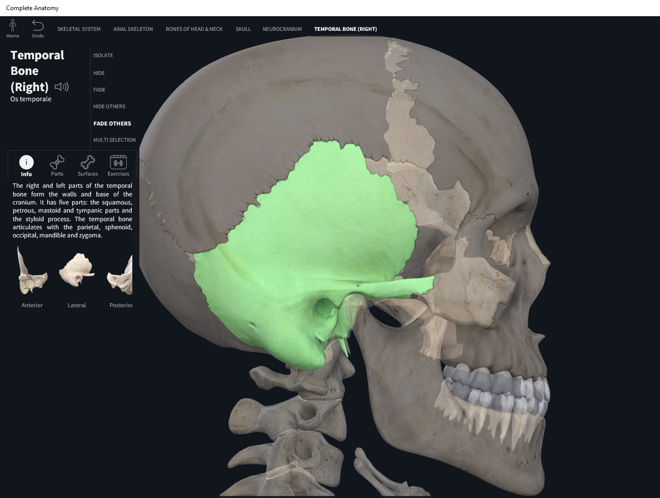
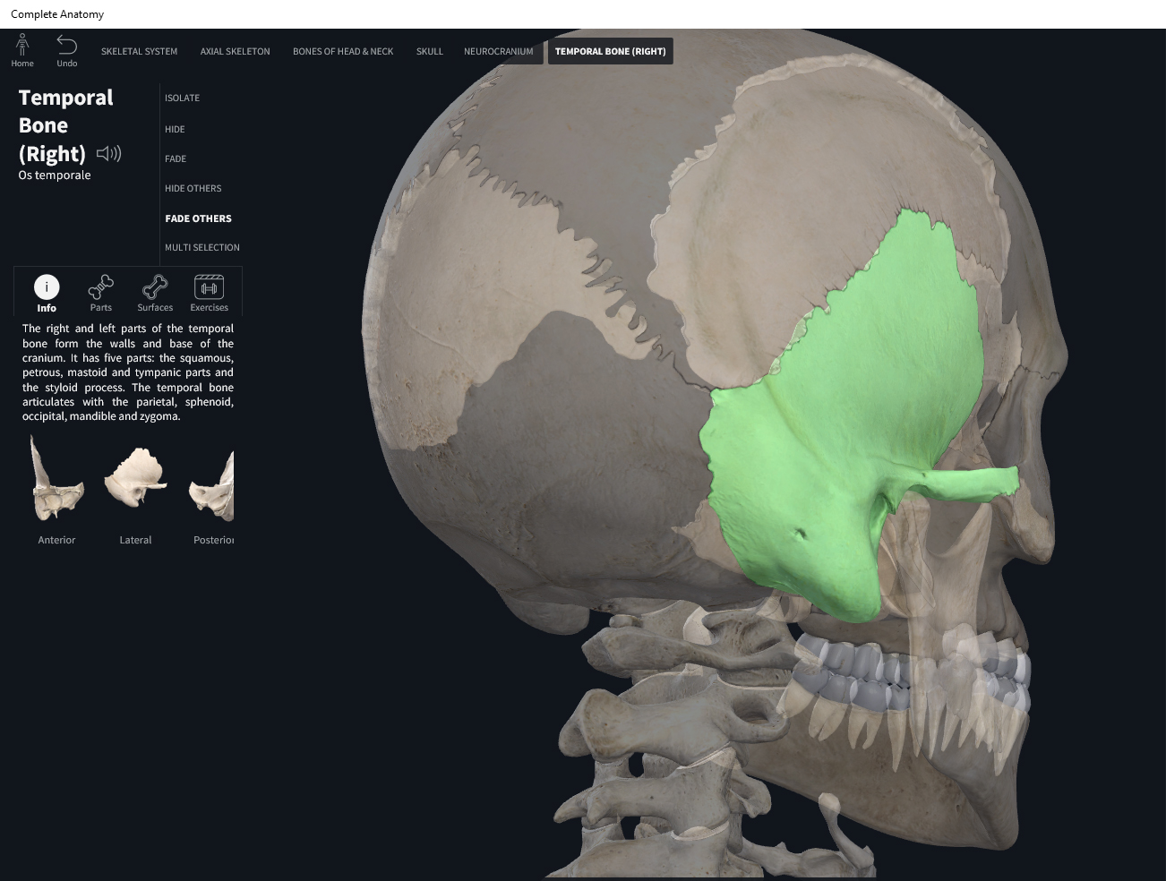
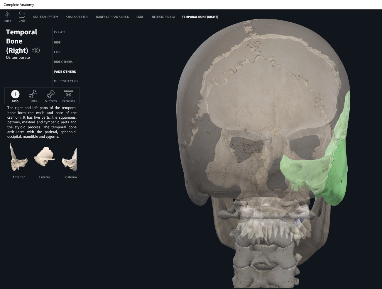
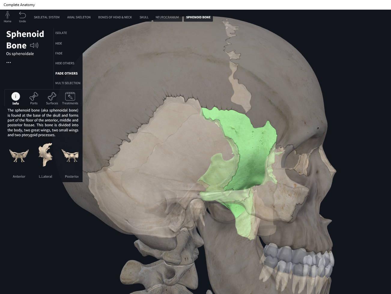
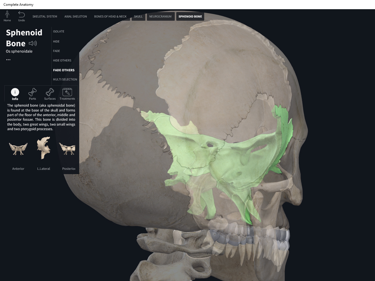
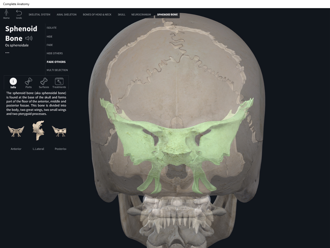
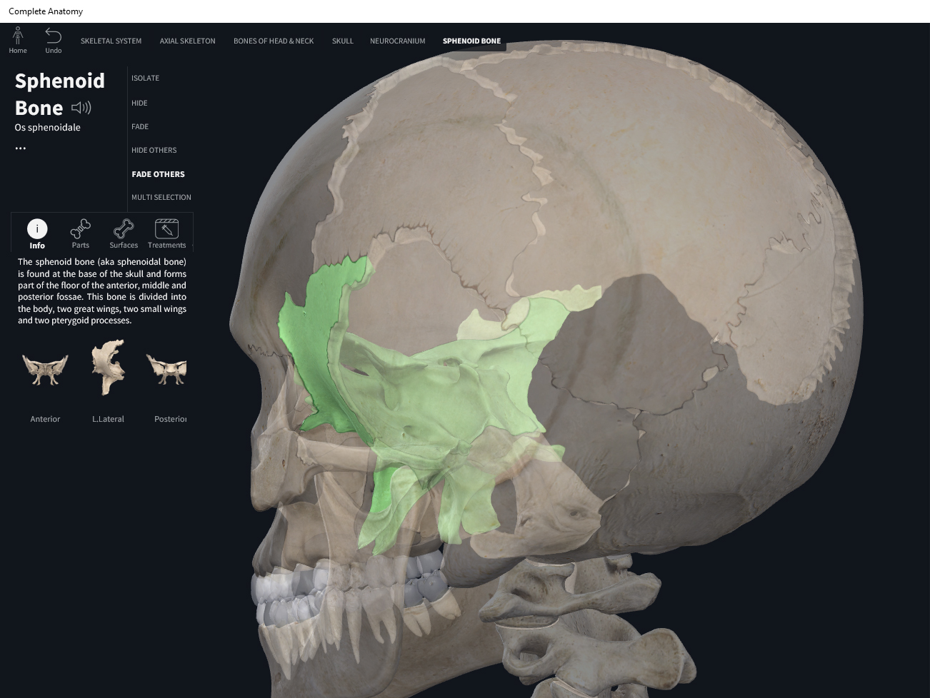
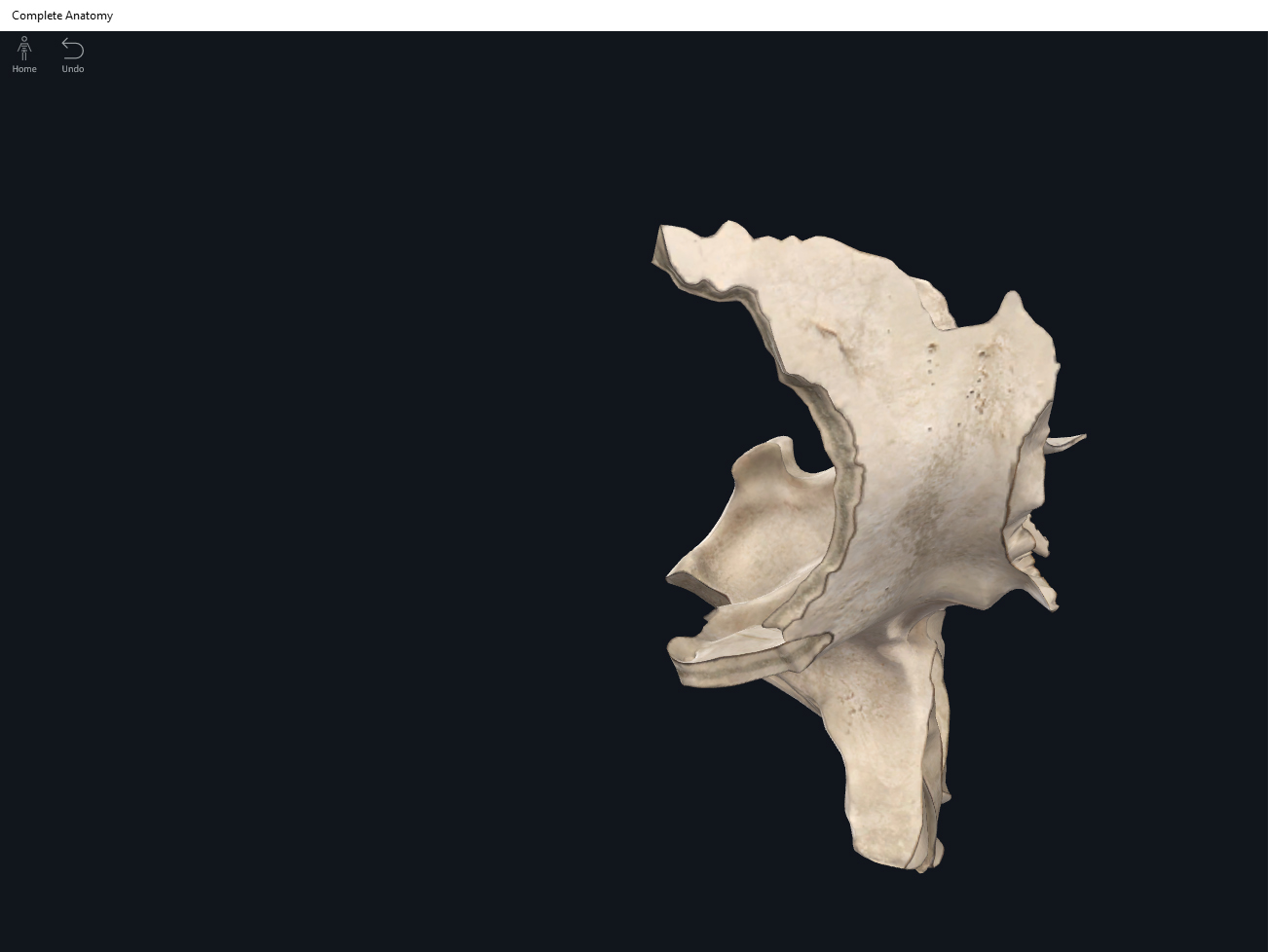
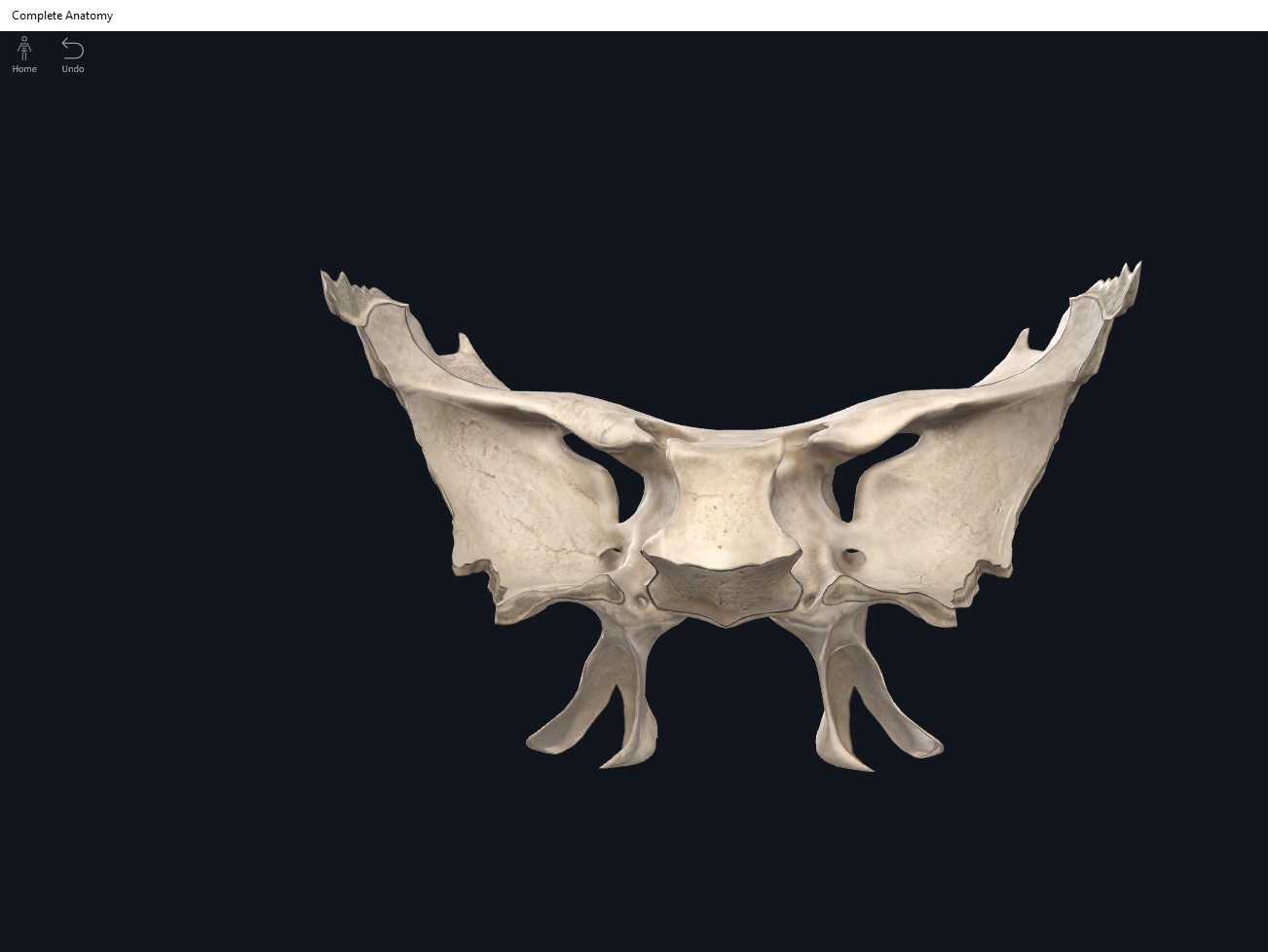
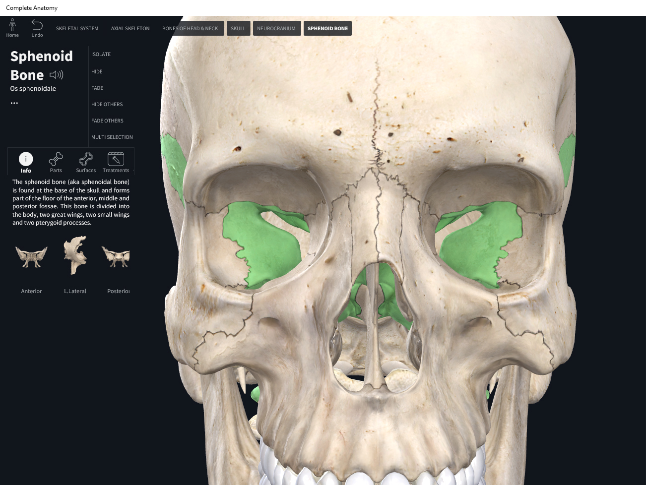
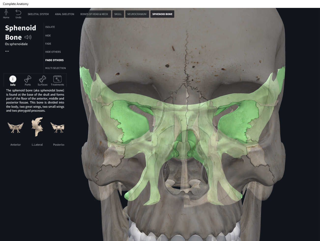


References

Biel, A. (2015). Trail guide to the body: A hands-on guide to locating muscles, bones and more.

Cedars-Sinai. (2018). Vertebrae of the spine. Retrieved from https://www.cedars-sinai.org/health-library/diseases-and-conditions/v/vertebrae-of-the-spine.html

Jenkins, G., & Tortora, G. J. (2012). Anatomy and Physiology: From Science to Life, 3rd Edition International Stu. John Wiley & Sons.

Muscolino, J. E. (2017). The muscular system manual: The skeletal muscles of the human body.

Posted on

Bones: Skull, zygomatic.

Download these notes.

Anatomy & Physiology: Bones—Skull, Zygomatic Bone.


Structure.

  • The pair of zygomatic bones form the cheekbones, and part of the lateral wall and floor of the orbits
  • Temporal process: articulates with the zygomatic process of the temporal bone forming the zygomatic arch.


Function.


Clinical Significance.


References

Biel, A. (2015). Trail guide to the body: A hands-on guide to locating muscles, bones and more.

Jenkins, G., & Tortora, G. J. (2012). Anatomy and Physiology: From Science to Life, 3rd Edition International Stu. John Wiley & Sons.

Muscolino, J. E. (2017). The muscular system manual: The skeletal muscles of the human body.

Posted on

Bones: Skull, vomer.

Download these notes.

Anatomy & Physiology: Bones—Skull, Vomer.


Structure.

  • Triangular shaped.
  • Forms part of the nasal cavity floor and nasal septum.
  • Articulates with perpendicular plate of the ethmoid, sphenoid, and both maxillae and palatine bones.


Function.


Clinical Significance.


References

Biel, A. (2015). Trail guide to the body: A hands-on guide to locating muscles, bones and more.

Jenkins, G., & Tortora, G. J. (2012). Anatomy and Physiology: From Science to Life, 3rd Edition International Stu. John Wiley & Sons.

Muscolino, J. E. (2017). The muscular system manual: The skeletal muscles of the human body.

Posted on

Bones: Skull, temporal.

Download these notes.

Anatomy & Physiology: Bones—Skull, Temporal Bone.


Structure.

  • Forms the inferior lateral side and part of the floor of the cranial cavity.
  • Zygomatic process: is a slender (and slightly inferior) part of the temporal bone that projects anteriorly to articulate with the temporal process of the zygomatic (cheek) bone. On its inferior and posterior surface, the zygomatic process has a socket (mandibular fossa) where it articulates with the mandible. Together they form the temporomandibular joint (TMJ).
  • Zygomatic arch: the complex formed by the zygomatic process of the temporal bone and zygomatic bone.
  • External auditory meatus: the ear canal which is inferior to the zygomatic process.
  • Mastoid process: a stubby, rounded protrusion inferior and posterior to the ear canal. It’s an important attachment point for many neck muscles.
  • Styloid process: is a small, thin, pointed (almost needlelike) projection (pointing anteriorly) inferior and anterior to the ear canal. It’s an attachment point for muscles and ligaments of the neck and tongue. The styloid process is better viewed from underneath the skull.
  • Stylomastoid foramin: located between the mastoid and styloid processes (also viewed from underneath the skull) is a hole for a nerve that controls facial muscles.
  • Foramin lacerum: formed (viewed from underneath the skull) between the temporal bone, sphenoid bone, and occipital bone.
  • Carotid foramin: on the underside of the skull.
  • Jugular foramin: on the underside of the skull, the temporal bone and occipital bone form this passageway.
  • Petrous portion: interior of the skull and on the cranial floor, there is a ridge (like a mountain range) bordering the temporal and occipital bones. This petrous portion contains the internal and middle ear structures—hearing and equilibrium. The internal auditory meatus is the hole in the petrous portion.


Function.


Clinical Significance.


References

Biel, A. (2015). Trail guide to the body: A hands-on guide to locating muscles, bones and more.

Jenkins, G., & Tortora, G. J. (2012). Anatomy and Physiology: From Science to Life, 3rd Edition International Stu. John Wiley & Sons.

Muscolino, J. E. (2017). The muscular system manual: The skeletal muscles of the human body.

Posted on

Bones: Skull, sutures.

Download these notes.

Anatomy & Physiology: Bones—Skull, Sutures.


Structure.

  • Suture are the “seams” between the skull bones.
  • In adults, they are immovable joints.
  • In infants/young children, the sutures are slightly moveable to allow for skull development.
  • Coronal suture: union of frontal bone with both parietal bones.
  • Sagittal suture: union of the two parietal bones along the midsagittal plane.
  • Lambdoid suture: union of the two parietal bones with the occipital bone.
  • Squamous sutures: bilateral; union of parietal and temporal bones.


Function.


Clinical Significance.


References

Biel, A. (2015). Trail guide to the body: A hands-on guide to locating muscles, bones and more.

Jenkins, G., & Tortora, G. J. (2012). Anatomy and Physiology: From Science to Life, 3rd Edition International Stu. John Wiley & Sons.

Muscolino, J. E. (2017). The muscular system manual: The skeletal muscles of the human body.

Posted on

Bones: Skull, sphenoid.

Download these notes.

Anatomy & Physiology: Bones—Skull, Sphenoid Bone.


Structure.

  • The name means “wedge” but it’s often called the butterfly bone of the skull. Easily the most intricate of the cranial bones.
  • Located in the middle of the base of skull, portion of the cranial floor; and is a keystone structure as it articulates with all the cranial bones—with the frontal bone anteriorly; with the parietal and temporal bones laterally; and with the occipital posteriorly. The sphenoid is posterior and superior to the nasal cavity. The sphenoid also forms part of the floor, sides and rear of the orbit.
  • The “body” (frontal plane, anterior view) of the butterfly has two cavities called the sphenoid sinuses which drain to the nasal cavity.
  • The “greater wings” (frontal plane, anterior view) are the large wings of the butterfly. Forms the anterolateral cranial floor and part of the lateral wall. There are 3 foramen: the foramen rotundum (round hole) allows passageway for the nerve for facial sensations; the foramen ovale (oval hole) a passage for the nerve that controls movement of the mandible; and the foramen spinosum (spinous hole) for the blood vessel supplying the membranes of the brain.
  • The “lesser wings” (frontal plane, anterior view) are superior to the greater wings and almost look like whiskers. The optic foramen may be found close to where the lesser wing joins to the body of the butterfly. The lesser wings form part of the cranial floor, and also the posterior of the orbit.
  • Sella turcica: “saddle” shaped smooth surface on the body of the sphenoid bone.
  • Hypophyseal fossa: the depression of the sella turcica housing the pituitary gland.
  • Pterygoid processes: two trailing wings (a pair on each side of the butterfly) inferior to the body. Serves as an attachment point for some of the muscles that help move the mandible.
  • Superior orbital fissure: laterally between the greater and lesser wings; triangular slit for nerves that control eye movement.


Function.


Clinical Significance.


References

Biel, A. (2015). Trail guide to the body: A hands-on guide to locating muscles, bones and more.

Jenkins, G., & Tortora, G. J. (2012). Anatomy and Physiology: From Science to Life, 3rd Edition International Stu. John Wiley & Sons.

Muscolino, J. E. (2017). The muscular system manual: The skeletal muscles of the human body.

Posted on

Bones: Skull, parietal.

Download these notes.

Anatomy & Physiology: Bones—Skull, Parietal Bone.

Structure.

  • Forms the side and roof of cranial cavity.
  • Inner surface is “rough” with “terrain” as attachment sites for vessels (of duramatter) and the brain’s superficial membrane.
  • Forms part of the coronal, squamous, and lambdoid sutures.

Function.

Clinical Significance.

References

Biel, A. (2015). Trail guide to the body: A hands-on guide to locating muscles, bones and more.

Jenkins, G., & Tortora, G. J. (2012). Anatomy and Physiology: From Science to Life, 3rd Edition International Stu. John Wiley & Sons.

Muscolino, J. E. (2017). The muscular system manual: The skeletal muscles of the human body.

Posted on

Bones: Skull, paranasal sinuses.

Download these notes.

Anatomy & Physiology: Bones—Skull, Paranasal Sinuses.


Structure.

  • Cavities near the nasal cavity that have a mucousal membrane lining that are continuous with nasal cavity lining.


Function.

  • Lighten the weight of the skull.
  • Increase area of nasal mucosa for filtering, warming, and moistening the air.


Clinical Significance.


References

Biel, A. (2015). Trail guide to the body: A hands-on guide to locating muscles, bones and more.

Jenkins, G., & Tortora, G. J. (2012). Anatomy and Physiology: From Science to Life, 3rd Edition International Stu. John Wiley & Sons.

Muscolino, J. E. (2017). The muscular system manual: The skeletal muscles of the human body.

Posted on

Bones: Skull, palatine bones.

Download these notes.

Anatomy & Physiology: Bones—Skull, Palatine Bones.


Structure.

  • Pair of bones that are L-shaped and form the posterior end of the hard palate, and part of the floor and side walls of the nasal cavity, and part of the orbital floor.


Function.


Clinical Significance.


References

Biel, A. (2015). Trail guide to the body: A hands-on guide to locating muscles, bones and more.

Jenkins, G., & Tortora, G. J. (2012). Anatomy and Physiology: From Science to Life, 3rd Edition International Stu. John Wiley & Sons.

Muscolino, J. E. (2017). The muscular system manual: The skeletal muscles of the human body.