Download these notes.
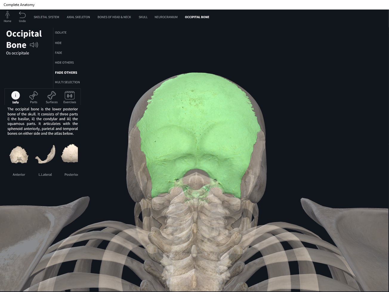
Anatomy & Physiology: Bones—Skull, Occipital Bone.
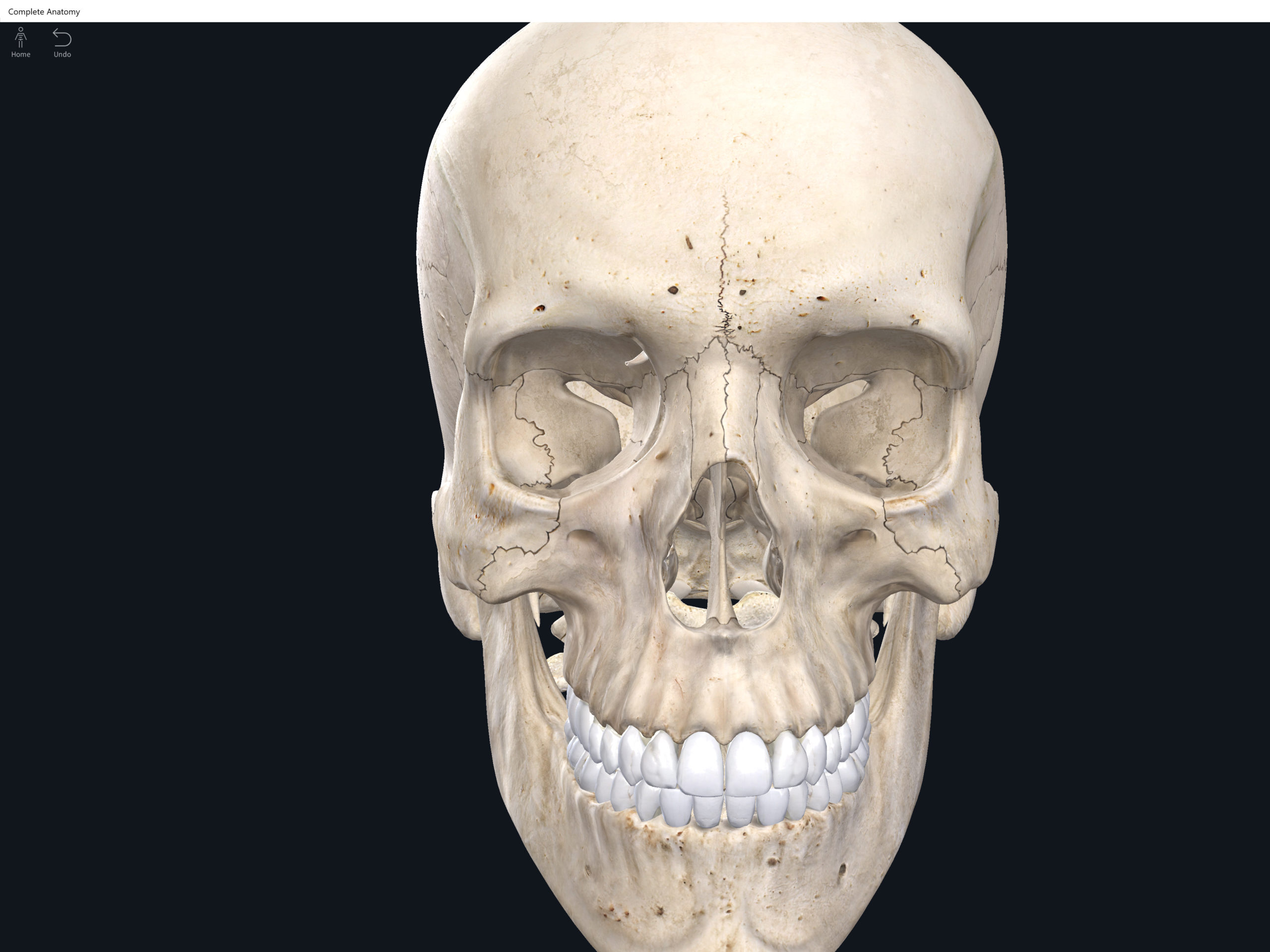
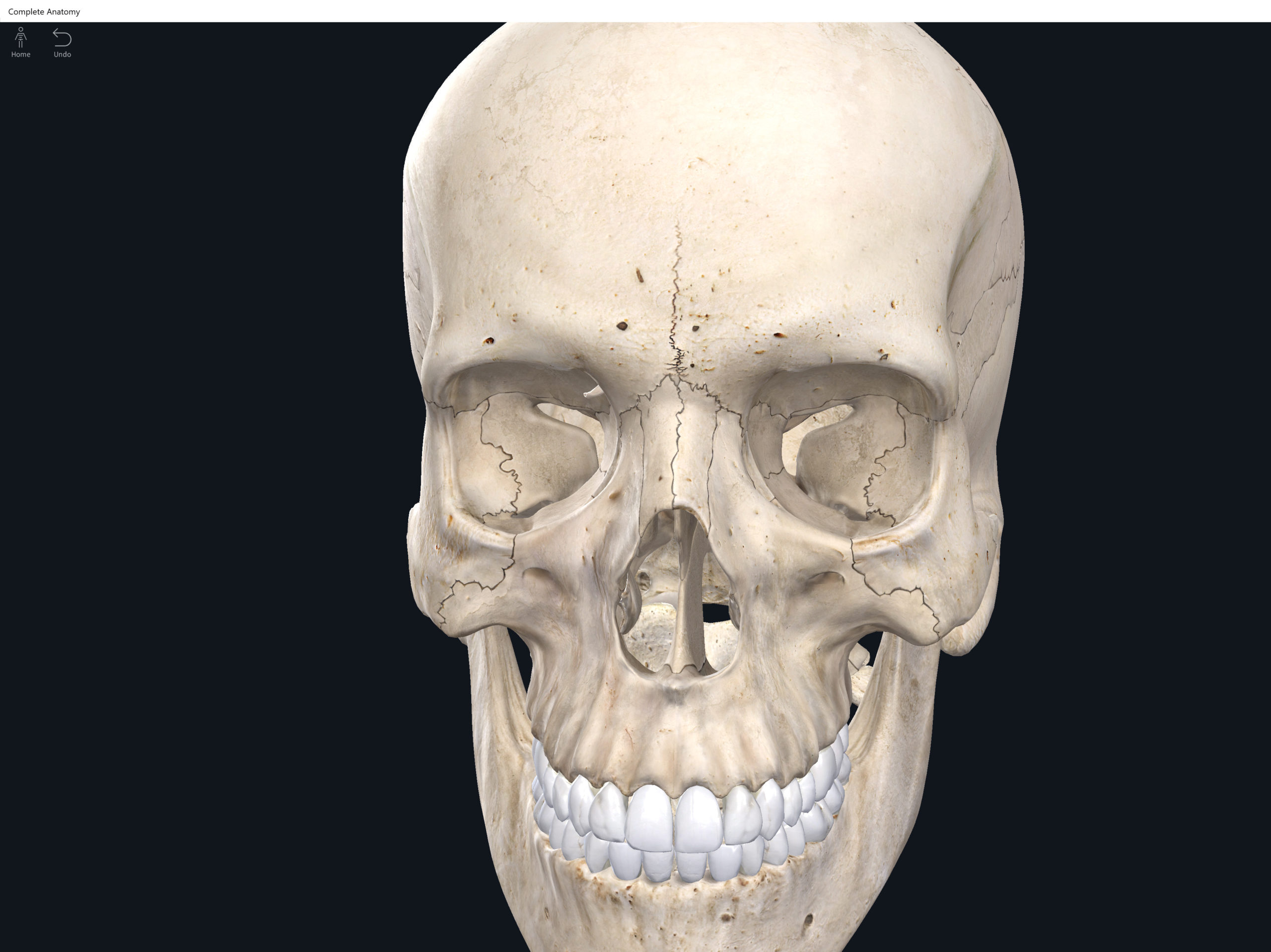
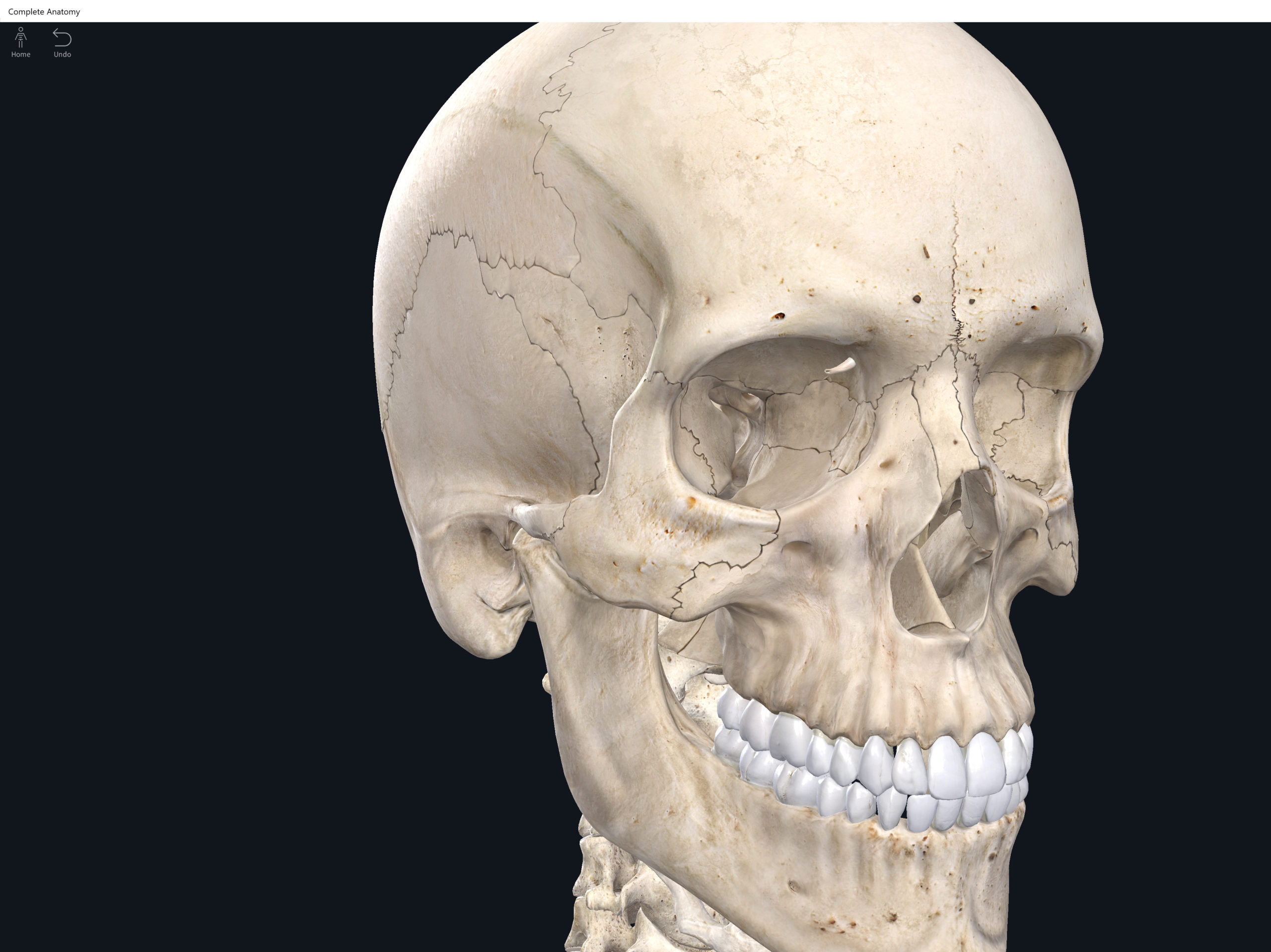
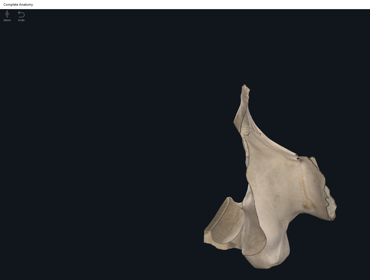
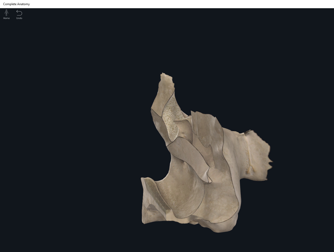
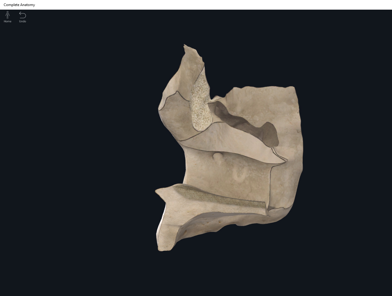
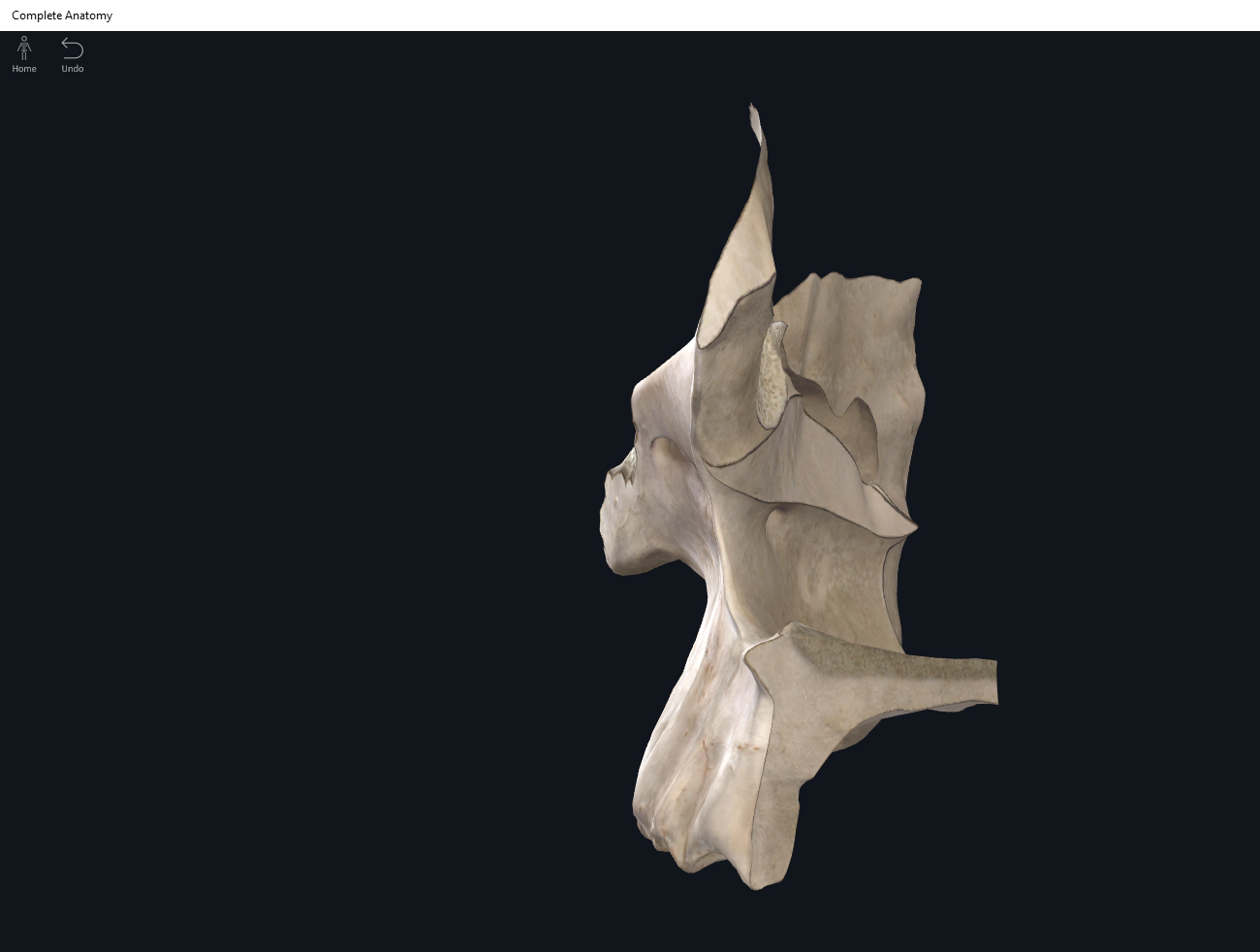
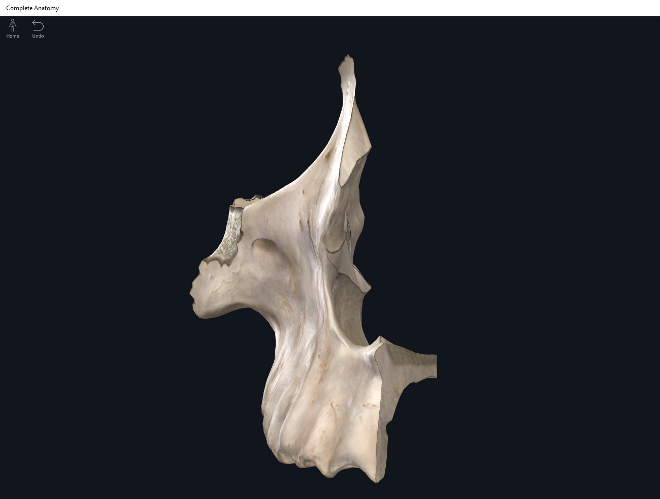
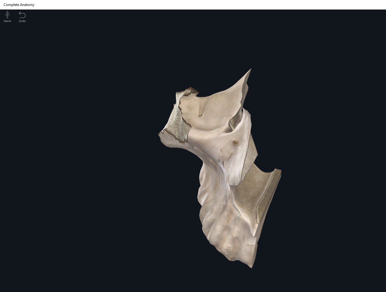
Structure.
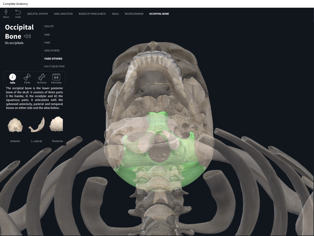
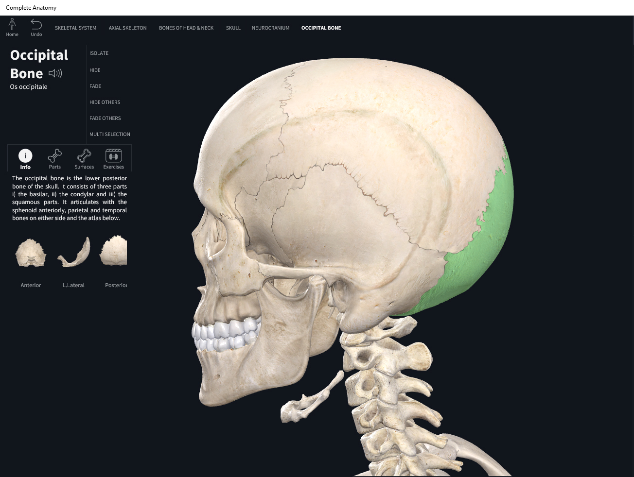
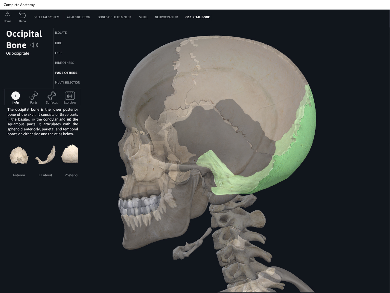
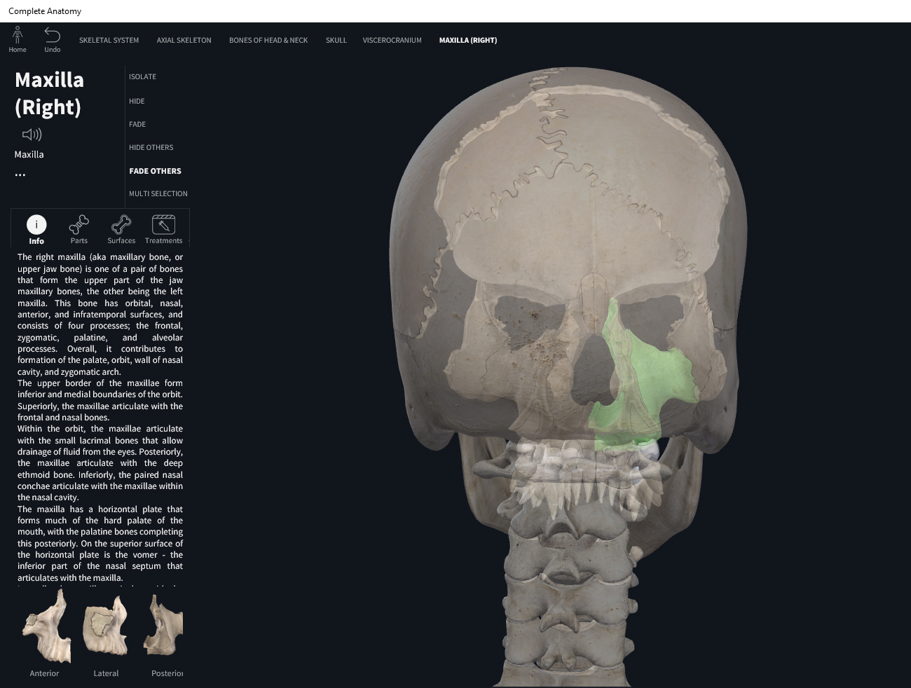
- Forms the back of the head, most of the cranial floor and foramin magnum (where the medulla oblongata connects with the spinal cord; vertebral and spinal arteries also pass through this hole).
- The occipital condyles are ovalish convex “stumps” that articulate with C1 (atlas; remember in Greek mythology that Atlas held up the sky or in this case, the skull). This articulation allows the head to nod (like when a person nods “yes”).
- Hypoglosssal canal: a foramen interior of the skull, superior to the occipital condyles, allowing for the hypoglossal nerve to pass through that controls tongue movement.
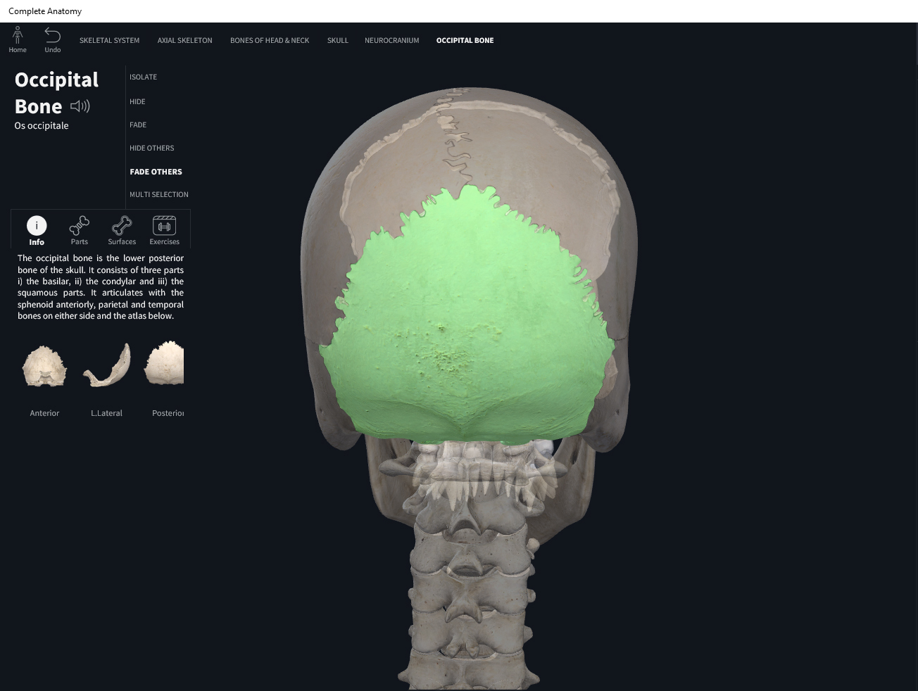
- External occipital protuberance: inferior to the lambdoid suture, the external occipital protuberance may feel like a “bump”. From a midsagittal cross-sectional view, it looks like a “gentle hill”; it is not sharp nor pointy. It is the thickest area of the occipital.
- Superior nuchal line: extending bilaterally from the external occipital protuberance, a horizontal “line”. Provides attachment points for muscles.
- Inferior nuchal line: similar to the superior nuchal line but inferior to it; extends bilaterally from the external occipital protuberance, a horizontal “line”. Provides attachment points for muscles.
Function.
Clinical Significance.
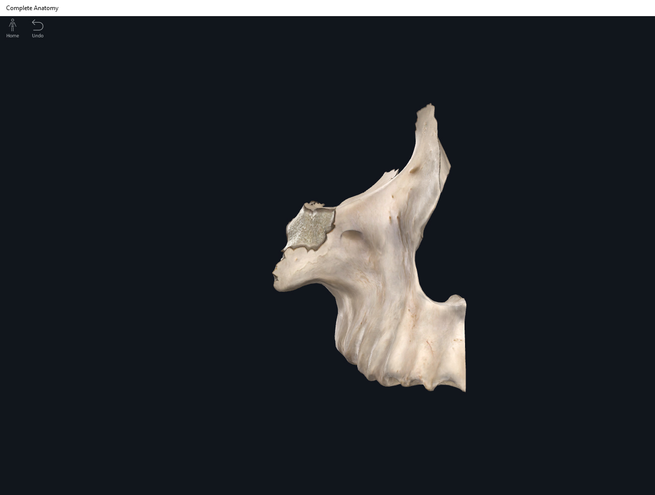
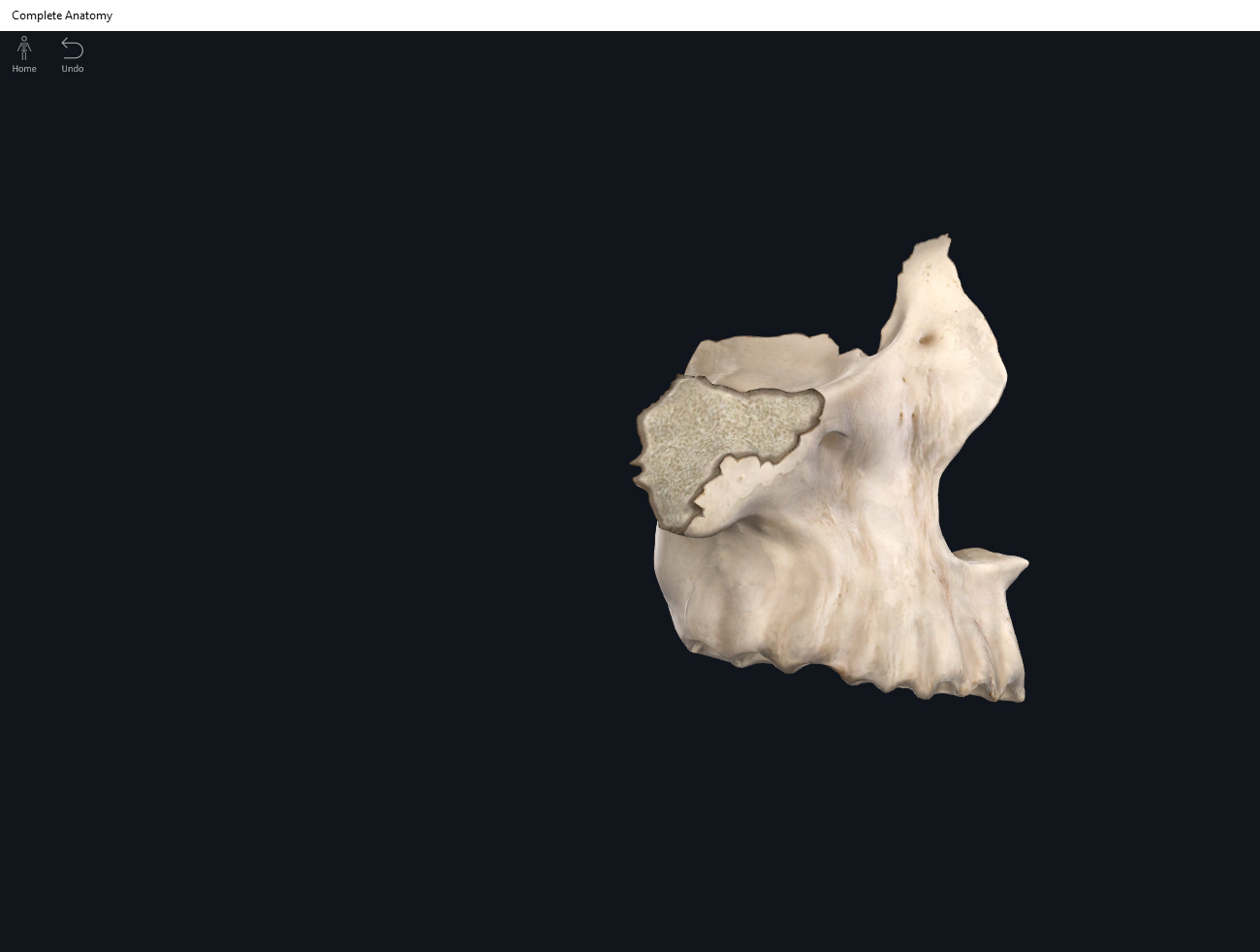
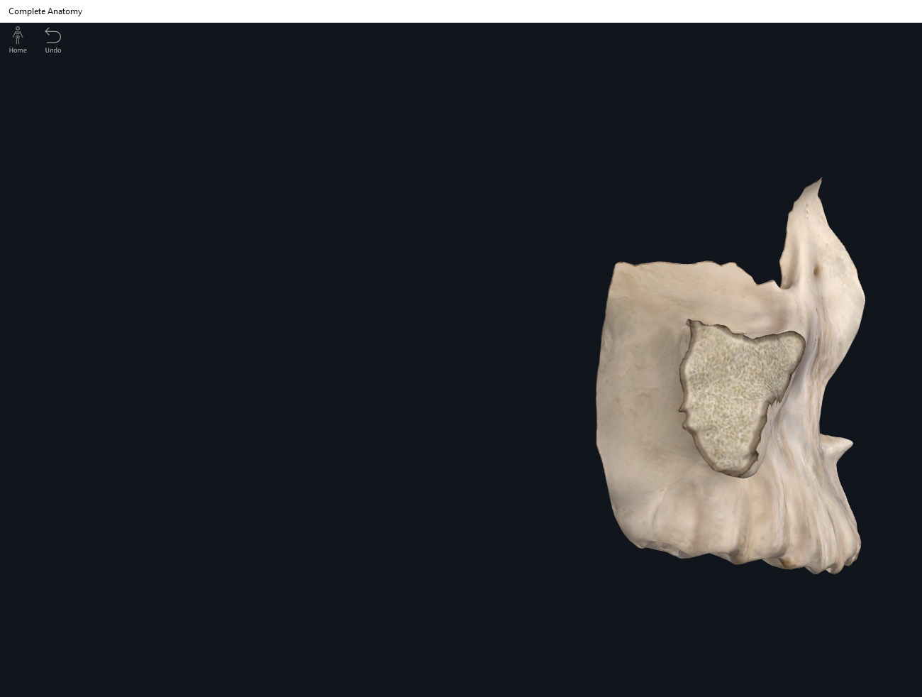
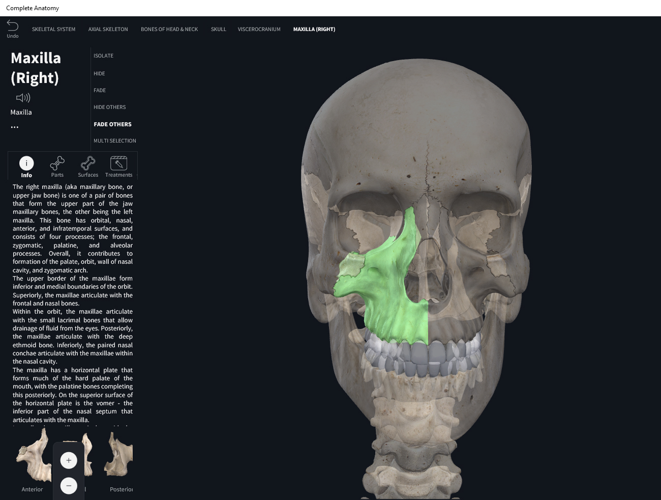
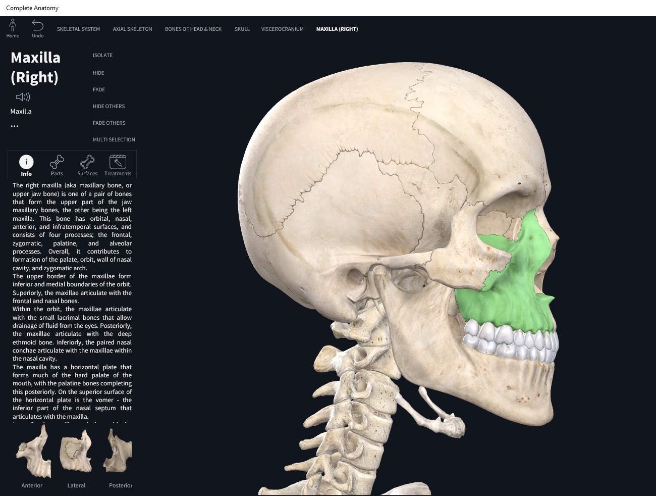
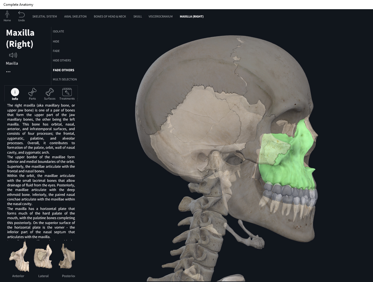
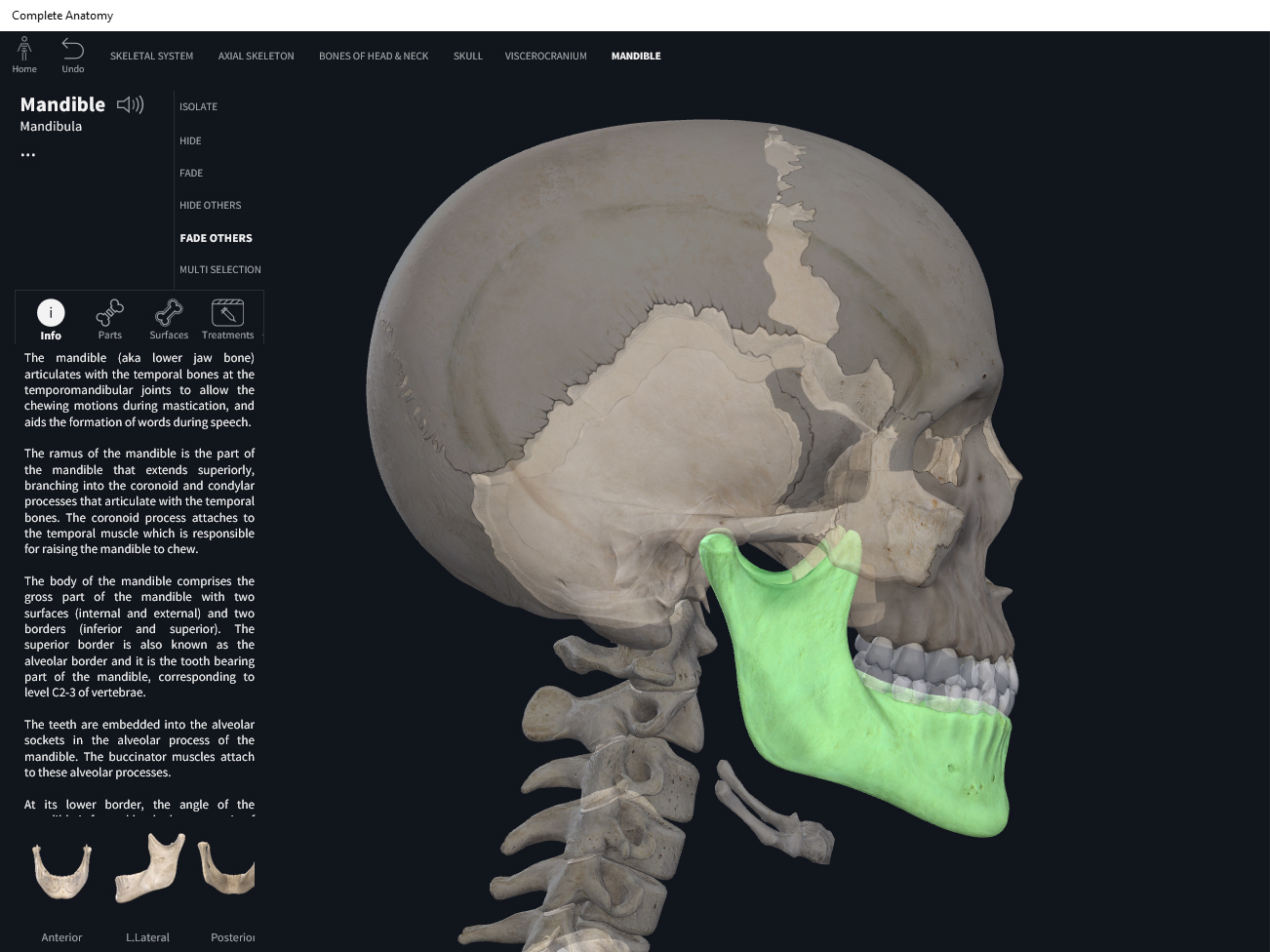
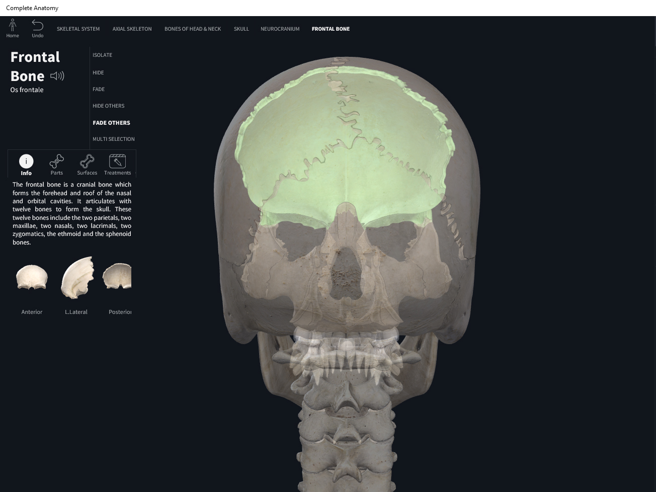
Occipital. Used with permission by 3D4Medical.
References
Biel, A. (2015). Trail guide to the body: A hands-on guide to locating muscles, bones and more.
Jenkins, G., & Tortora, G. J. (2012). Anatomy and Physiology: From Science to Life, 3rd Edition International Stu. John Wiley & Sons.
Muscolino, J. E. (2017). The muscular system manual: The skeletal muscles of the human body.