Posted on

Bones: Skull.

Download these notes.

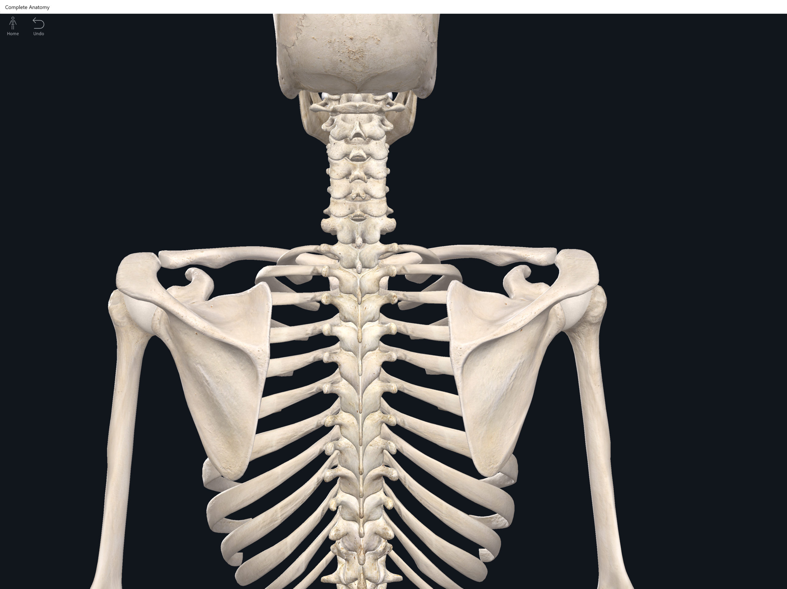
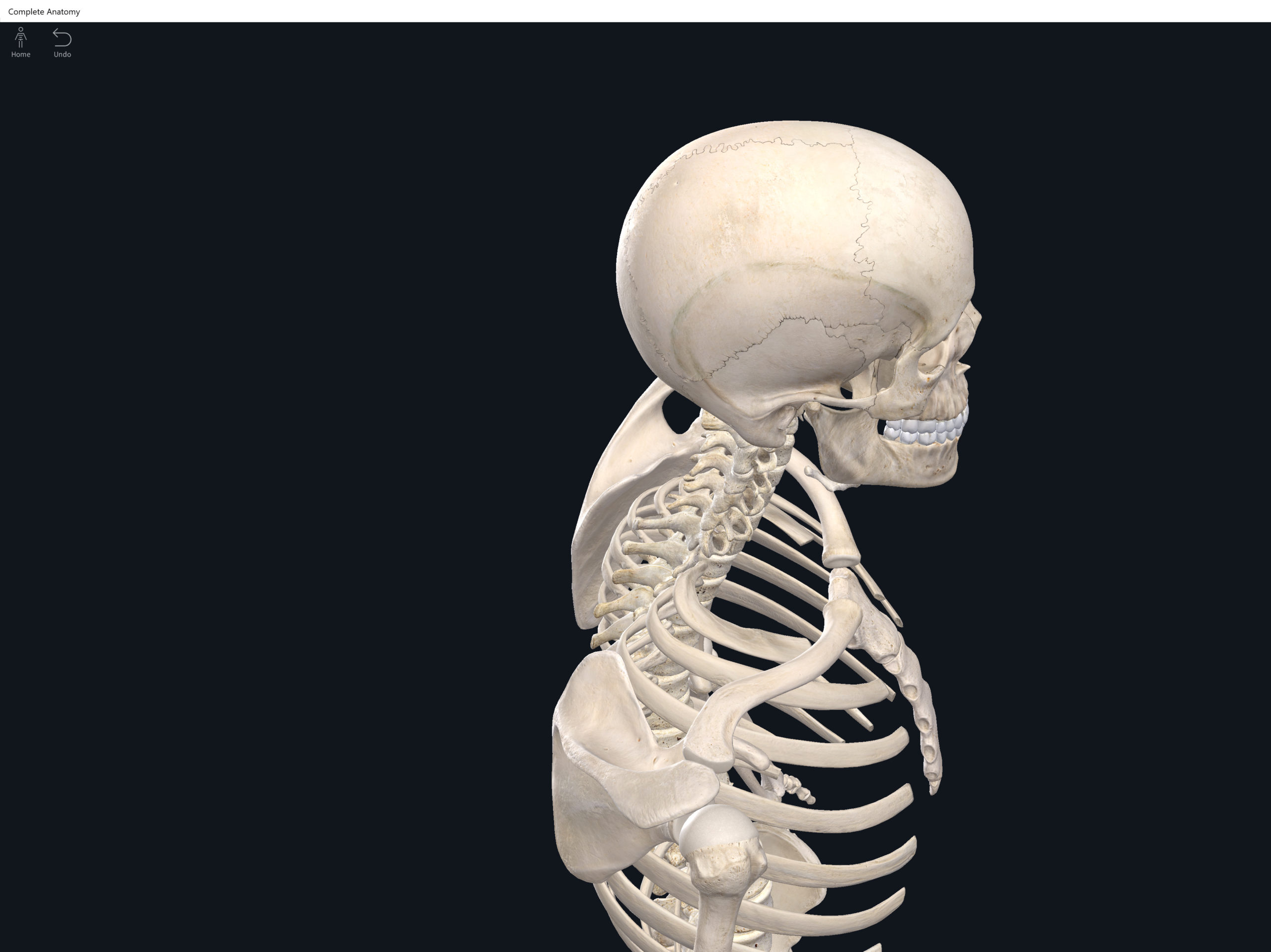
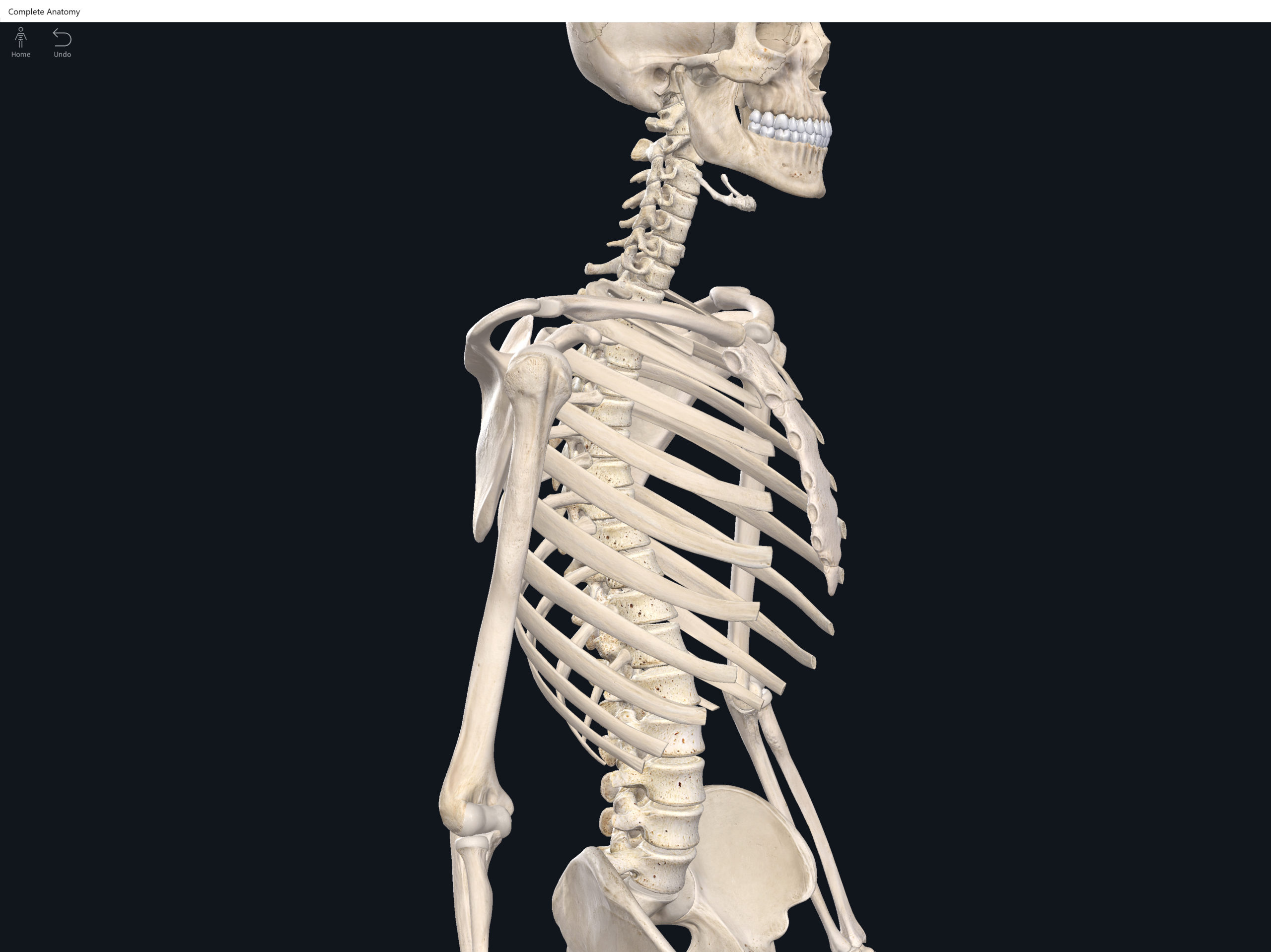
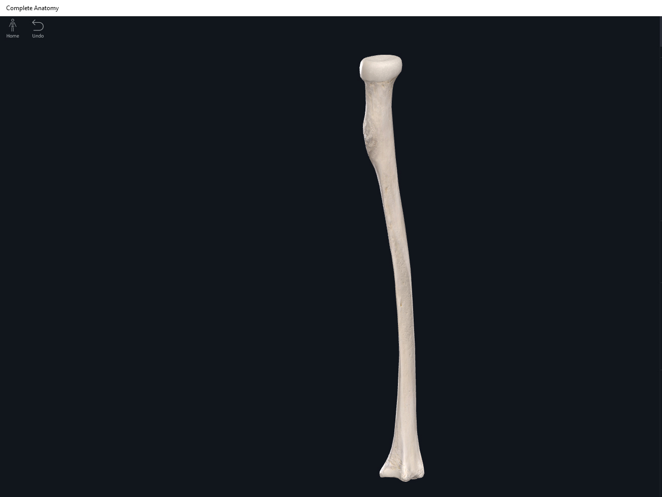
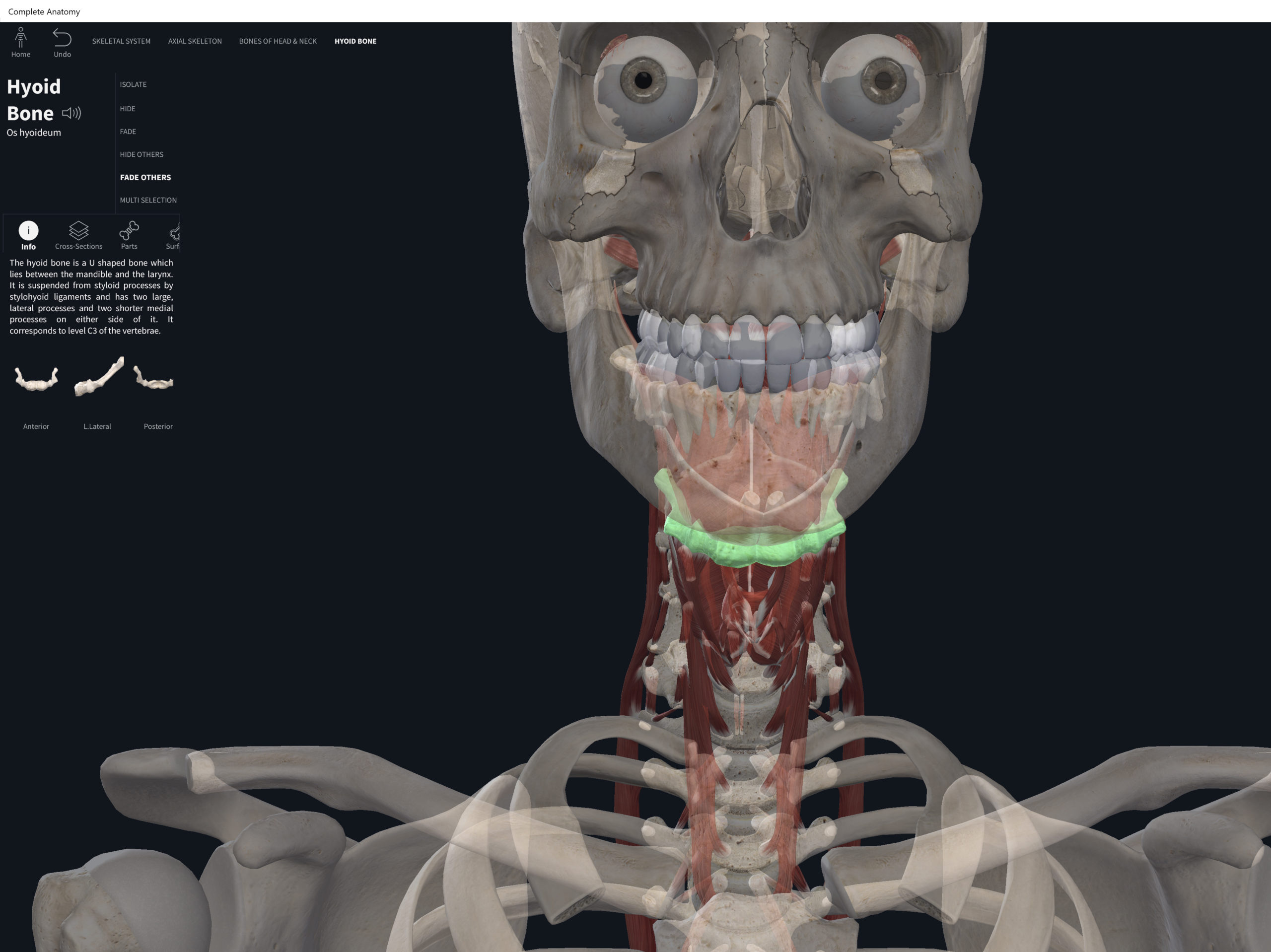
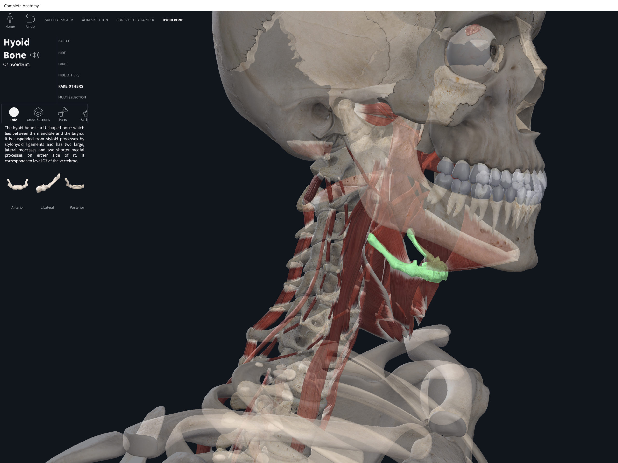
Anatomy & Physiology: Bones—Skull.


Structure.

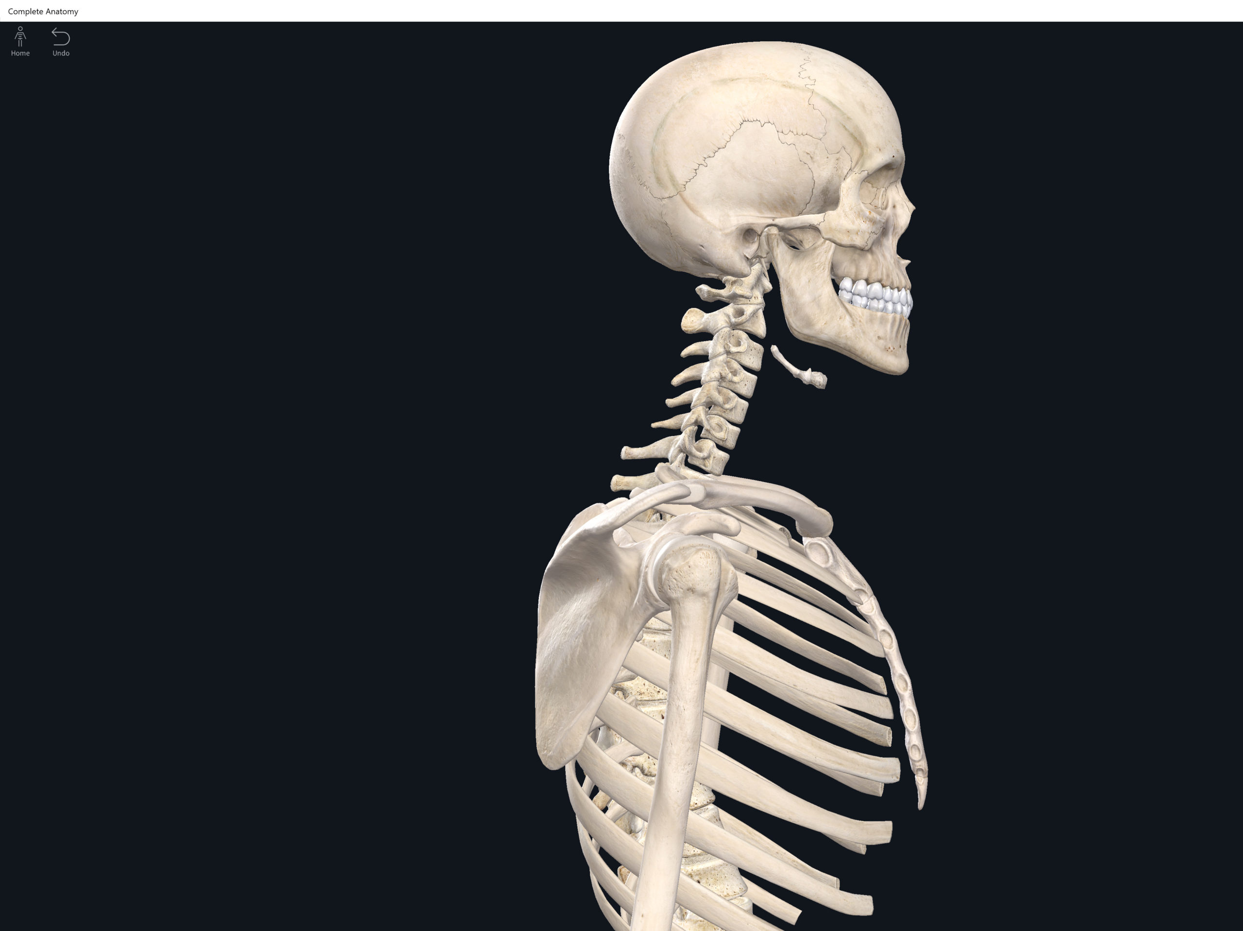
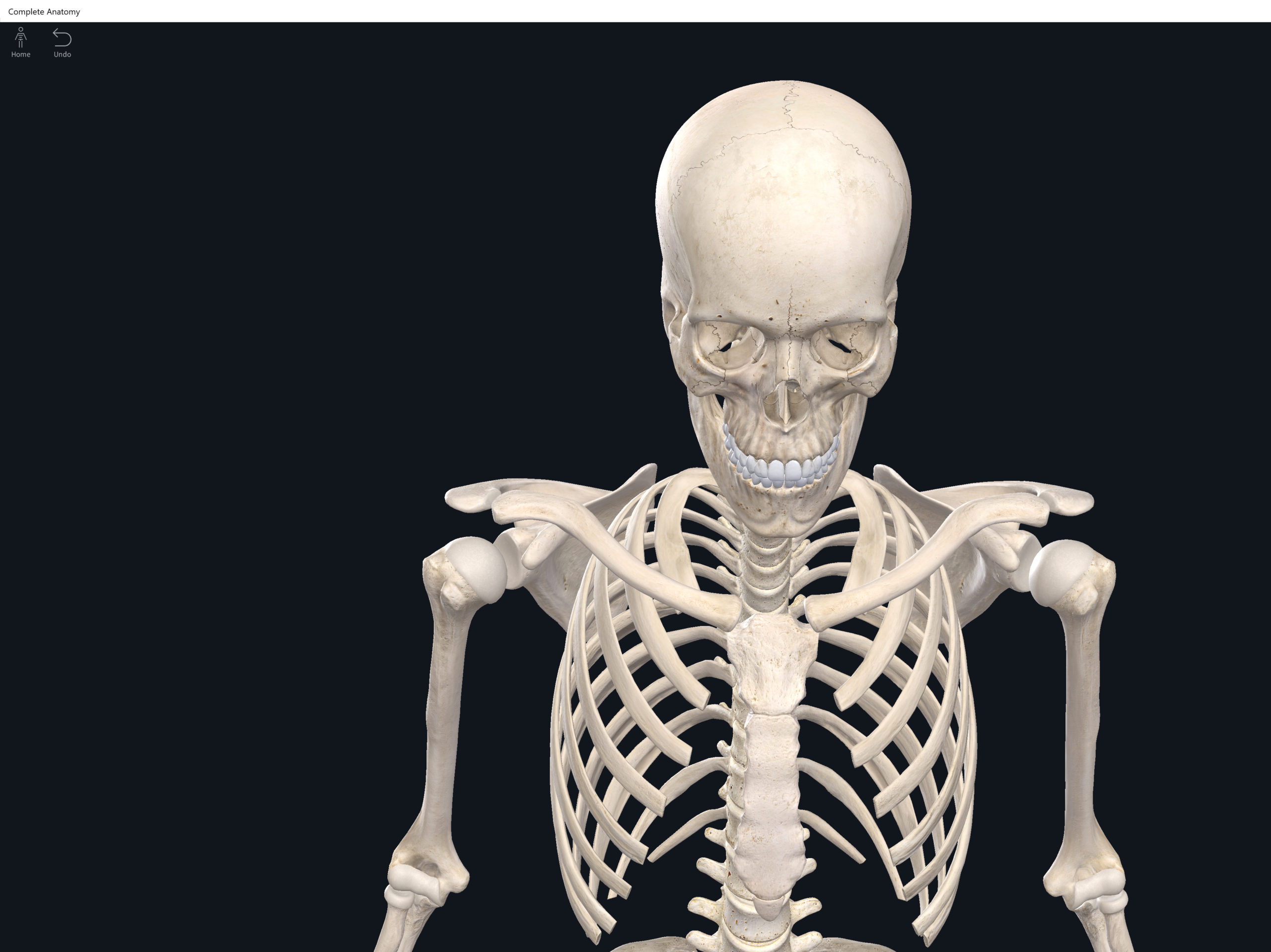
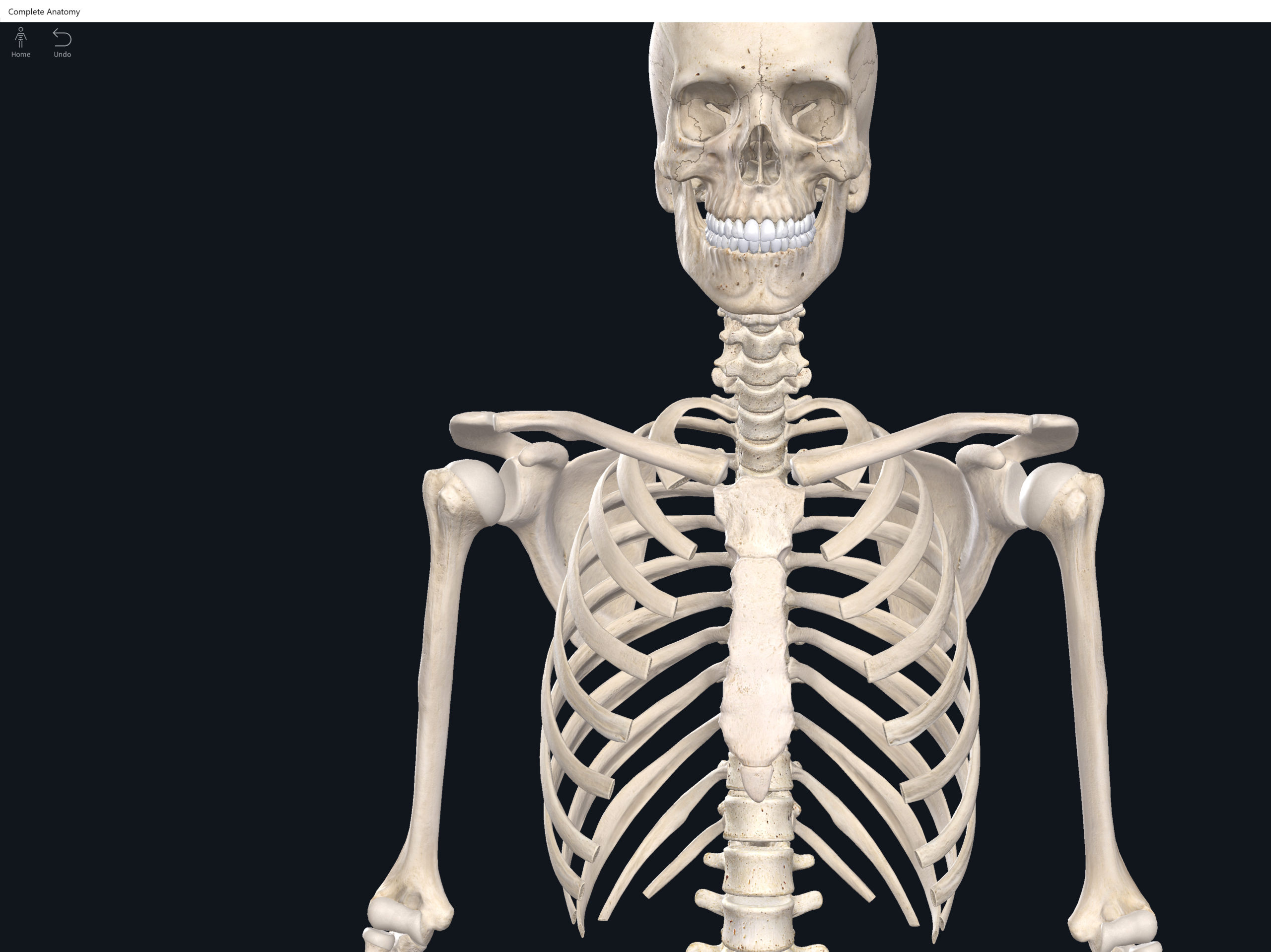
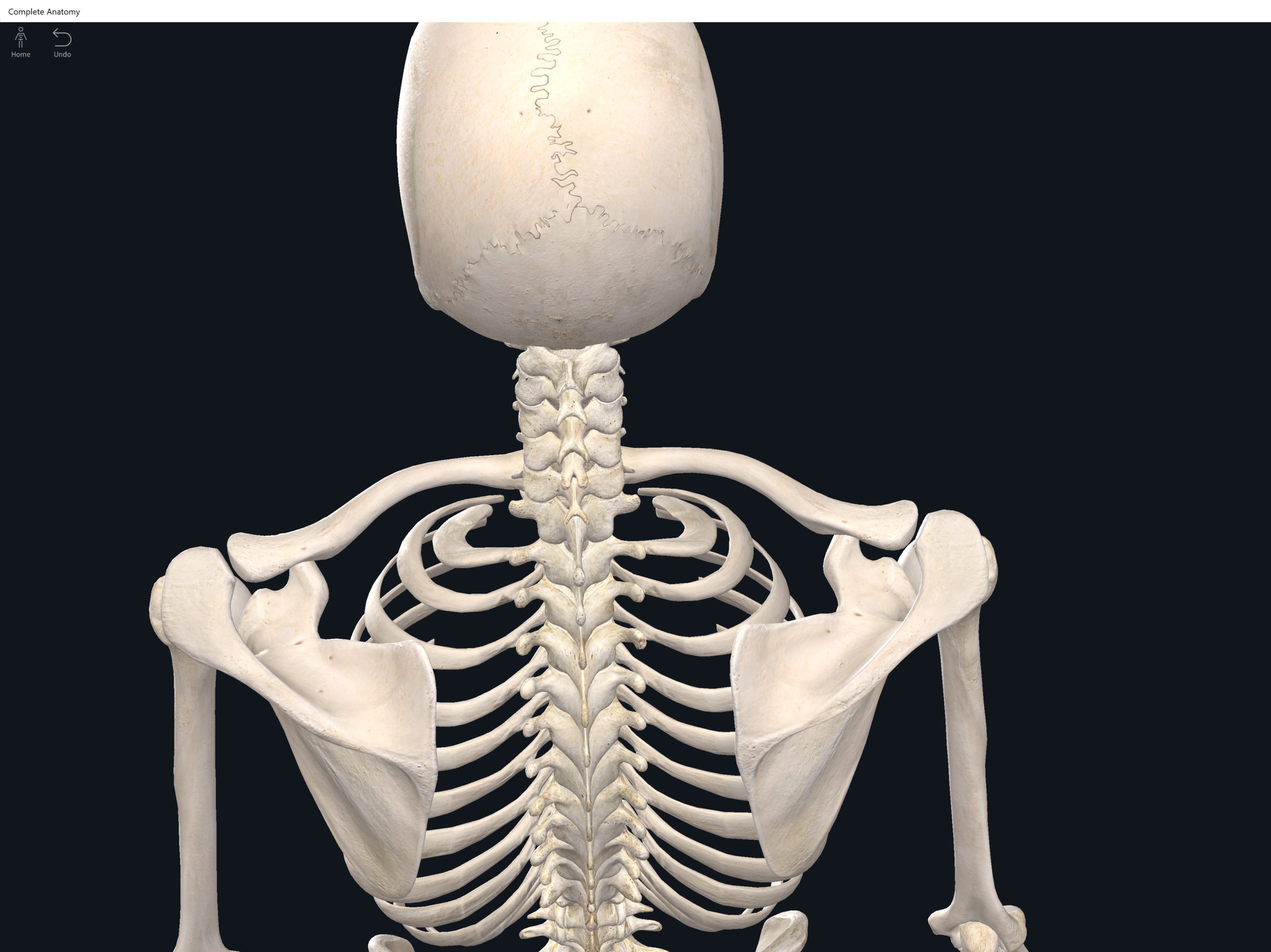
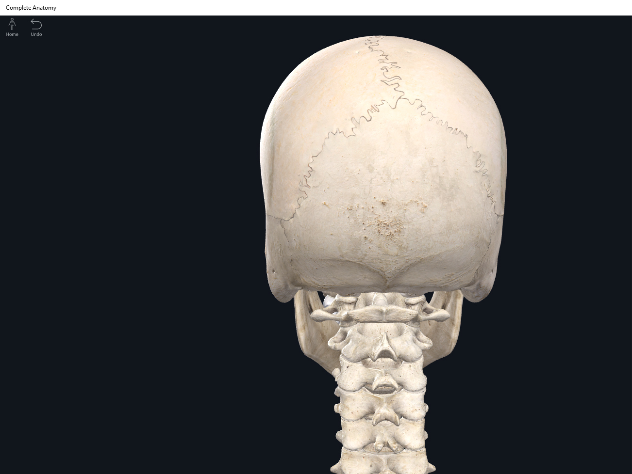
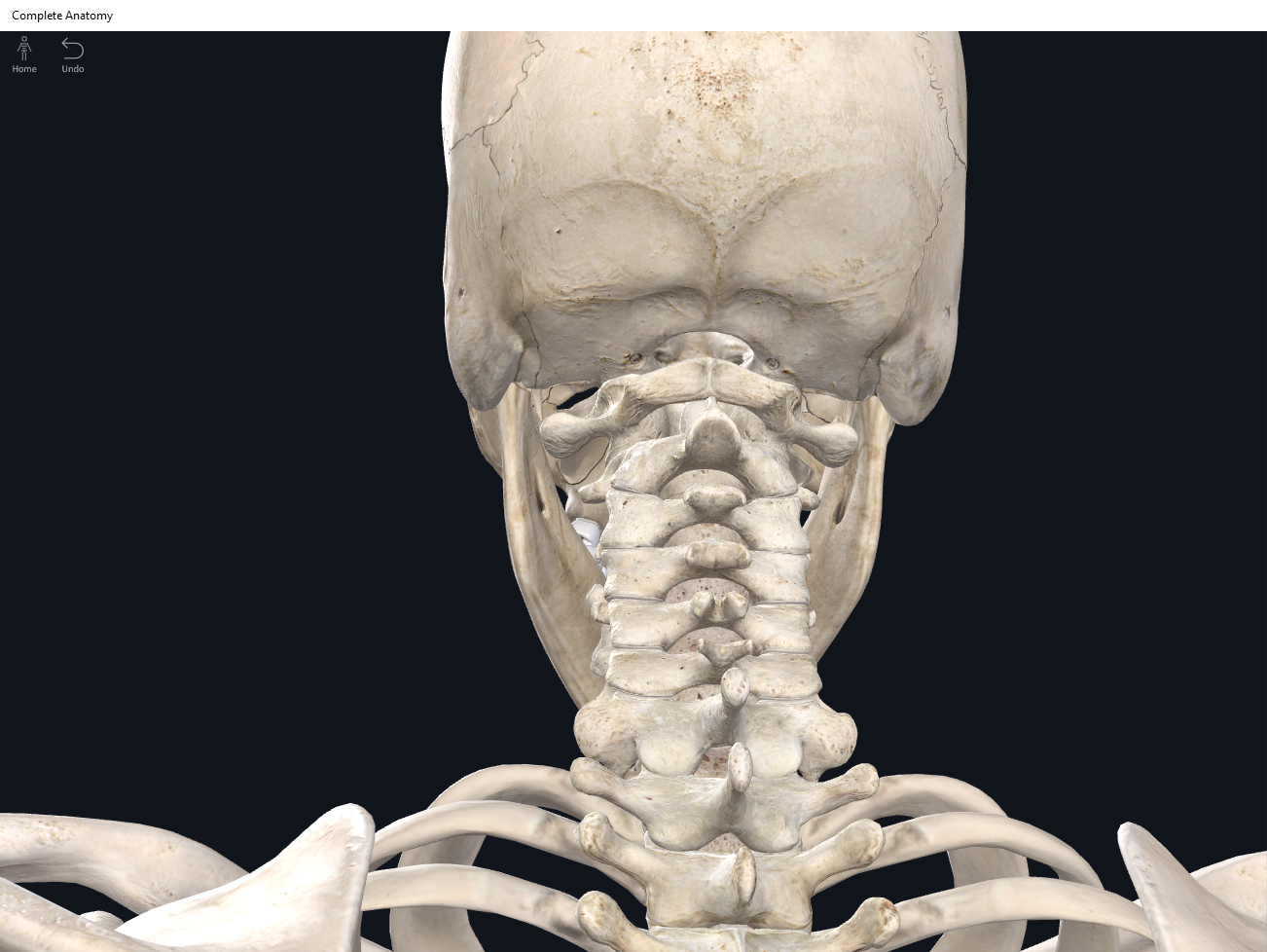
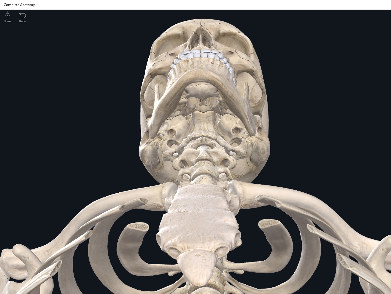
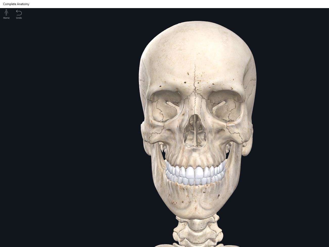
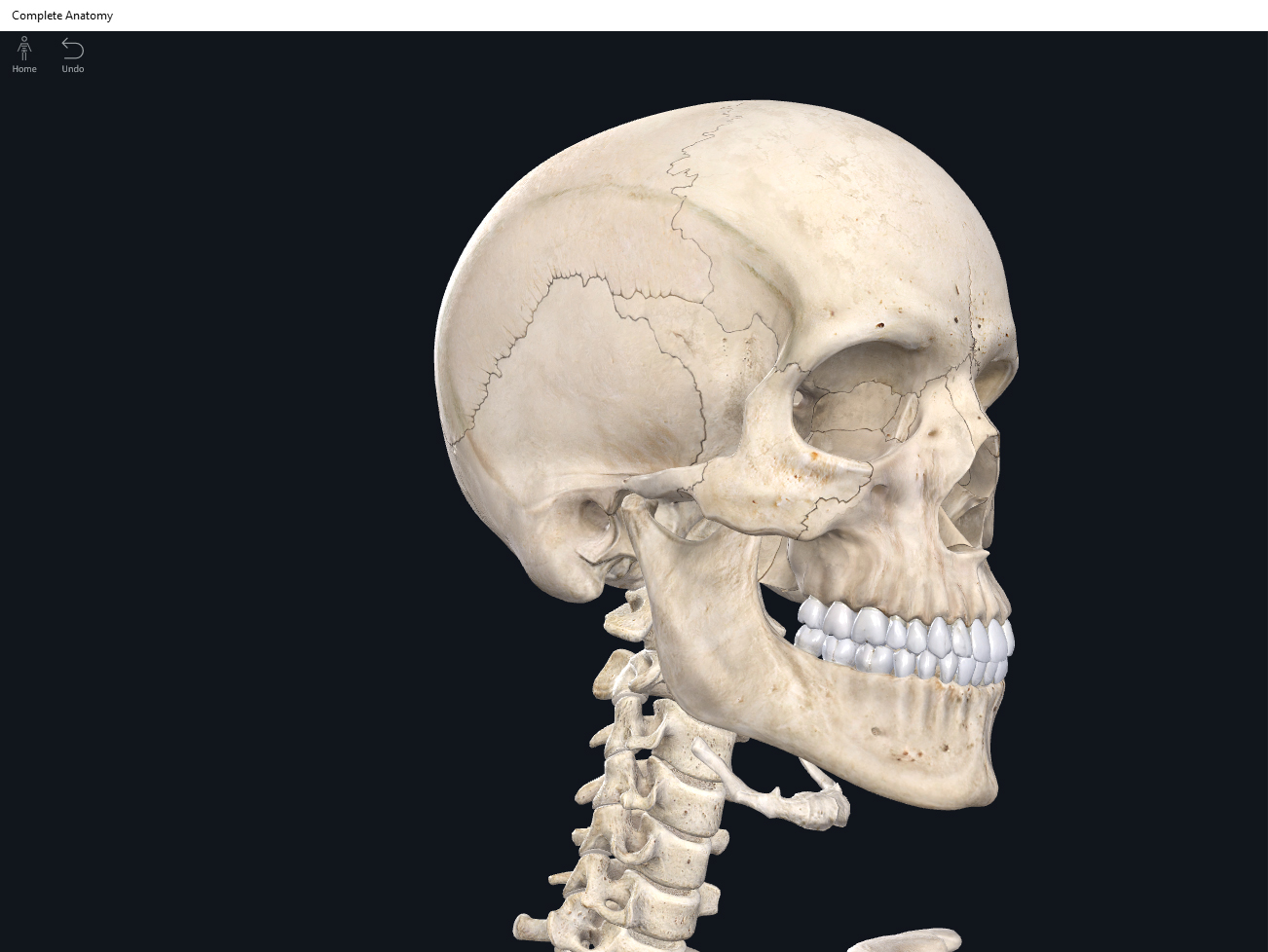
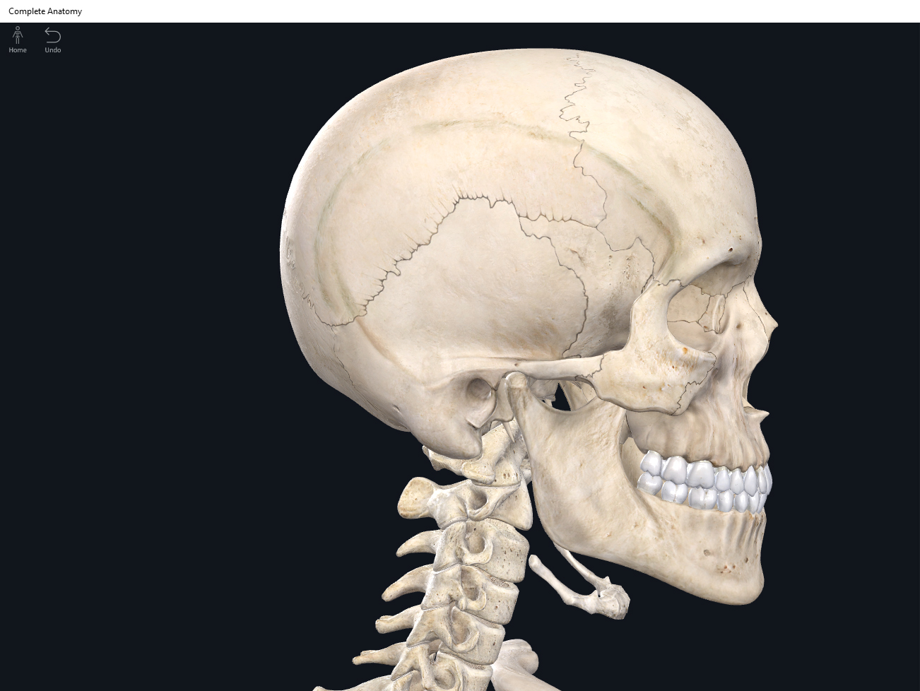
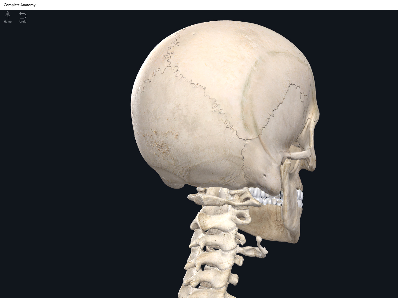
Cranial bones: frontal bone, 2 parietal bones, 2 temporal bones, occipital bone, sphenoid bone, ethmoid bone.

Facial bones: 2 nasal bones, 2 maxillae, 2 zygomatic bones, mandible, 2 lacrimal bones, 2 palatine bones, 2 inferior nasal conchae, vomer.

  • Except for the auditory ossicles, the mandible is the only moveable bone of the skull.
  • Sutures: immoveable joints that hold the skull together.


Function.


  • Protects, supports, and stabilizes brain, sense organs, nerves, and vessels.
  • Serves as attachment sites for muscles and membranes (meninges).


Clinical Significance.


References

Biel, A. (2015). Trail guide to the body: A hands-on guide to locating muscles, bones and more.

Jenkins, G., & Tortora, G. J. (2012). Anatomy and Physiology: From Science to Life, 3rd Edition International Stu. John Wiley & Sons.

Muscolino, J. E. (2017). The muscular system manual: The skeletal muscles of the human body.

Posted on

Bones: Shoulder Girdle.

Download these notes.

Anatomy & Physiology: Bones—Shoulder Girdle.


Structure.

  • The shoulder girdles (aka pectoral girdles) attach the upper limbs to the vertebral skeleton.
  • Each shoulder girdle consists of the clavicle and scapula.
  • The clavicle articulates with the manubrium.
  • The scapula articulates with the clavicle and humerus.
  • The shoulder girdles are attached to the axial skeleton and stabilized via muscles which allow for movement congruency and controlled movement.


Function.


Clinical Significance.


References

Biel, A. (2015). Trail guide to the body: A hands-on guide to locating muscles, bones and more.

Cedars-Sinai. (2018). Vertebrae of the spine. Retrieved from https://www.cedars-sinai.org/health-library/diseases-and-conditions/v/vertebrae-of-the-spine.html

Jenkins, G., & Tortora, G. J. (2012). Anatomy and Physiology: From Science to Life, 3rd Edition International Stu. John Wiley & Sons.

Muscolino, J. E. (2017). The muscular system manual: The skeletal muscles of the human body.

Posted on

Bones: Scapula.

Download these notes.

Anatomy & Physiology: Bones—Scapula.


Structure.

  • The scapula is a triangle shaped flat bone that articulates with the clavicle and humerus.
  • It is one of the most complex structures.
  • Scapular spine: posteriorly, is a prominent/obvious ridge running diagonally from the medial border to the superior lateral corner to flatten out forming the acromion. Note that this easily orients the scapula’s posterior side as the anterior side has no such ridge, allowing the scapula to lay and move congruently with the posterior rib cage.
  • Acromion: a process that is the high point of the shoulder and is easily palpateable. The acromion articulates with the clavicle’s acromial end.
  • Glenoid cavity: a slight/shallow depression that articulates with the head of the humerus.
  • Coracoid process: (name meaning like a “beak”) seen from the anterior view and inferior to the acromion, it is a substantial hook-shaped projection “pointing” (with an opening) laterally.
  • Superior angle.
  • Superior border.
  • Scapular notch.
  • Supraspinous fossa.
  • Infraspinous fossa.
  • Subscapular fossa.
  • Medial border: thinner than the lateral border.
  • Inferior angle.
  • Lateral border: thicker than the medial border.


Function.


Clinical Significance.


References

Biel, A. (2015). Trail guide to the body: A hands-on guide to locating muscles, bones and more.

Cedars-Sinai. (2018). Vertebrae of the spine. Retrieved from https://www.cedars-sinai.org/health-library/diseases-and-conditions/v/vertebrae-of-the-spine.html

Jenkins, G., & Tortora, G. J. (2012). Anatomy and Physiology: From Science to Life, 3rd Edition International Stu. John Wiley & Sons.

Muscolino, J. E. (2017). The muscular system manual: The skeletal muscles of the human body.

Posted on

Bones: Ribs.

Download these notes.

Anatomy & Physiology: Bones—Ribs.


Structure.

  • There are 12 pairs of ribs.
  • Rib length increases from rib 1 to rib 7; rib length decreases from rib 8 to rib 12.
  • Ribs articulate posteriorly with the thoracic vertebrae.
  • Ribs 1-7: called “true ribs” because they directly articulate anteriorly with the sternum via costal cartilage (hyaline).
  • Ribs 8-12: called “false ribs” because they do not directly articulate with the sternum via costal cartilage. Instead, they are indirectly attached to the sternum via costal cartilage or are not at all attached to the sternum.
  • Ribs 11-12: called “floating ribs” because they do not attach to the sternum at all via their costal cartilage.
  • Head of rib: the end that attaches posteriorly to its corresponding thoracic vertebra via facet of the vertebral body or demifacets of two neighboring vertebrae.
  • Superior and inferior facets.
  • Neck: slightly narrower and lateral to the head.
  • Tubercle: knob-like. Where the head joins the rib’s body.
  • Facet of tubercle: articulates with the facet of a transverse processes of the thoracic vertebrate.
  • Body: the main shaft.
  • Costal angle: at the curve from posterior to anterior.
  • Costal groove: attachment site and protects vessels and nerves.


Function.


Clinical Significance.

  • Intercostal space: space between ribs; occupied by muscles, nerves, and vessesls. This space usually needs to be breached in order to access the internal organs.


References

Biel, A. (2015). Trail guide to the body: A hands-on guide to locating muscles, bones and more.

Cedars-Sinai. (2018). Vertebrae of the spine. Retrieved from https://www.cedars-sinai.org/health-library/diseases-and-conditions/v/vertebrae-of-the-spine.html

Jenkins, G., & Tortora, G. J. (2012). Anatomy and Physiology: From Science to Life, 3rd Edition International Stu. John Wiley & Sons.

Muscolino, J. E. (2017). The muscular system manual: The skeletal muscles of the human body.

Posted on

Bones: Radius.

Download these notes.

Anatomy & Physiology: Bones—Radius.


Structure.

  • The forearm bone on the thumb side. You can remember this as “Thumbs up for Rad!”
  • It is smaller and shorter than the ulna.
  • Head: radial head at the proximal end articulates with both the capitulum of the humerus and the radial notch of the ulna.
  • (of radius)
  • Radial tuberosity.
  •  
  • Ulnar notch: at the distal end, medial side, articulates with the ulnar head.
  • Styloid process: at the distal end, lateral side. Easily palpateable.
  • Radius (distal end) also articulates with 3 bones of the wrist: lunate, scaphoid, and triquetrum.


Function.


Clinical Significance.


References

Biel, A. (2015). Trail guide to the body: A hands-on guide to locating muscles, bones and more.

Cedars-Sinai. (2018). Vertebrae of the spine. Retrieved from https://www.cedars-sinai.org/health-library/diseases-and-conditions/v/vertebrae-of-the-spine.html

Jenkins, G., & Tortora, G. J. (2012). Anatomy and Physiology: From Science to Life, 3rd Edition International Stu. John Wiley & Sons.

Muscolino, J. E. (2017). The muscular system manual: The skeletal muscles of the human body.

Posted on

Bones: Patella.

Download these notes.

Anatomy & Physiology: Bones—Patella.


Structure.

  • The kneecap bone.
  • Small, triangular sesamoid.
  • Base: the superior portion; widest portion of the triangle.
  • Apex: the inferior “tip” of the triangle.
  • Articular facets: posterior view; two facets (medial and lateral) allow for articulation with the femoral condyle.


Function.


Clinical Significance.

  • Increases the leverage of quadraceps femoris.


References

Biel, A. (2015). Trail guide to the body: A hands-on guide to locating muscles, bones and more.

Cedars-Sinai. (2018). Vertebrae of the spine. Retrieved from https://www.cedars-sinai.org/health-library/diseases-and-conditions/v/vertebrae-of-the-spine.html

Jenkins, G., & Tortora, G. J. (2012). Anatomy and Physiology: From Science to Life, 3rd Edition International Stu. John Wiley & Sons.

Muscolino, J. E. (2017). The muscular system manual: The skeletal muscles of the human body.

Posted on

Bones: Lumbo-Pelvic Hip Complex.

Download these notes.

Anatomy & Physiology: Bones—Lumbo-Pelvic Hip Complex (Pelvic Girdle).


Structure.

  • The lumbo-pelvic hip complex (LPHC) is a keystone structure in the human body as it transmits forces up through the lower kinetic chain to the upper kinetic chain.
  • The LPHC consists of: 2 coxal (hip) bones joined anteriorly at the pubic symphasis; the sacrum; and the coccyx.
  • Sacroiliac joint: where the auricular surface of the ilium articulate with the auricular surface of the sacrum.
  • Coxal bone (hip bone): consists of 3 bones (initially separated by cartilage but fuses together around age 23 yrs): ilium, pubis; ischium (“Your hip is I.P.I. or ippy”).
  • Ilium: superior and largest of the hip bones.
    • Ala (“wing”).
    • Acetabulum: forms part of the hip socket.
    •  
    • Iliac crest: superior border, extending anteriorly to form the anterior superior iliac spine. Palpateable.
    • Anterior superior iliac spine (ASIS): palpateable landmark.
    • Anterior inferior iliac spine (AIIS): palateable landmark.
    • Posterior superior iliac spine (PSIS): palpateable landmark.
    • Posterior inferior iliac spine (PIIS): palpateable landmark.
    • Greater sciatic notch: inferior to the PIIS, through which the sciatic nerve passes (sciatic nerve is longest nerve in human body).
    • Iliac fossa.
    • Auricular surface: roughened area and articulates with the auricular surface of the sacrum to form the SI joint.
    • Arcuate line.
    • Posterior gluteal line (lateral surface).
    • Anterior gluteal line (lateral surface).
    • Inferior gluteal line (lateral surface).
  • Ischium: inferior and posterior portion of the coxal bone.
    •  
    • Ramus: fuses with the pubis.
    • Ischial spine.
    • Lesser sciatic notch.
    • Ischial tuberosity: rough and thickened area.
    • Obturator foramen: formed from the ischium and pubis. Largest foramen in the skeleton. Almost totally closed off by fibrous obturator membrane.
  • Pubis: inferior and anterior portion of the coxal bone.
    • Superior ramus.
    • Inferior ramus: the inferior rami of the two coxal bones, form the pubic arch.
    •  
    • Pubic tubercle.
    • Pubic crest.
    • Pubic symphasis: joint between two hip bones with fibrocartilage inbetween. In pregnant women, relaxin (hormone) increases the flexibility of the pubic symphasis.
    • Obturator foramen.
  • Acetabulum: hip socket of hip joint formed by ilium, ischium, and pubis.
  • False vs. True Pelvis.
    • Pelvic brim: defines the superior and inferior pelvis. Higher posteriorly than anteriorly due to tilt.
    • False pelvis: the portion of the pelvis superior to the pelvic brim.
    • True pelvis: the portion of the pelvis inferior to the pelvic brim.
    • Pelvic inlet: superior opening of the true pelvis.
    • Pelvic outlet: inferior opening of the true pelvis.
    • Pelvic axis.
  • Male pelvis: tend to be larger, heavier, with more surface markings.
  • Female pelvis: tend to be shallower and wider, more spacious true pelvis.

Function.

Clinical Significance.


References

Biel, A. (2015). Trail guide to the body: A hands-on guide to locating muscles, bones and more.

Cedars-Sinai. (2018). Vertebrae of the spine. Retrieved from https://www.cedars-sinai.org/health-library/diseases-and-conditions/v/vertebrae-of-the-spine.html

Jenkins, G., & Tortora, G. J. (2012). Anatomy and Physiology: From Science to Life, 3rd Edition International Stu. John Wiley & Sons.

Muscolino, J. E. (2017). The muscular system manual: The skeletal muscles of the human body.

Posted on

Bones: Hyoid.

Download these notes.

Anatomy & Physiology: Bones—Hyoid.


Structure.

  • Unique as the hyoid doesn’t articulate with any other bone. Ligaments and muscles suspend the hyoid between the mandible and larynx.
  • U-shaped.
  •  
  • Greater horns: paired; the rounded end of the side of the “U”.
  • Lesser horns: paired; the small triangular bump (fang-shaped) lateral to the body.

Function.

  • Supports tongue.
  • Serves as attachment points for the tongue muscles and muscles for the pharynx and larynx.

Clinical Significance.

  • In strangulation and hangings, the hyoid is often fractured.


References

Biel, A. (2015). Trail guide to the body: A hands-on guide to locating muscles, bones and more.

Jenkins, G., & Tortora, G. J. (2012). Anatomy and Physiology: From Science to Life, 3rd Edition International Stu. John Wiley & Sons.

Muscolino, J. E. (2017). The muscular system manual: The skeletal muscles of the human body.

Posted on

Bones: Humerus.

Download these notes.

Anatomy & Physiology: Bones—Humerus.


Structure.

  • The largest and longest upper limb bone.
  • The humerus articulates with the scapula, radius, and ulna.
  • Head: the proximal end is the head which articulates with the scapula at the glenoid fossa. This forms the shoulder joint.
  • Anatomical neck.
  • Surgical neck.
  • Greater tubercle.
  • Lesser tubercle.
  • Intertubercular sulcus (groove).
  • Body (diaphysis/shaft).
  • Deltoid tuberosity: where the tendons of the deltoid attach.
  • Capitulum: “capit” means head; a rounded knob-like structure on the lateral side. Articulates with the radial head.
  • Radial fossa: anterior depression superior to the capitulum. The radial head gets tucked into this depression when the elbow joint is bent. Most lateral fossa.
  • Trochlea: medial to the capitulum. Articulates with the ulnar trochlear notch.
  • Coronoid fossa: anterior depression that allows the coronoid process of the ulna to tuck into this depression when the elbow is bent. This fossa is medial to the radial fossa.
  • Olecranon fossa: posterior deep depression that allows the olecranon of the ulna to “tuck” into when the elbow is extended.
  • Medial epicondyle.
  • Lateral epicondyle.


Function.


Clinical Significance.


References

Biel, A. (2015). Trail guide to the body: A hands-on guide to locating muscles, bones and more.

Cedars-Sinai. (2018). Vertebrae of the spine. Retrieved from https://www.cedars-sinai.org/health-library/diseases-and-conditions/v/vertebrae-of-the-spine.html

Jenkins, G., & Tortora, G. J. (2012). Anatomy and Physiology: From Science to Life, 3rd Edition International Stu. John Wiley & Sons.

Muscolino, J. E. (2017). The muscular system manual: The skeletal muscles of the human body.

Posted on

Bones: Foot and Ankle.

Download these notes.

Anatomy & Physiology: Bones—Foot and Ankle.


Structure.

  • Tarsus: the ankle; proximally located. Consists of: talus (ankle bone); calcaneous (heel bone, largest and strongest of the tarsal bones); navicular (medial, forms the “platform” for the 3 cuneiform bones); cuneiform (x3, lateral, intermediate, medial); cuboid (lateral, cube-shaped).
  • Metatarsals: intermediate region of the foot and numbered I-V starting with the great toe (hallux as I). Articulate with cuboid and cuneiforms.
  • Phalanges: proximal, middle and distal; 3 phalanges per toe except for the great toe (hallux) which only has 2. Phalanges are numbered medial to lateral starting with 1 at the great toe.

Function.


Clinical Significance.


References

Biel, A. (2015). Trail guide to the body: A hands-on guide to locating muscles, bones and more.

Cedars-Sinai. (2018). Vertebrae of the spine. Retrieved from https://www.cedars-sinai.org/health-library/diseases-and-conditions/v/vertebrae-of-the-spine.html

Jenkins, G., & Tortora, G. J. (2012). Anatomy and Physiology: From Science to Life, 3rd Edition International Stu. John Wiley & Sons.

Muscolino, J. E. (2017). The muscular system manual: The skeletal muscles of the human body.