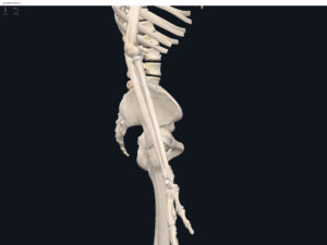
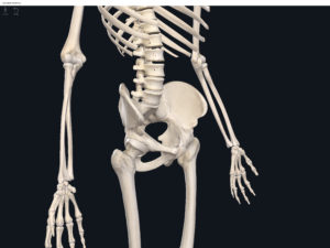
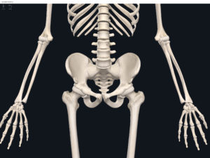
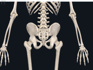
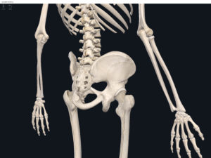
Anatomy & Physiology: Bones—Lumbo-Pelvic Hip Complex (Pelvic Girdle).
Structure.
- The lumbo-pelvic hip complex (LPHC) is a keystone structure in the human body as it transmits forces up through the lower kinetic chain to the upper kinetic chain.
- The LPHC consists of: 2 coxal (hip) bones joined anteriorly at the pubic symphasis; the sacrum; and the coccyx.
- Sacroiliac joint: where the auricular surface of the ilium articulate with the auricular surface of the sacrum.
- Coxal bone (hip bone): consists of 3 bones (initially separated by cartilage but fuses together around age 23 yrs): ilium, pubis; ischium (“Your hip is I.P.I. or ippy”).
- Ilium: superior and largest of the hip bones.
- Ala (“wing”).
- Acetabulum: forms part of the hip socket.
- Iliac crest: superior border, extending anteriorly to form the anterior superior iliac spine. Palpateable.
- Anterior superior iliac spine (ASIS): palpateable landmark.
- Anterior inferior iliac spine (AIIS): palateable landmark.
- Posterior superior iliac spine (PSIS): palpateable landmark.
- Posterior inferior iliac spine (PIIS): palpateable landmark.
- Greater sciatic notch: inferior to the PIIS, through which the sciatic nerve passes (sciatic nerve is longest nerve in human body).
- Iliac fossa.
- Auricular surface: roughened area and articulates with the auricular surface of the sacrum to form the SI joint.
- Arcuate line.
- Posterior gluteal line (lateral surface).
- Anterior gluteal line (lateral surface).
- Inferior gluteal line (lateral surface).
- Ischium: inferior and posterior portion of the coxal bone.
- Ramus: fuses with the pubis.
- Ischial spine.
- Lesser sciatic notch.
- Ischial tuberosity: rough and thickened area.
- Obturator foramen: formed from the ischium and pubis. Largest foramen in the skeleton. Almost totally closed off by fibrous obturator membrane.
- Pubis: inferior and anterior portion of the coxal bone.
- Superior ramus.
- Inferior ramus: the inferior rami of the two coxal bones, form the pubic arch.
- Pubic tubercle.
- Pubic crest.
- Pubic symphasis: joint between two hip bones with fibrocartilage inbetween. In pregnant women, relaxin (hormone) increases the flexibility of the pubic symphasis.
- Obturator foramen.
- Acetabulum: hip socket of hip joint formed by ilium, ischium, and pubis.
- False vs. True Pelvis.
- Pelvic brim: defines the superior and inferior pelvis. Higher posteriorly than anteriorly due to tilt.
- False pelvis: the portion of the pelvis superior to the pelvic brim.
- True pelvis: the portion of the pelvis inferior to the pelvic brim.
- Pelvic inlet: superior opening of the true pelvis.
- Pelvic outlet: inferior opening of the true pelvis.
- Pelvic axis.
- Male pelvis: tend to be larger, heavier, with more surface markings.
- Female pelvis: tend to be shallower and wider, more spacious true pelvis.
Function.
Clinical Significance.
References
Biel, A. (2015). Trail guide to the body: A hands-on guide to locating muscles, bones and more.
Cedars-Sinai. (2018). Vertebrae of the spine. Retrieved from https://www.cedars-sinai.org/health-library/diseases-and-conditions/v/vertebrae-of-the-spine.html
Jenkins, G., & Tortora, G. J. (2012). Anatomy and Physiology: From Science to Life, 3rd Edition International Stu. John Wiley & Sons.
Muscolino, J. E. (2017). The muscular system manual: The skeletal muscles of the human body.
