Anatomy & Physiology: Bones—Skull, Ethmoid Bone.
Structure.
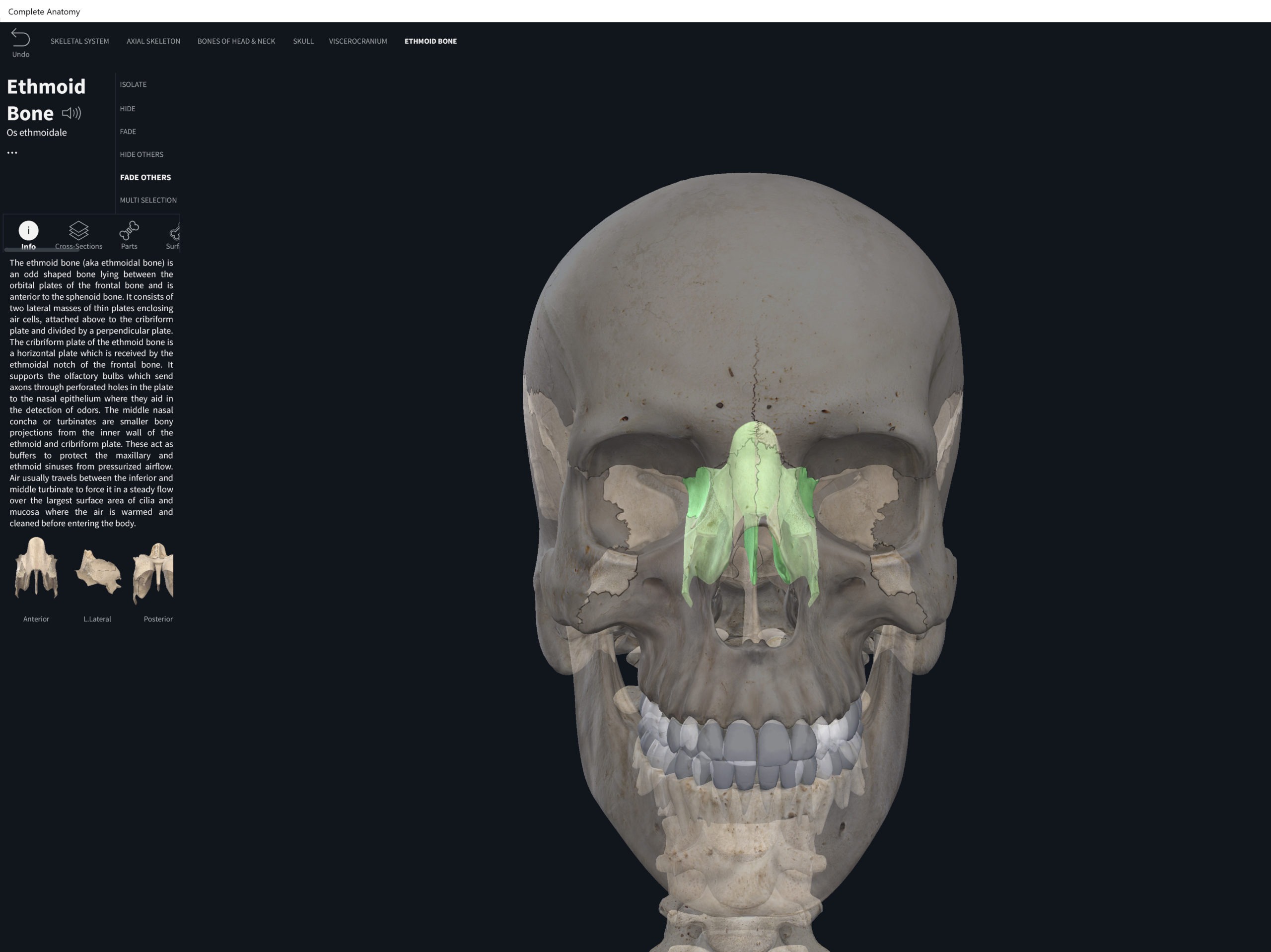
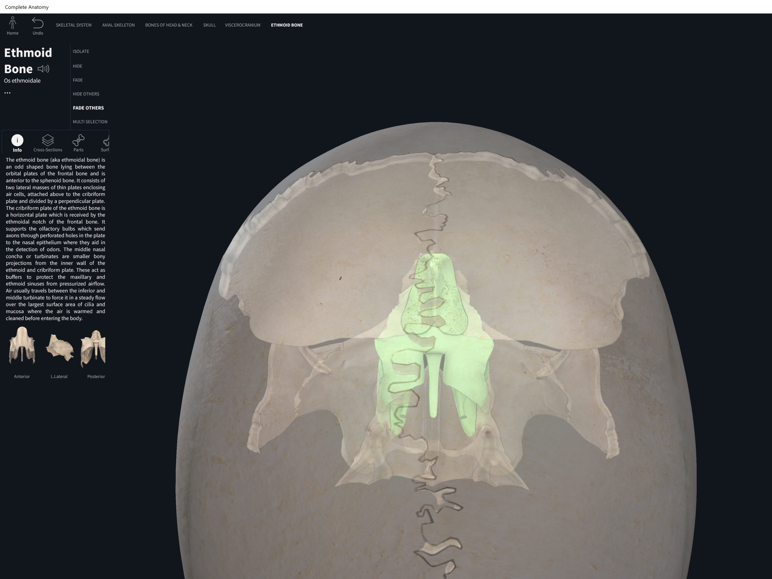
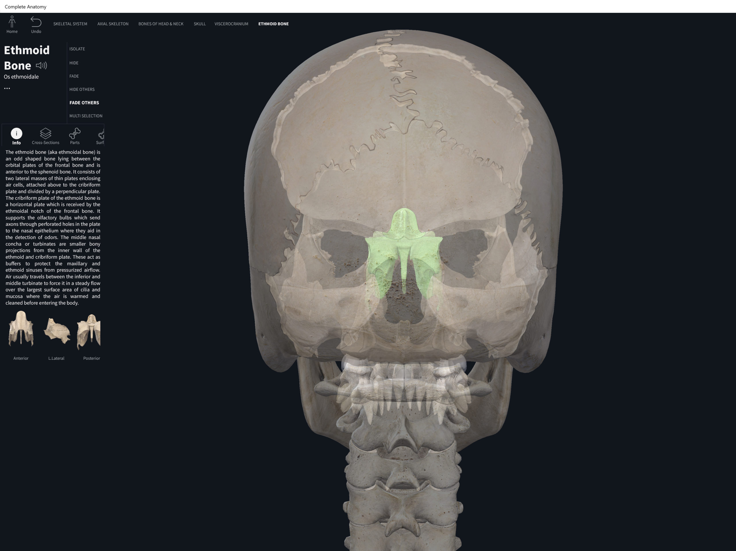
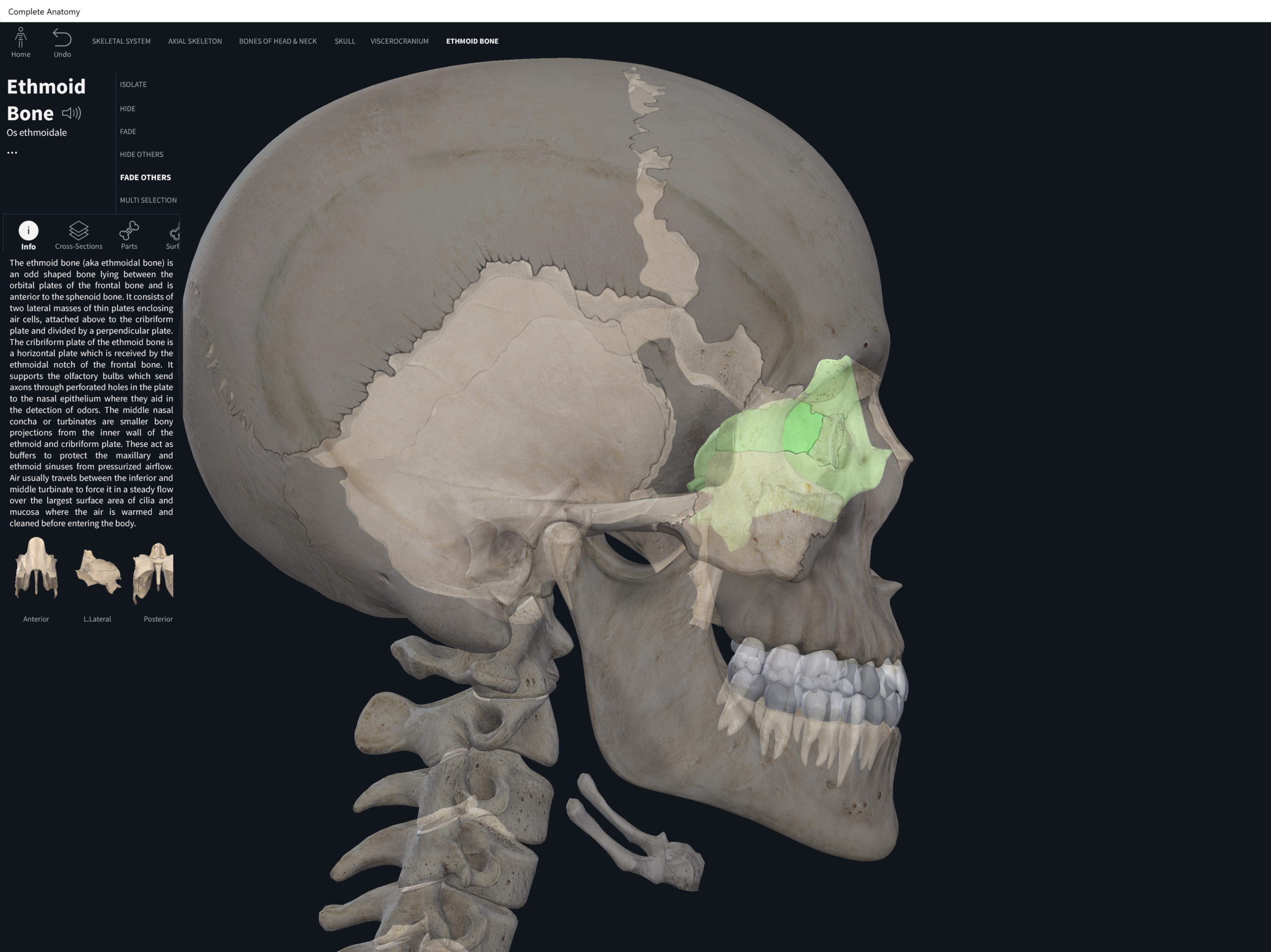
- Forms part of the anterior cranial floor and is a slender bone “wedged” midsagittally between the orbits (forms the medial wall of the orbits); transversely between the frontal and sphenoid bones; and posterior to the nasal bones thus forming part of the nasal septum and the nasal side walls.
- The name “ethmoid” means a sieve, sponge-like.
- Cribriform plate: forms the roof of the nasal cavity and contains the olfactory foramina where scents/smells get transmitted to the brain.
- Crista galli: superior to the cribriform plate, triangular shaped. It is an important landmark because it is an attachment point for membranes separating the two halves of the brain.
- Perpendicular plate: inferior to the cribriform plate, and forms the upper part of the nasal septum.
- The ethmoid contains 3-18 “air spaces”. These are called the ethmoidal sinuses.
Function.
- Superior and middle nasal concha: scroll-like formations on either side of the ethmoid. The concha increase surface area and helps “swirl” the air (create currents); warm and moisten the air; and helps to “clean” the air as foreign particles might stick to the mucous.
Clinical Significance.
Ethmoid. Used with permission by 3D4Medical.
References
Biel, A. (2015). Trail guide to the body: A hands-on guide to locating muscles, bones and more.
Jenkins, G., & Tortora, G. J. (2012). Anatomy and Physiology: From Science to Life, 3rd Edition International Stu. John Wiley & Sons.
Muscolino, J. E. (2017). The muscular system manual: The skeletal muscles of the human body.
