Posted on

Muscles: Rotatores.

Download these notes.

Anatomy & Physiology: Muscles—Rotatores.

Structure.

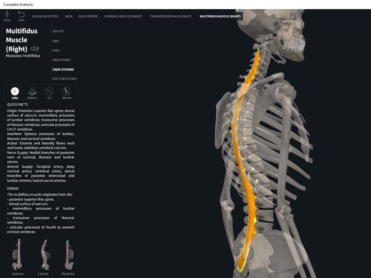
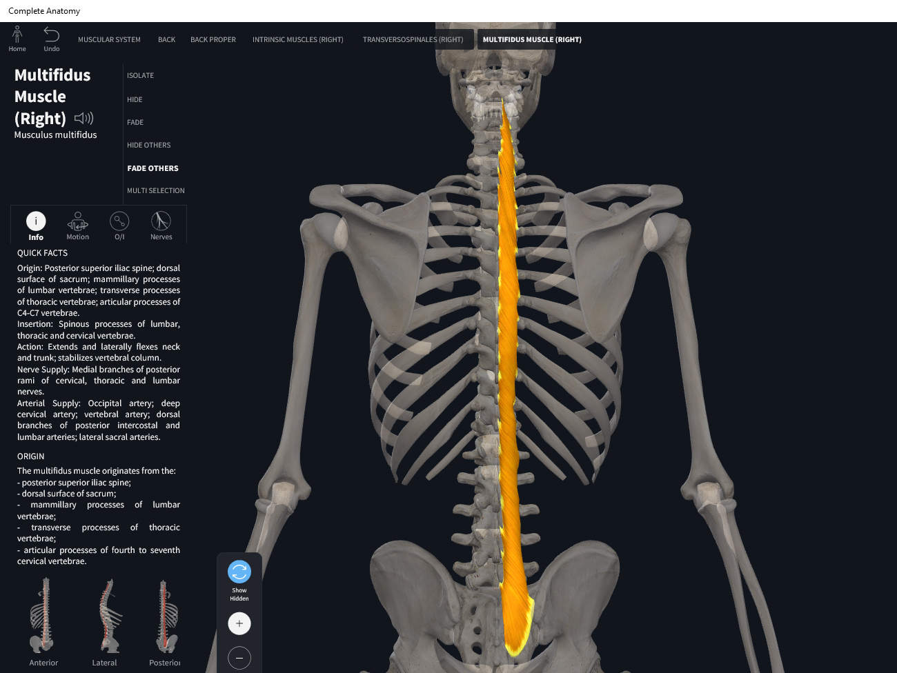
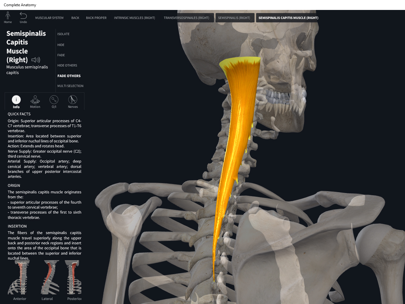
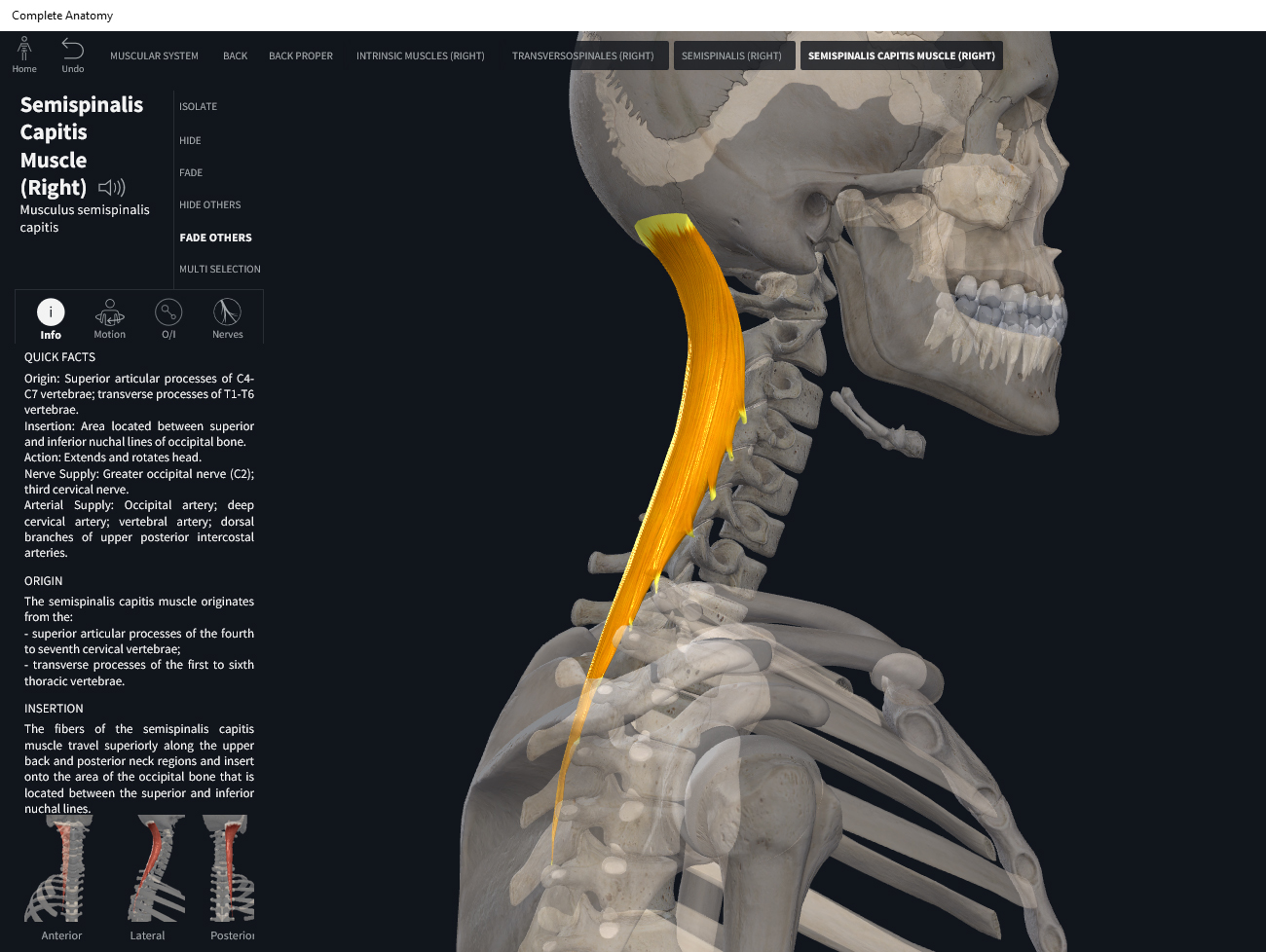
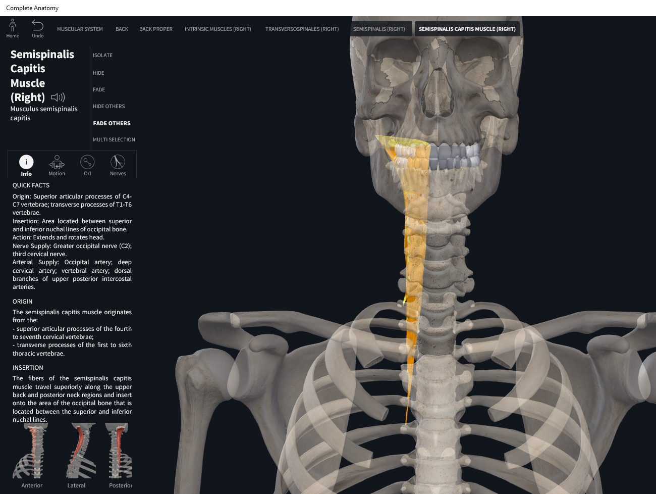
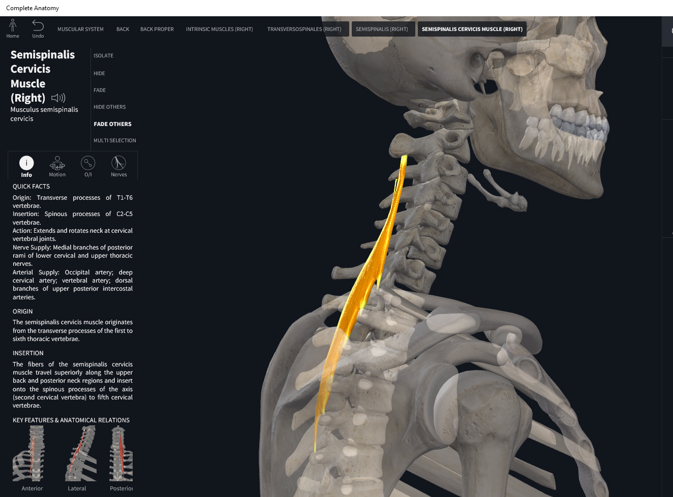
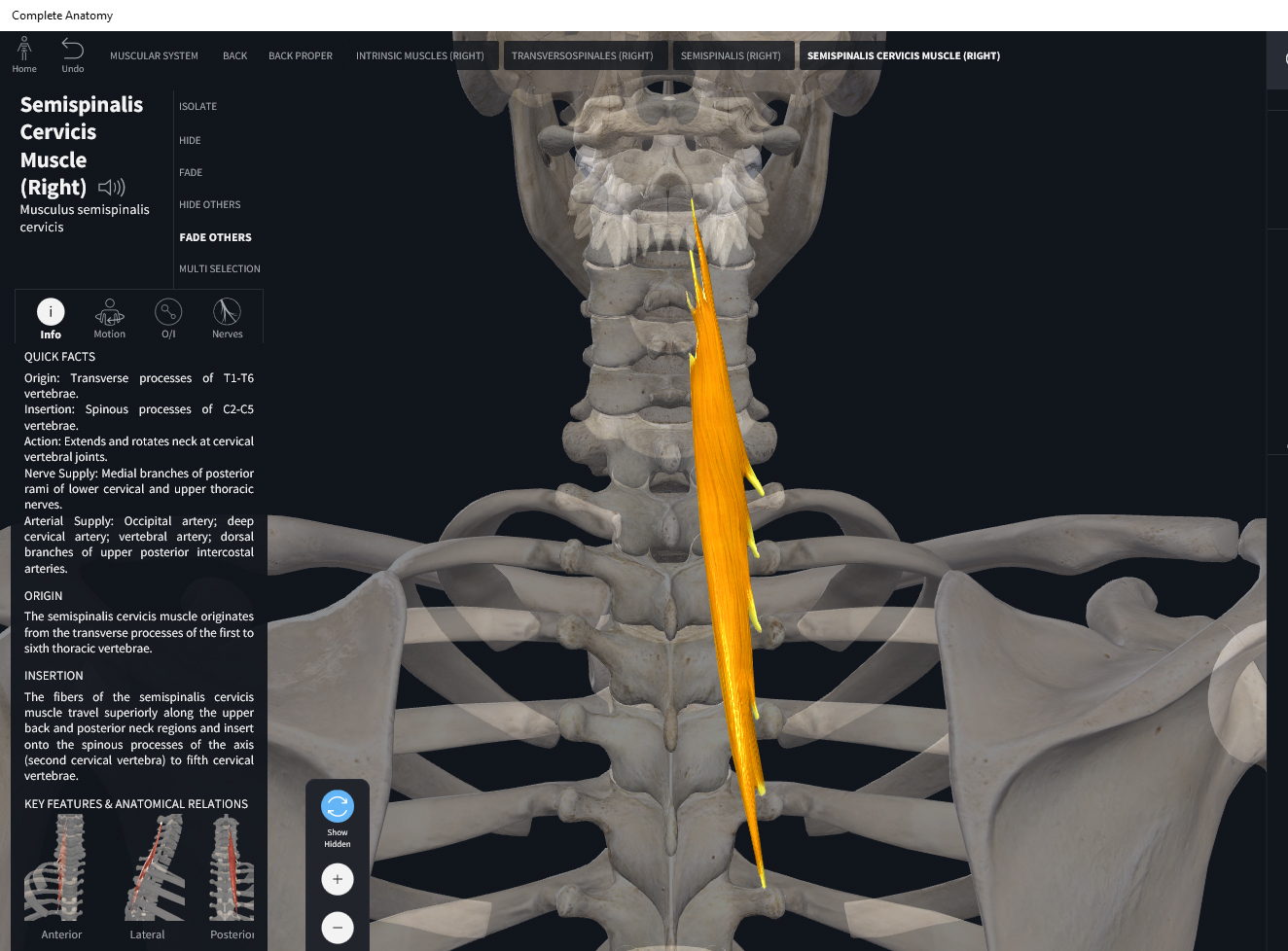
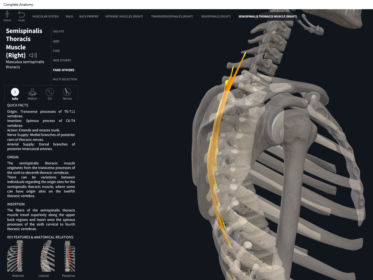
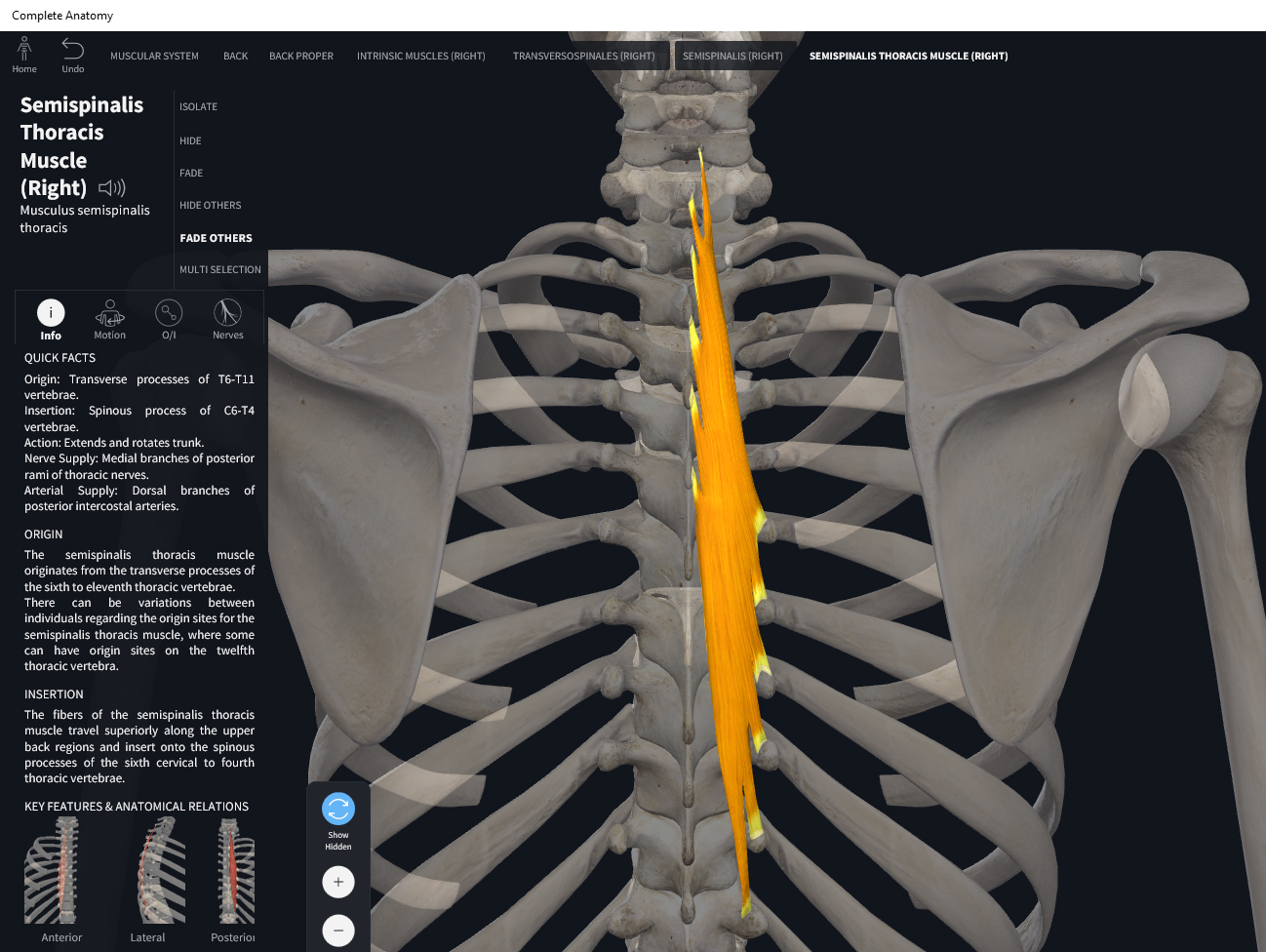
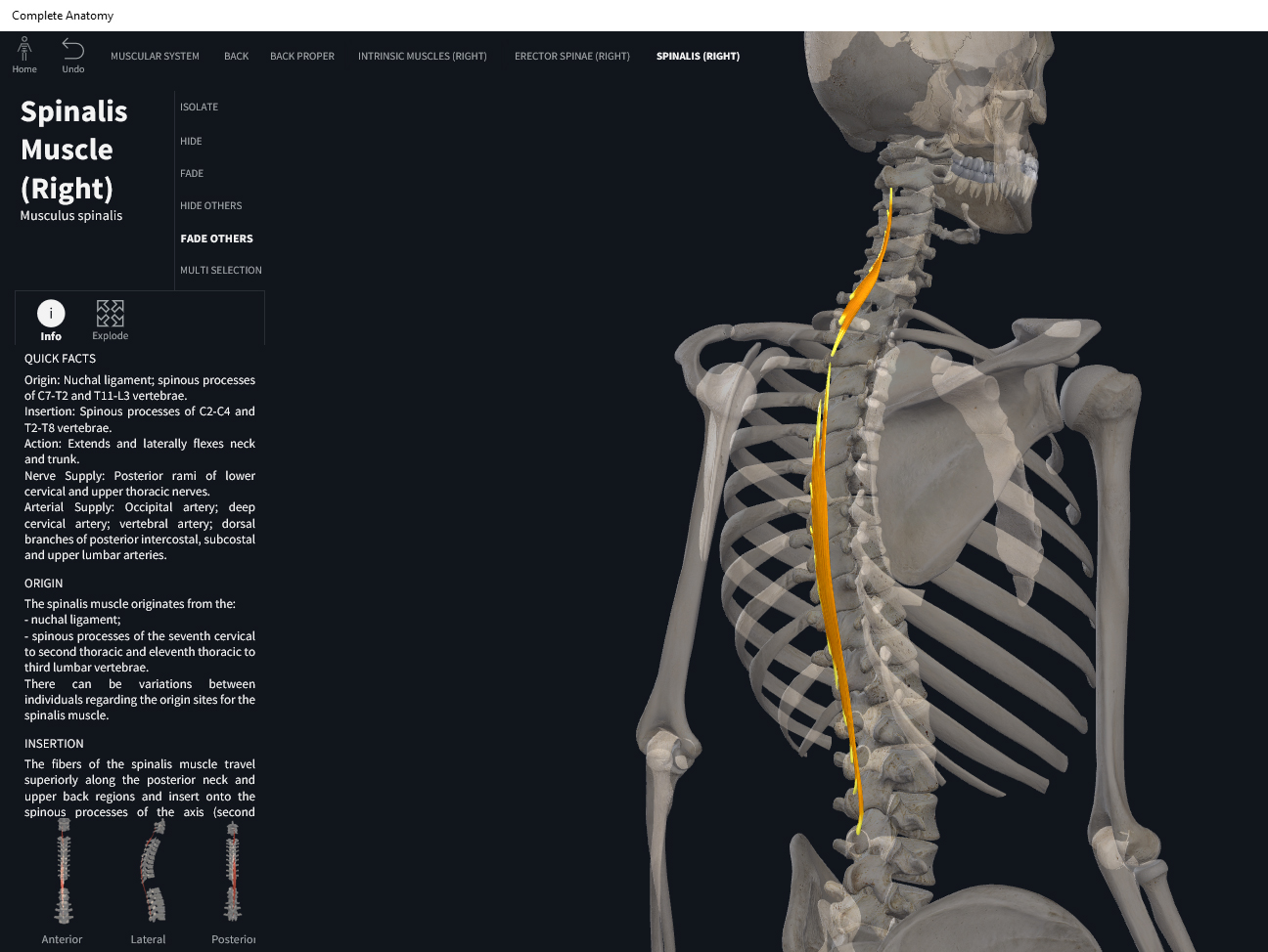
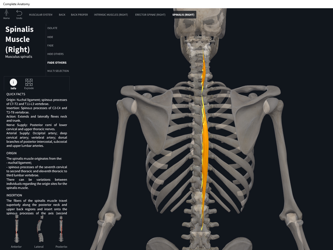
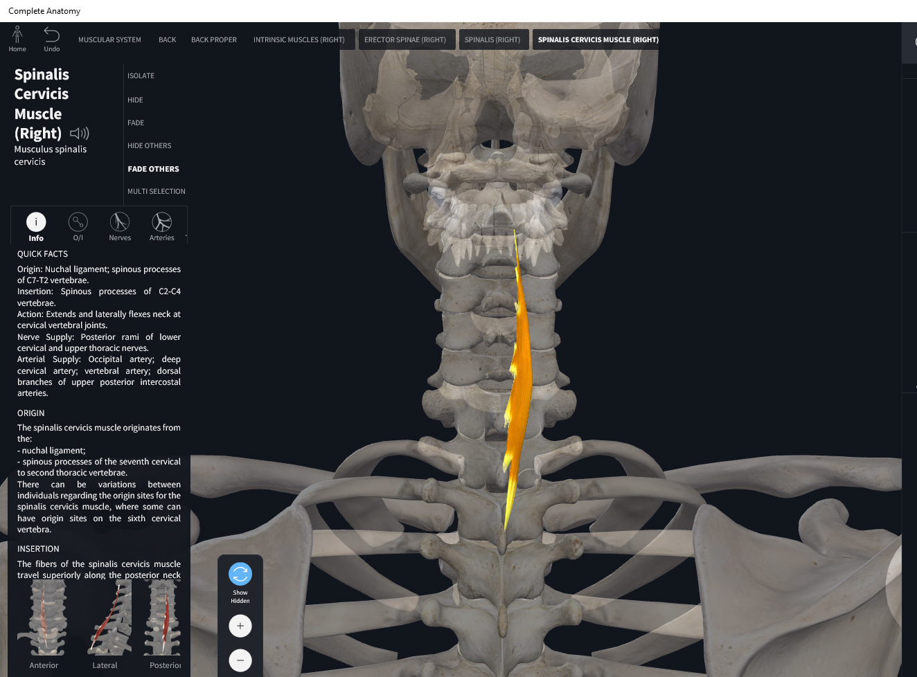
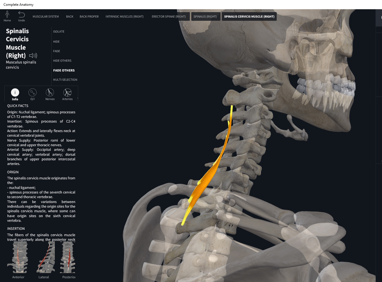
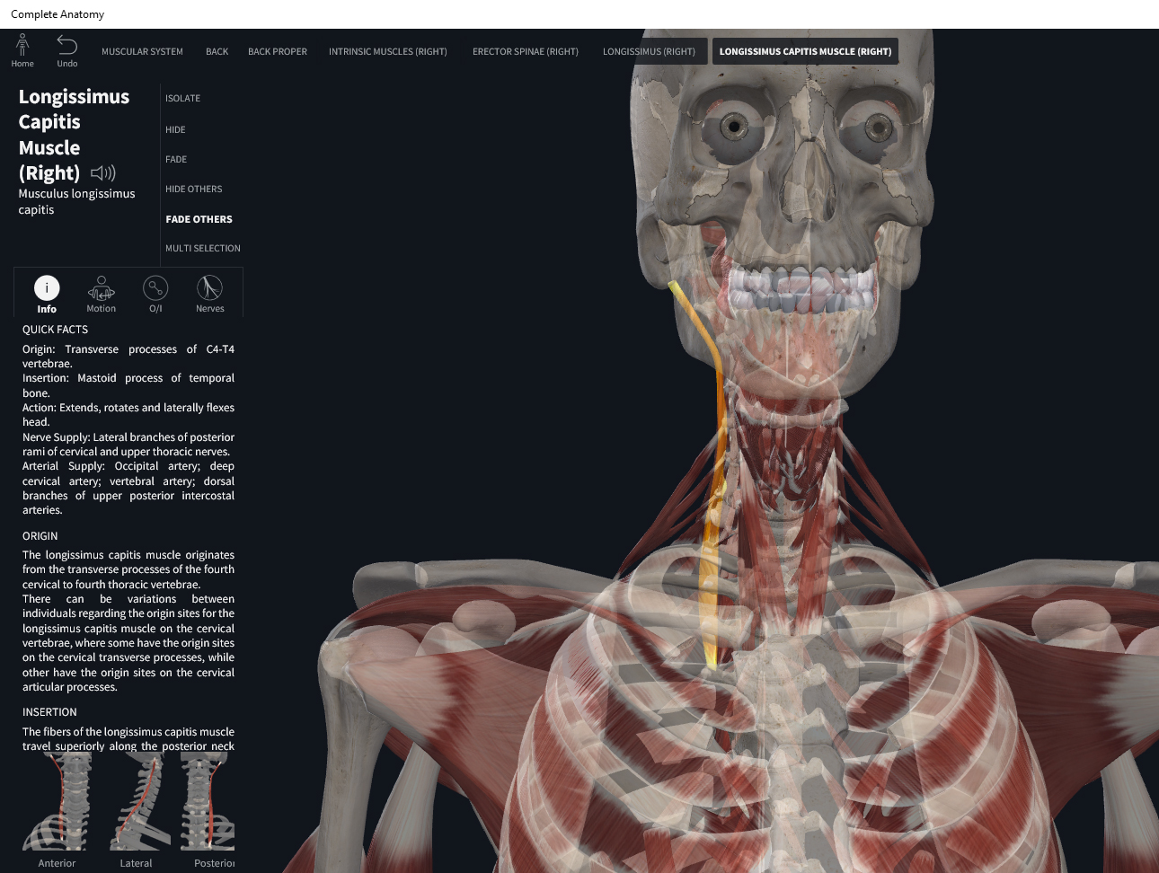
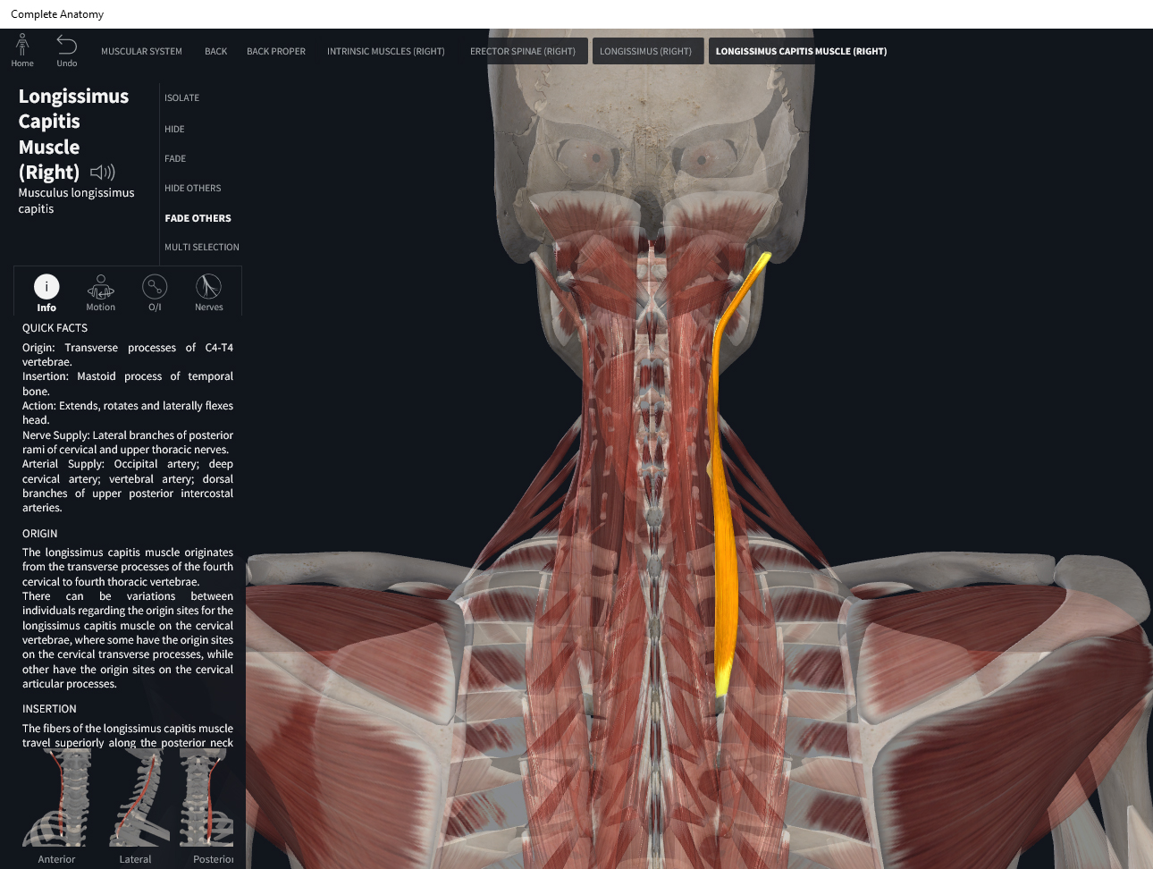
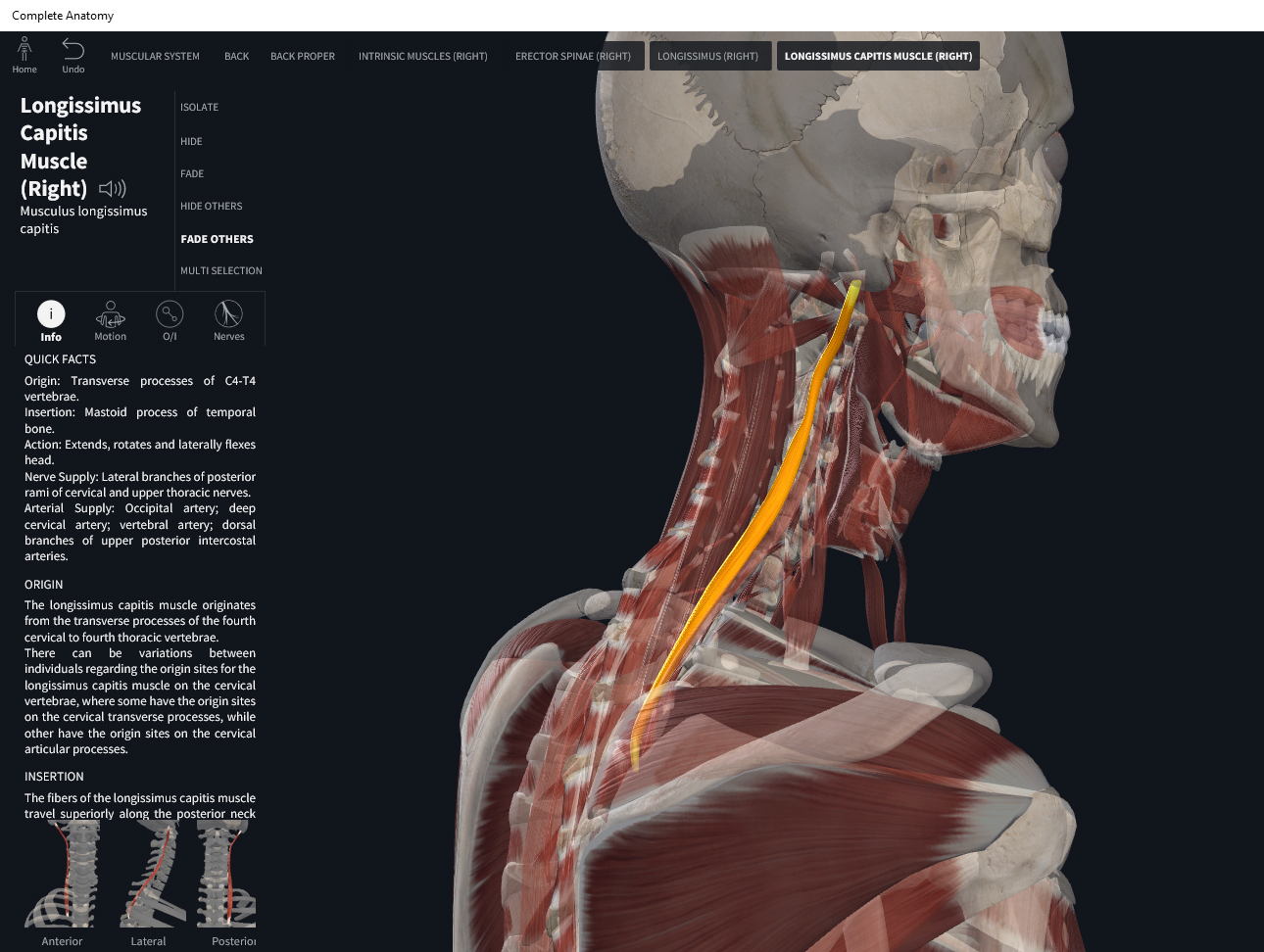
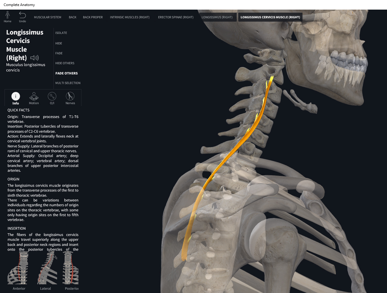
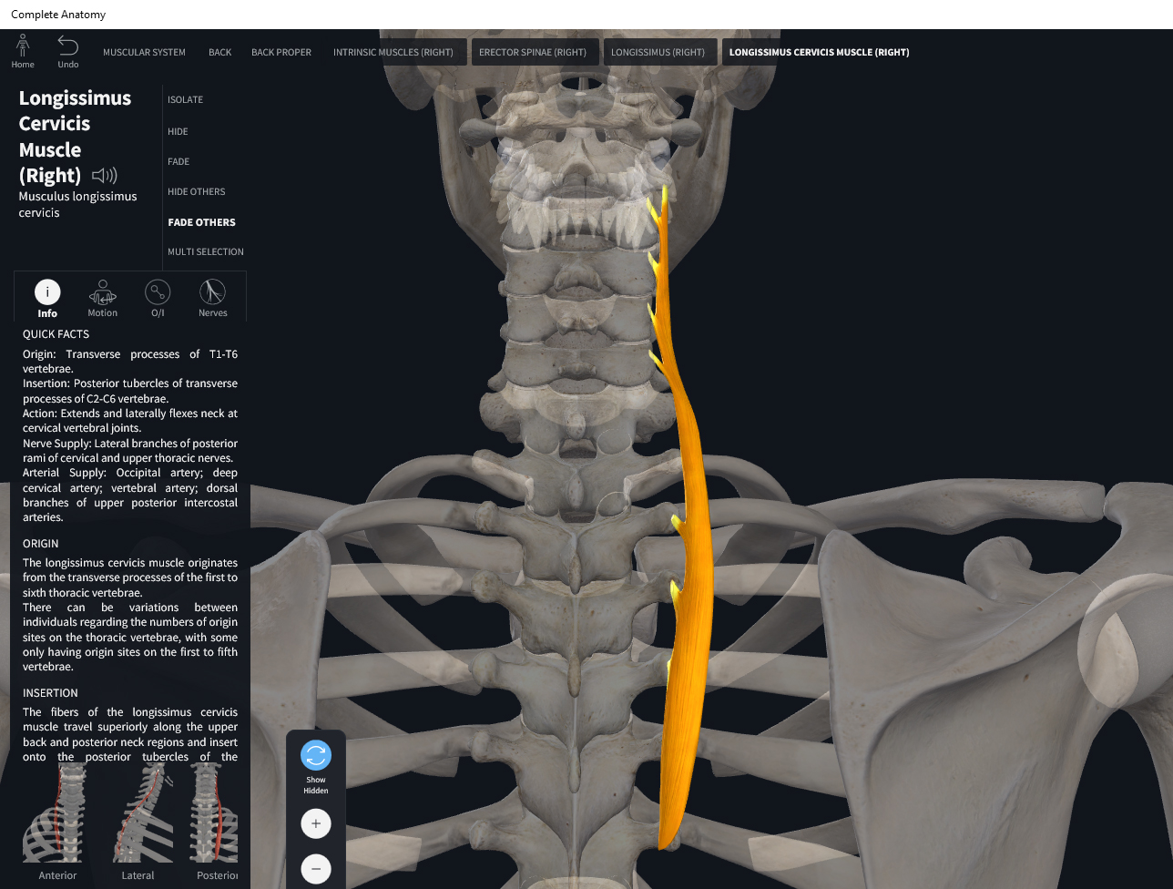
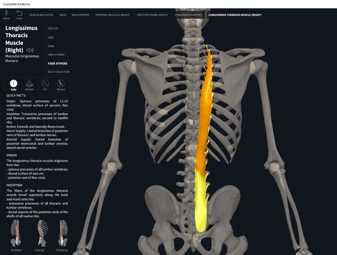
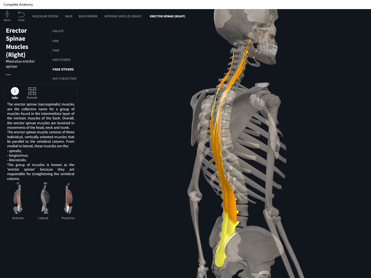
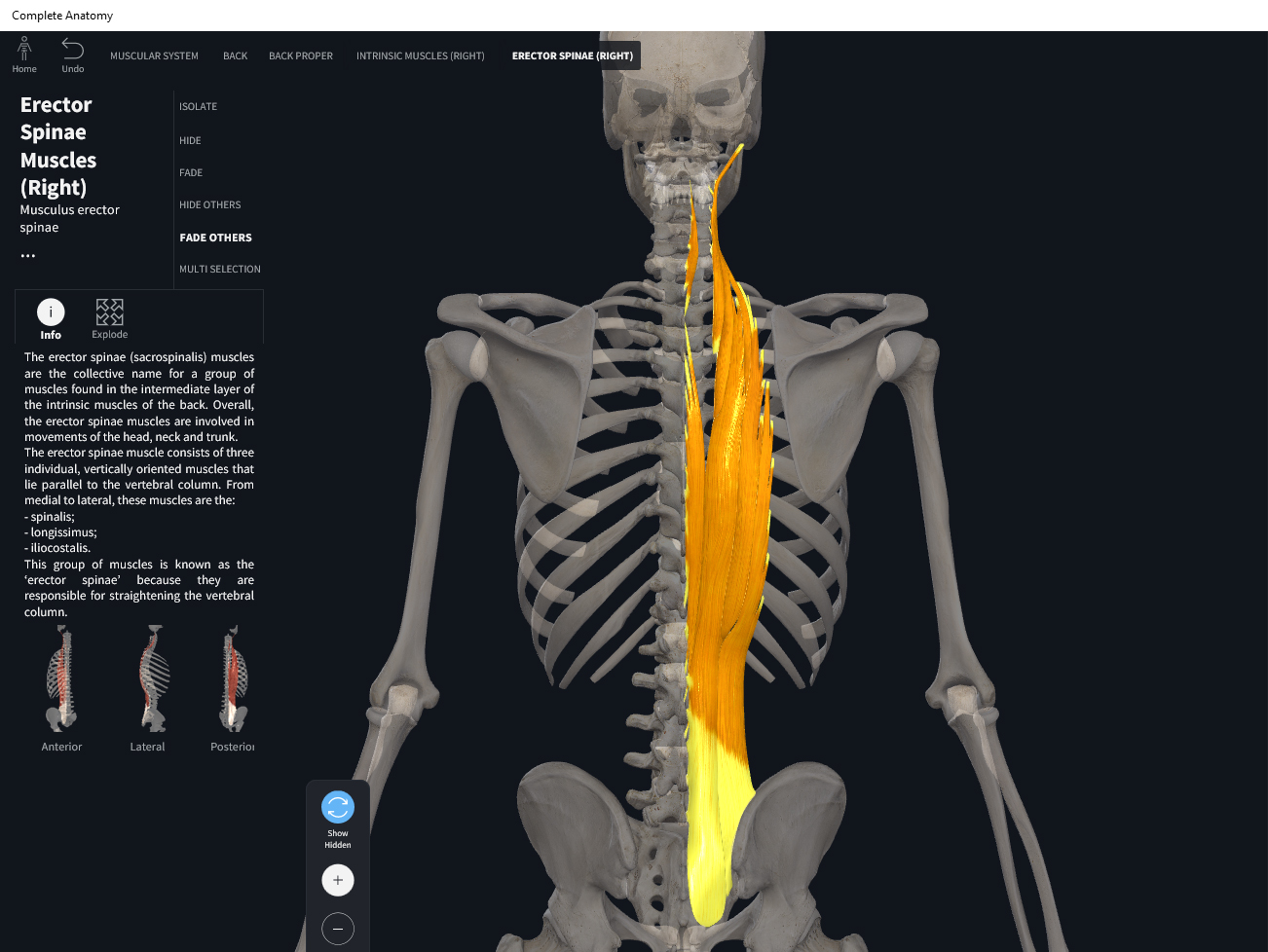
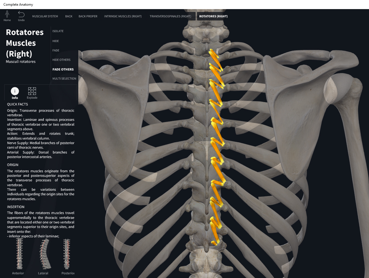
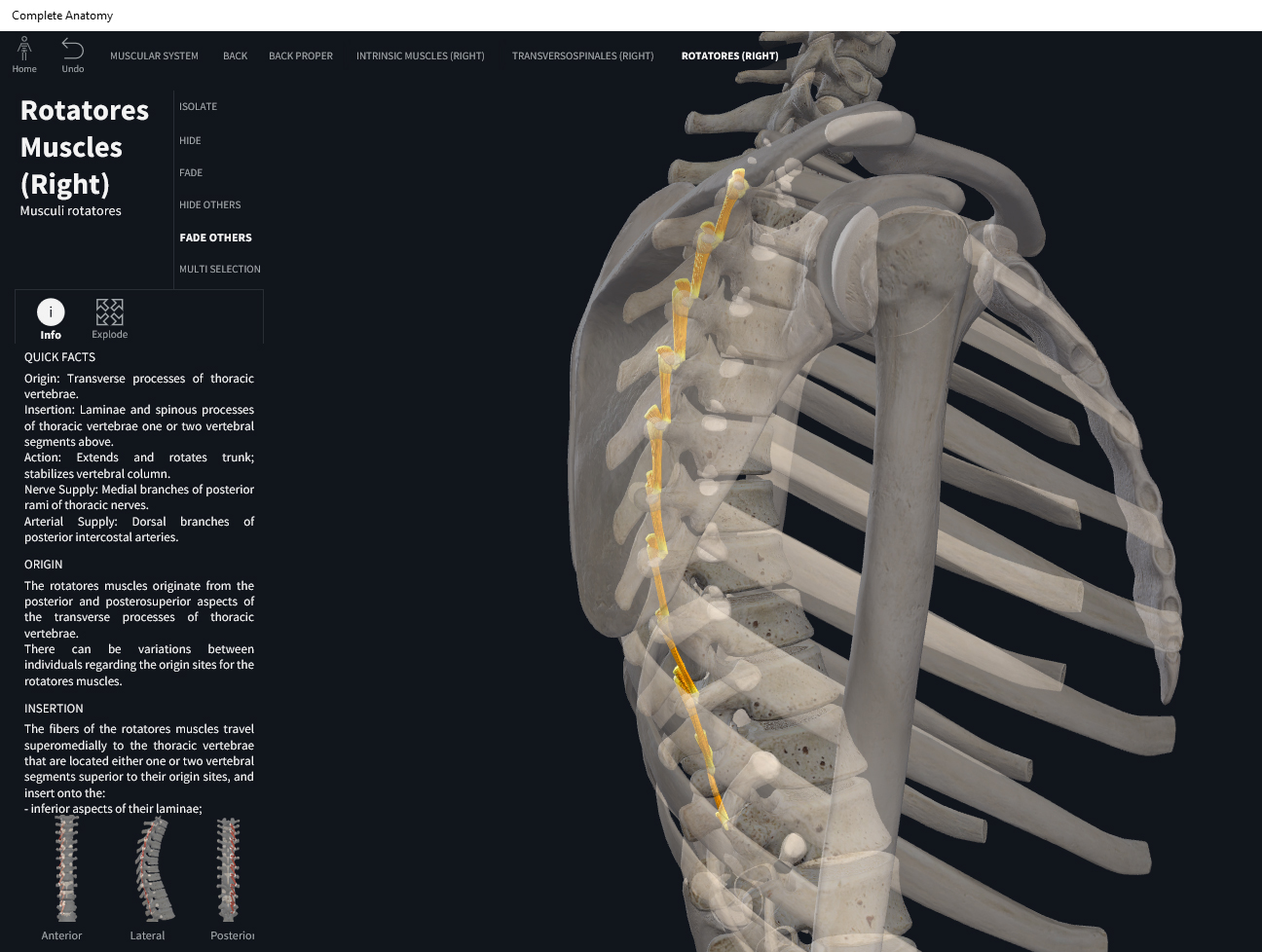
  • Origin: transverse processes of all vertebra.
  • Insertion: spinous processes above the origin.

Function.

  • Concentric action: lateral flexion, spinal extension, and contralateral rotation of neck, and spine.
  • Reverse mover action: ipsilateral rotation, extension, and lateral flexion of lower spine.
  • Eccentric action: controls/restrains/slows ipsilateral rotation, flexion, and contralateral lateral flexion of neck and trunk; and contralateral rotation of lower spine.
  • Isometric action: stabilization of the spine.
  • Innervation: corresponding cervical, thoracic, and lumbar spinal nerves.
  • Arterial supply: dorsal branches of posterior intercostal and lumbar arteries.

Clinical Significance.


References

Biel, A. (2015). Trail guide to the body: A hands-on guide to locating muscles, bones and more.

Cedars-Sinai. (2018). Vertebrae of the spine. Retrieved from https://www.cedars-sinai.org/health-library/diseases-and-conditions/v/vertebrae-of-the-spine.html

Clark, M., Lucett, S., Sutton, B. G., & National Academy of Sports Medicine. (2014). NASM essentials of corrective exercise training. Burlington, MA: Jones & Bartlett Learning.

Jenkins, G., & Tortora, G. J. (2012). Anatomy and Physiology: From Science to Life, 3rd Edition International Stu. John Wiley & Sons.

Muscolino, J. E. (2017). The muscular system manual: The skeletal muscles of the human body.

Posted on

Muscles: Multifidus.

Download these notes.

Anatomy & Physiology: Muscles—Multifidus, of transversospinalis group.

Structure.

  • Origin: posterior aspect of sacrum, ilium; transverse processes of L1-L5, T1-T12, and C1-C4.
  • Insertion: spinous processes above the origin.

Function.

  • Concentric action: neck and spinal extension, lateral flexion, and contralateral flexion.
  • Reverse mover action: anterior til of pelvis, lower spine extension. Lesser: ipsilateral elevation of pelvis, lateral flexion of lower spine, ipsilateral rotation of pelvis.
  • Eccentric action: controls/restrains/slows spinal flexion and rotation.
  • Isometric action: stabilization of the spine.
  • Innervation: corresponding cervical, thoracic, and lumbar spinal nerves.
  • Arterial supply: dorsal branches of posterior intercostal and lumbar arteries.

Clinical Significance.


References

Biel, A. (2015). Trail guide to the body: A hands-on guide to locating muscles, bones and more.

Cedars-Sinai. (2018). Vertebrae of the spine. Retrieved from https://www.cedars-sinai.org/health-library/diseases-and-conditions/v/vertebrae-of-the-spine.html

Clark, M., Lucett, S., Sutton, B. G., & National Academy of Sports Medicine. (2014). NASM essentials of corrective exercise training. Burlington, MA: Jones & Bartlett Learning.

Jenkins, G., & Tortora, G. J. (2012). Anatomy and Physiology: From Science to Life, 3rd Edition International Stu. John Wiley & Sons.

Muscolino, J. E. (2017). The muscular system manual: The skeletal muscles of the human body.

Posted on

Muscles: Semispinalis.

Download these notes.

Anatomy & Physiology: Muscles—Semispinalis, of transversospinalis group.

Structure.

  • Semispinalis Capitis
    • Origin: transverse processes of C7-T6, articular processes of C4-C6.
    • Insertion: occipital bone between the superior and inferior nuchal lines.
  • Semispinalis Cervicis
    • Origin: transverse processes of T1-T5.
    • Insertion: spinous processes of C2-C5.
  • Semispinalis Thoracis
    • Origin: transverse processes of T6-T10.
    • Insertion: spinous processes of C6-T4.

Function.

  • Concentric action: head, neck, and trunk extension; lateral flexion of head, neck, and trunk; rotate head to side opposing contracting muscle.
  • Reverse mover action: lower spine extension; lower spine lateral flexion; ipsilateral rotation of the lower spine.
  • Eccentric action: controls/restrains/slows flexion and contralateral lateral flexion of head, neck, and trunk; ipsilateral rotation of neck and trunk; contralateral rotation of the lower spine.
  • Isometric action: stabilization of spine and head.
  • Innervation: cervical and thoracic spinal nerves.
  • Arterial supply: occipital artery, dorsal branches of posterior intercostal arteries, deep cervical artery.

Clinical Significance.


References

Biel, A. (2015). Trail guide to the body: A hands-on guide to locating muscles, bones and more.

Cedars-Sinai. (2018). Vertebrae of the spine. Retrieved from https://www.cedars-sinai.org/health-library/diseases-and-conditions/v/vertebrae-of-the-spine.html

Clark, M., Lucett, S., Sutton, B. G., & National Academy of Sports Medicine. (2014). NASM essentials of corrective exercise training. Burlington, MA: Jones & Bartlett Learning.

Jenkins, G., & Tortora, G. J. (2012). Anatomy and Physiology: From Science to Life, 3rd Edition International Stu. John Wiley & Sons.

Muscolino, J. E. (2017). The muscular system manual: The skeletal muscles of the human body.

Posted on

Muscles: Transversospinalis Group.

Download these notes.

Anatomy & Physiology: Muscles—Transversospinalis Group.

Structure.

  • Transversospinalis Capitis
    • Origin: transverse process T6-C7; articular process of C6-C4.
    • Insertion: nuchal line of occipital bone.
  • Transversospinalis Cervicis
    • Origin: transverse process T6-C4.
    • Insertion: spinous process C5-C2.
  • Transversospinalis Thoracis
    • Origin: transverse process T12-T7.
    • Insertion: spinous process T4-C6.

Function.

  • Concentric action: extension of trunk, neck, and head; lateral flexion of trunk, neck, and head; contralateral rotation of trunk and neck at spinal joints.
  • Reverse mover action: anterior tilt of pelvis; lower spine extension. Lesser: ipsilateral elevation of pelvis; lateral flexion of lower spine; ipsilateral rotation of the pelvis and lower spine.
  • Eccentric action: decelerates lateral flexion of spine, flexion and contralateral rotation of head.
  • Isometric action: stabilization of the spine.
  • Innervation: dorsal rami C1-T6 spinal nerves.
  • Arterial supply: occipital artery, dorsal branches of posterior intercostal and lumbar arteries; deep cervical artery.

Clinical Significance.

References

Biel, A. (2015). Trail guide to the body: A hands-on guide to locating muscles, bones and more.

Cedars-Sinai. (2018). Vertebrae of the spine. Retrieved from https://www.cedars-sinai.org/health-library/diseases-and-conditions/v/vertebrae-of-the-spine.html

Clark, M., Lucett, S., Sutton, B. G., & National Academy of Sports Medicine. (2014). NASM essentials of corrective exercise training. Burlington, MA: Jones & Bartlett Learning.

Jenkins, G., & Tortora, G. J. (2012). Anatomy and Physiology: From Science to Life, 3rd Edition International Stu. John Wiley & Sons.

Muscolino, J. E. (2017). The muscular system manual: The skeletal muscles of the human body.

Posted on

Muscles: Spinalis.

Download these notes.

Anatomy & Physiology: Muscles—Spinalis, of erector spinae group.

Structure.

  • Spinalis Capitis
    • Origin: ligamentum nuchae and spinous process of C7; considered to be the medial portion of semispinalis capitis.
    • Insertion: spinous process of axis.
  • Spinalis Cervicis
    • Origin: inferior nuchal ligament and spinous process of C7.
    • Insertion: spinous process of C2.
  • Spinalis Thoracis
    • Origin: spinous processes of T11-L2.
    • Insertion: spinous processes of T4-T8.

Function.

  • Concentric action: trunk and neck extension at spinal joints; lateral flexion of the trunk and neck at spinal joints.
  • Reverse mover action: lower spine extension; lateral flexion of lower spine.
  • Eccentric action: controls/restrains/slows flexion and contralateral flexion of trunk and neck.
  • Isometric action: stabilization of the spine.
  • Innervation: inferior cervical and thoracic spinal nerves.
  • Arterial supply: dorsal branches of posterior intercostal and lumbar arteries.

Clinical Significance.


References

Biel, A. (2015). Trail guide to the body: A hands-on guide to locating muscles, bones and more.

Cedars-Sinai. (2018). Vertebrae of the spine. Retrieved from https://www.cedars-sinai.org/health-library/diseases-and-conditions/v/vertebrae-of-the-spine.html

Clark, M., Lucett, S., Sutton, B. G., & National Academy of Sports Medicine. (2014). NASM essentials of corrective exercise training. Burlington, MA: Jones & Bartlett Learning.

Jenkins, G., & Tortora, G. J. (2012). Anatomy and Physiology: From Science to Life, 3rd Edition International Stu. John Wiley & Sons.

Muscolino, J. E. (2017). The muscular system manual: The skeletal muscles of the human body.

Posted on

Muscles: Longissimus.

Download these notes.

Anatomy & Physiology: Muscles—Longissimus, of erector spinae group.

Structure.

  • Longissimus Capitis
    • Origin: transverse processes of T1-T5; articular processes of C4-C7.
    • Insertion: mastoid process of temporal bone.
  • Longissimus Cervicus
    • Origin: transverse processes of T1-T5.
    • Insertion: transverse processes of C2-C6.
  • Longissimus Thoracis
    • Origin: medial iliac crest, posterior sacrum, transverse processes and spinous processes of L1-L5.
    • Insertion: transverse processes of all thoracic vertebrae and lower nine ribs.

Function.

  • Concentric action: trunk, neck, and head extension at spinal joints; lateral flexion of trunk, neck, and head at spinal joints. Lesser: ipsilateral rotation of trunk, head, and neck.
  • Reverse mover action: anterior tilt of pelvis at the LS joint; lower spine extension. Lesser: ipsilateral elevation of pelvis and lateral flexion of lower spine; contralateral rotation of the pelvis and lower spine.
  • Eccentric action: controls/restrains/slows flexion, contralateral lateral flexion, and contralateral rotation of trunk, neck, and head; controls/restrains/slows posterior tilt, ipsilateral rotation, and ipsilateral depression of pelvis.
  • Isometric action: stabilization of the spine, ribs, and SI joint.
  • Innervation: middle and inferior cervical and spinal nerves.
  • Arterial supply: dorsal branches of posterior intercostal and lumbar arteries.

Clinical Significance.


References

Biel, A. (2015). Trail guide to the body: A hands-on guide to locating muscles, bones and more.

Cedars-Sinai. (2018). Vertebrae of the spine. Retrieved from https://www.cedars-sinai.org/health-library/diseases-and-conditions/v/vertebrae-of-the-spine.html

Clark, M., Lucett, S., Sutton, B. G., & National Academy of Sports Medicine. (2014). NASM essentials of corrective exercise training. Burlington, MA: Jones & Bartlett Learning.

Jenkins, G., & Tortora, G. J. (2012). Anatomy and Physiology: From Science to Life, 3rd Edition International Stu. John Wiley & Sons.

Muscolino, J. E. (2017). The muscular system manual: The skeletal muscles of the human body.

Posted on

Muscles: Iliocostalis.

Download these notes.

Anatomy & Physiology: Muscles—Iliocostalis,  of erector spinae group.

Structure.

  • Iliocostalis Cervicis
    • Origin: angles of ribs 3-6.
    • Insertion: transverse processes of C4-C6.
  • Iliocostalis Thoracis
    • Origin: angles of ribs 7-12.
    • Insertion: angles of ribs 1-6 and transverse process of C7.
  • Iliocostalis Lumborum
    • Origin: medial iliac crest and medial and lateral sacral crests.
    • Insertion: angles of ribs 7-12.

Function.

  • Concentric action: spinal extension, rotation and lateral flexion; maintain erect position.
  • Reverse mover action: anterior tilt of pelvis at lumbosacral joint; extension of lower spine relative to upper spine; ipsilateral elevation of pelvis at LS and lateral flexion of lower spine; contralateral rotation of the pelvis at LS and lower spine contralateral rotation.
  • Eccentric action: controls/restrains/slows spinal flexion, rotation, and lateral flexion of trunk and neck; also the posterior tilt and ipsilateral depression of pelvis.
  • Isometric action: stabilization of the spine, ribs, and sacroiliac joint.
  • Innervation: cervical and thoracic spinal nerves.
  • Arterial supply: dorsal branches of posterior intercostal and lumbar arteries; thoracodorsal artery.

Clinical Significance.


References

Biel, A. (2015). Trail guide to the body: A hands-on guide to locating muscles, bones and more.

Cedars-Sinai. (2018). Vertebrae of the spine. Retrieved from https://www.cedars-sinai.org/health-library/diseases-and-conditions/v/vertebrae-of-the-spine.html

Clark, M., Lucett, S., Sutton, B. G., & National Academy of Sports Medicine. (2014). NASM essentials of corrective exercise training. Burlington, MA: Jones & Bartlett Learning.

Jenkins, G., & Tortora, G. J. (2012). Anatomy and Physiology: From Science to Life, 3rd Edition International Stu. John Wiley & Sons.

Muscolino, J. E. (2017). The muscular system manual: The skeletal muscles of the human body.

Posted on

Muscles: Erector Spinae Group.

Download these notes.

Anatomy & Physiology: Muscles—Erector Spinae Group.

Structure.

  • Origin: pelvis.
  • Insertion: spine, rib cage, and head.

Function.

  • Concentric action: trunk, neck, and head extension at the spinal joints; lateral flexion of the trunk, neck, and head at the spinal joints. Lesser: ipsilateral rotation of the trunk, neck, and head at spinal joints.
  • Reverse mover action: pelvic anterior tilt at lumbosacral joint; lower spine extension relative to upper spine. Lesser: ipsilateral elevation of pelvis at LS joint; lateral flexion of lower spine relative to upper spine; contralateral rotation of pelvis and lower spine.
  • Eccentric action: spinal flexion, rotation, and lateral flexion.
  • Isometric action: stabilization of the spine.
  • Innervation: cervical and thoracic spinal nerves.
  • Arterial supply: dorsal branches of posterior intercostal and lumbar arteries; thoracodorsal artery.

Clinical Significance.


References

Biel, A. (2015). Trail guide to the body: A hands-on guide to locating muscles, bones and more.

Cedars-Sinai. (2018). Vertebrae of the spine. Retrieved from https://www.cedars-sinai.org/health-library/diseases-and-conditions/v/vertebrae-of-the-spine.html

Clark, M., Lucett, S., Sutton, B. G., & National Academy of Sports Medicine. (2014). NASM essentials of corrective exercise training. Burlington, MA: Jones & Bartlett Learning.

Jenkins, G., & Tortora, G. J. (2012). Anatomy and Physiology: From Science to Life, 3rd Edition International Stu. John Wiley & Sons.

Muscolino, J. E. (2017). The muscular system manual: The skeletal muscles of the human body.

Posted on

Muscles: Palmaris Brevis.

Download these notes.

Anatomy & Physiology: Muscles—Palmaris Brevis.

Structure.

  • Origin: flexor retinaculum and palmar aponeurosis.
  • Insertion: Skin of palm into dermis.

Function.

  • Concentric action: wrinkles the skin of the palm.
  • Reverse mover action: wrinkles the skin of the palm.
  • Eccentric action: controls/restrains/slows stretching of the skin of the palm.
  • Isometric action: little to no stabilization functions.
  • Innervation: ulnar nerve.
  • Arterial supply: ulnar artery.

Clinical Significance.

References

Biel, A. (2015). Trail guide to the body: A hands-on guide to locating muscles, bones and more.

Cedars-Sinai. (2018). Vertebrae of the spine. Retrieved from https://www.cedars-sinai.org/health-library/diseases-and-conditions/v/vertebrae-of-the-spine.html

Clark, M., Lucett, S., Sutton, B. G., & National Academy of Sports Medicine. (2014). NASM essentials of corrective exercise training. Burlington, MA: Jones & Bartlett Learning.

Jenkins, G., & Tortora, G. J. (2012). Anatomy and Physiology: From Science to Life, 3rd Edition International Stu. John Wiley & Sons.

Muscolino, J. E. (2017). The muscular system manual: The skeletal muscles of the human body.

Posted on

Muscles: Dorsal Interossei Manus.

Download these notes.

Anatomy & Physiology: Muscles—Dorsal Interossei.

Structure.

  • Origin: adjacent sides of metacarpals.
  • Insertion: proximal phalanx of each finger.

Function.

  • Concentric action: abduct fingers 2-4 at metacarpophalangeal joints; flex fingers 2-4 at metacarpophalangeal joints; and extends fingers at interphalangeal joints.
  • Reverse mover action: abduct metacarpals of fingers 2-4; flex and adduct thumb at CMC; flex metacarpals of fingers 2-4 at MCP; extend proximal phalanges of fingers 2-4 at PIP and DIP.
  • Eccentric action: controls/restrains/slows adduction, flexion and extension of fingers 2-4 at MCP.
  • Isometric action: stabilize CMC of thumb, and MCP and IP for fingers 2-4.
  • Innervation: ulnar nerve.
  • Arterial supply: branches of radial and ulnar arteries.

Clinical Significance.

More.

References

Biel, A. (2015). Trail guide to the body: A hands-on guide to locating muscles, bones and more.

Cedars-Sinai. (2018). Vertebrae of the spine. Retrieved from https://www.cedars-sinai.org/health-library/diseases-and-conditions/v/vertebrae-of-the-spine.html

Clark, M., Lucett, S., Sutton, B. G., & National Academy of Sports Medicine. (2014). NASM essentials of corrective exercise training. Burlington, MA: Jones & Bartlett Learning.

Jenkins, G., & Tortora, G. J. (2012). Anatomy and Physiology: From Science to Life, 3rd Edition International Stu. John Wiley & Sons.

Muscolino, J. E. (2017). The muscular system manual: The skeletal muscles of the human body.