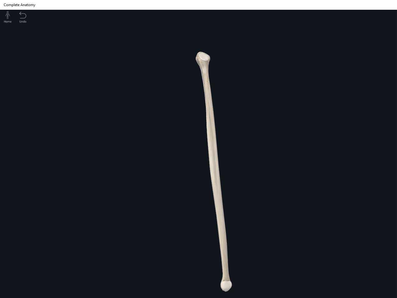
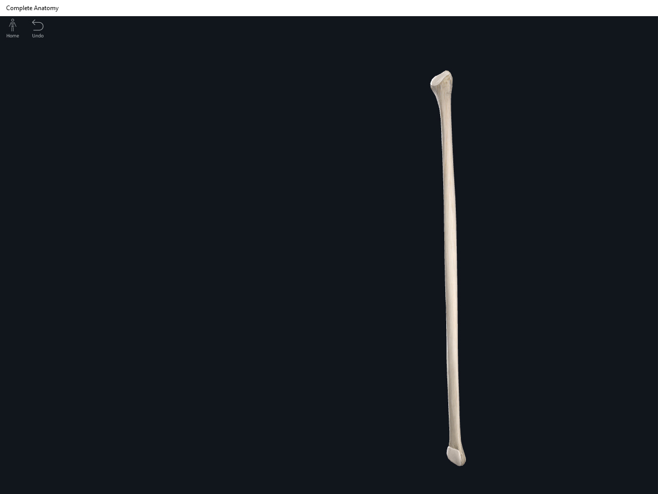
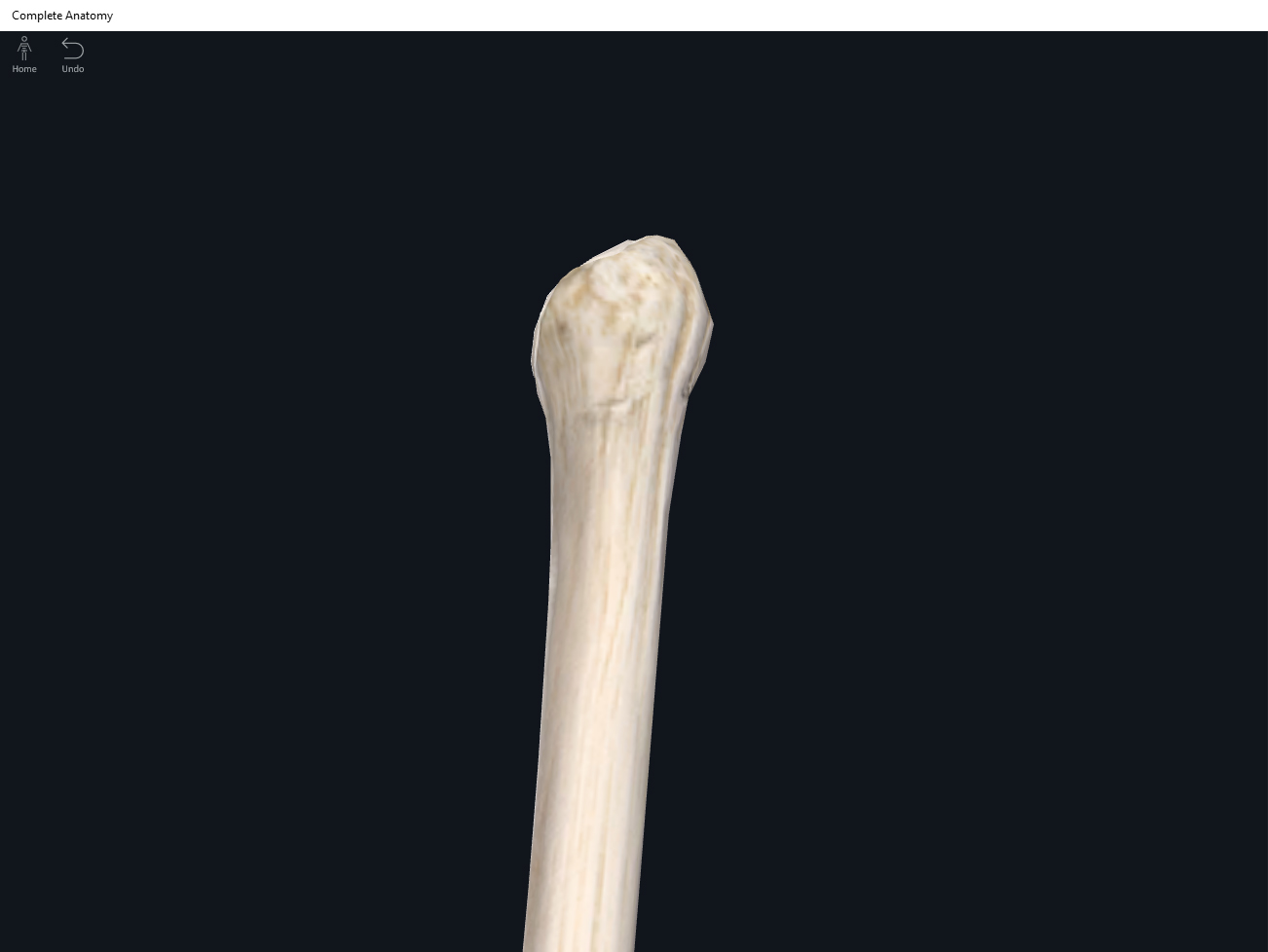
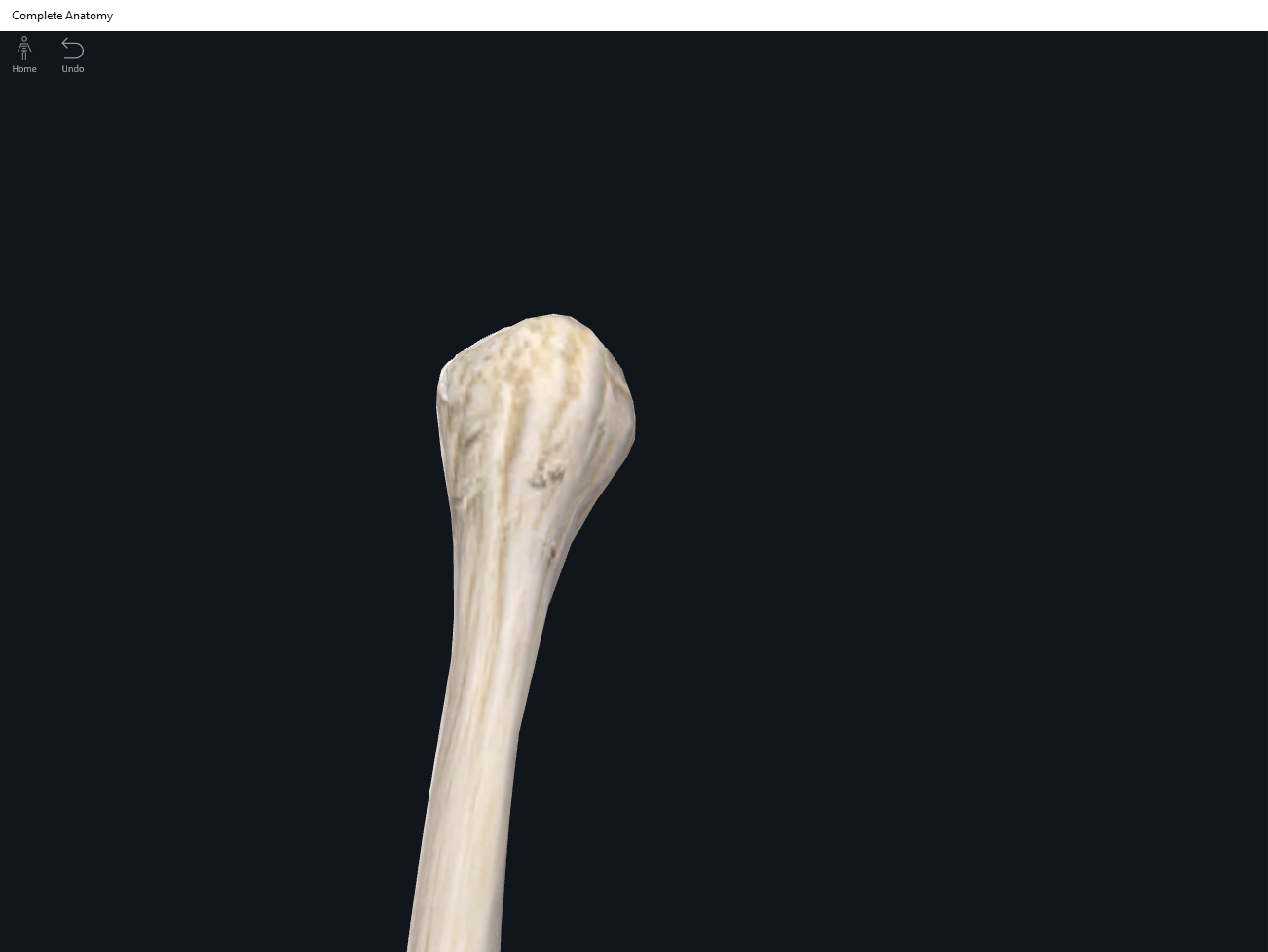
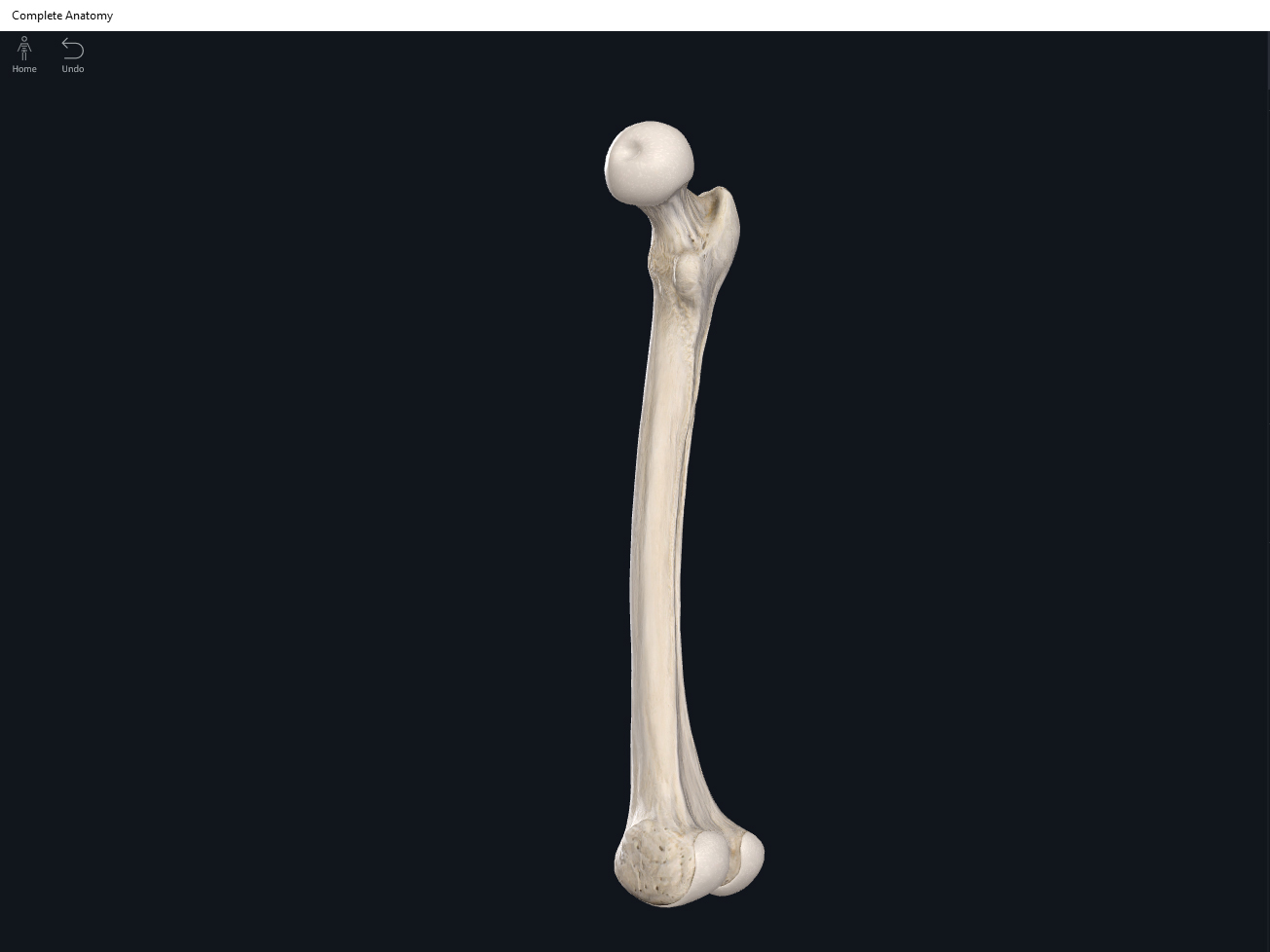
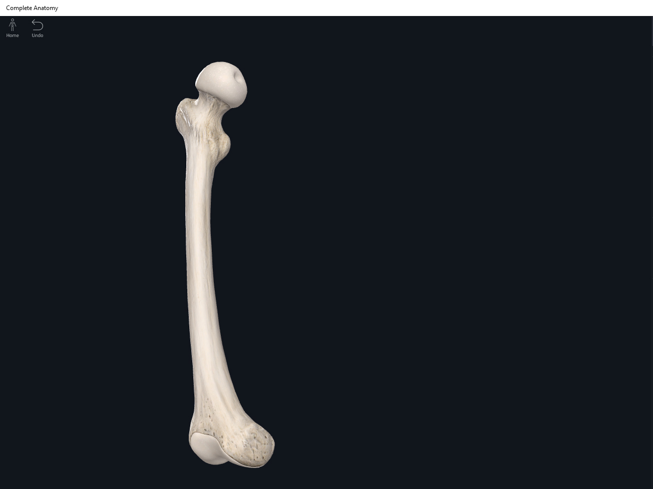
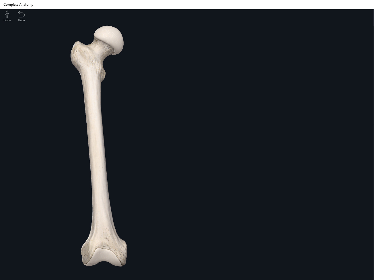
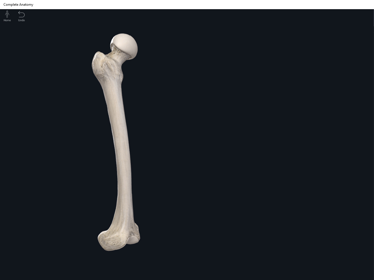
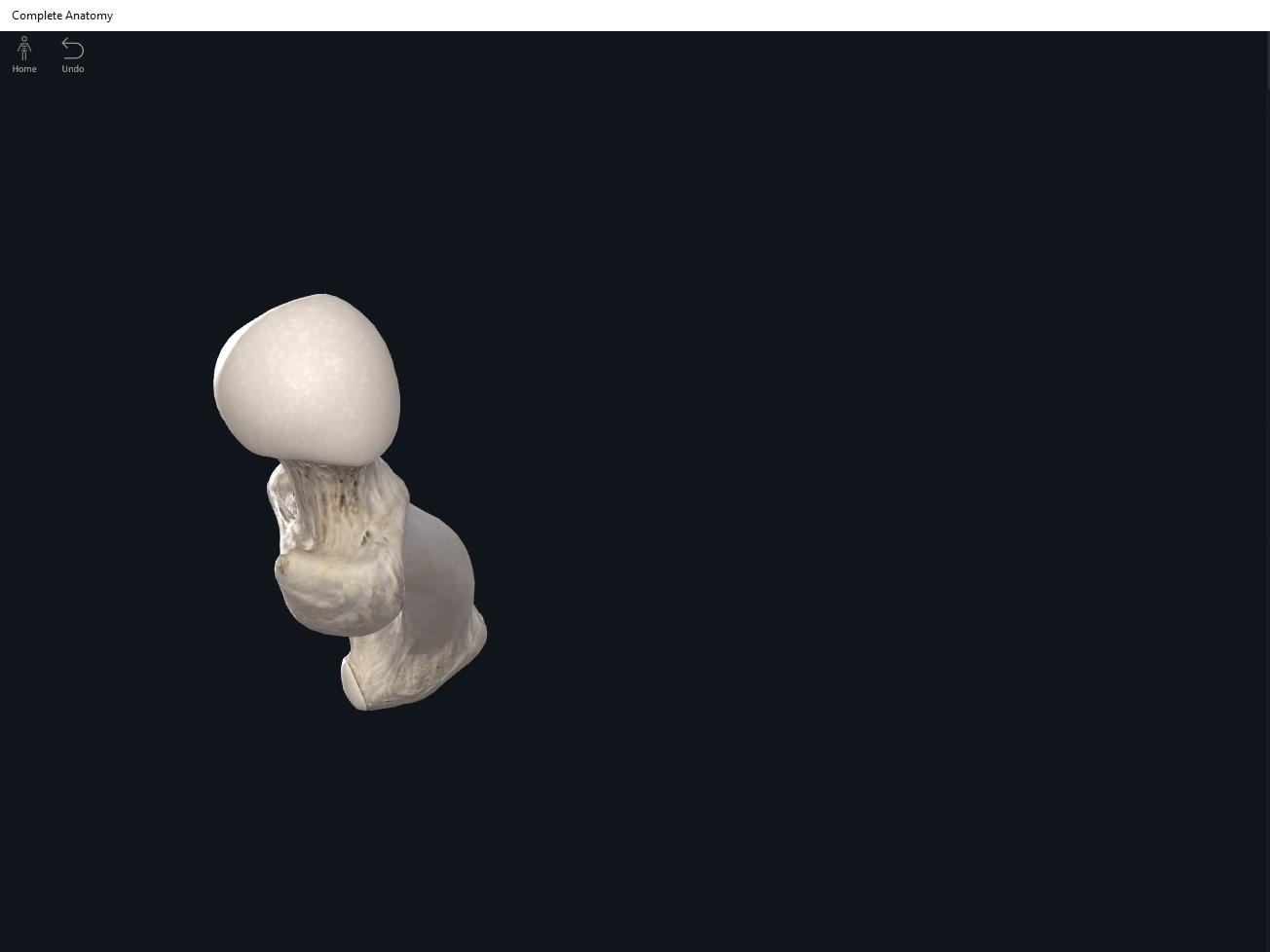
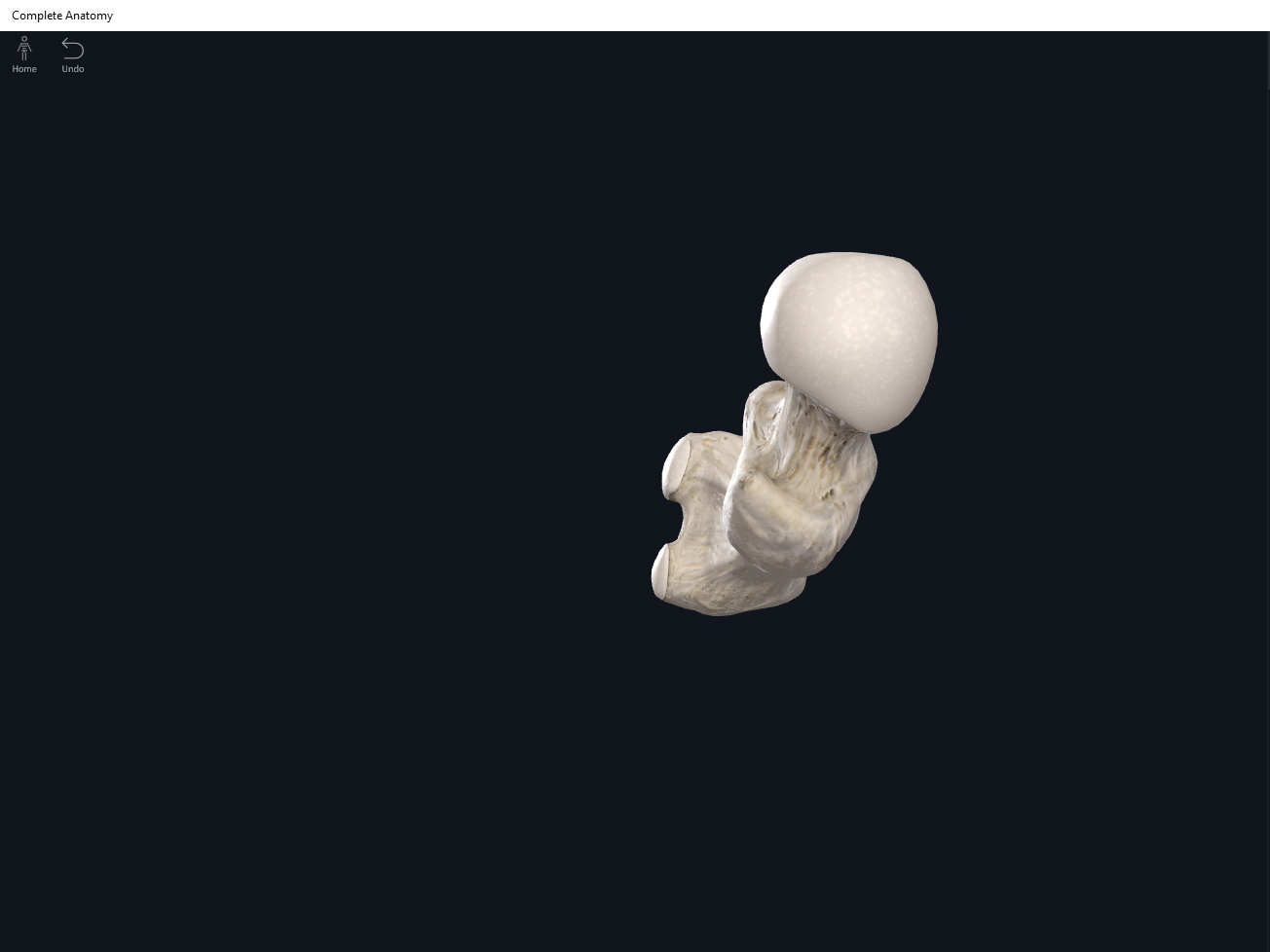
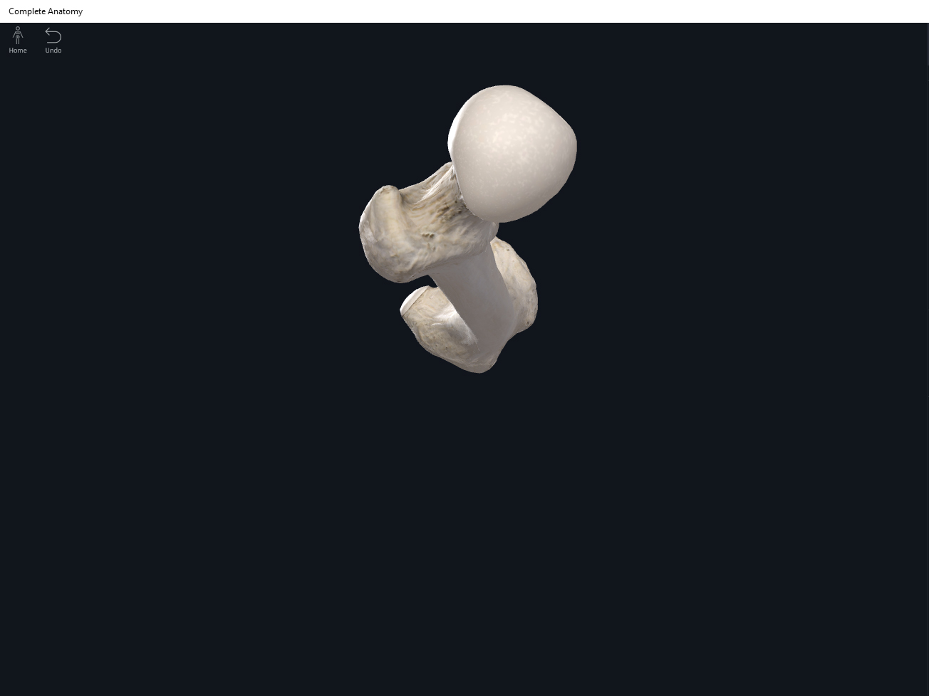
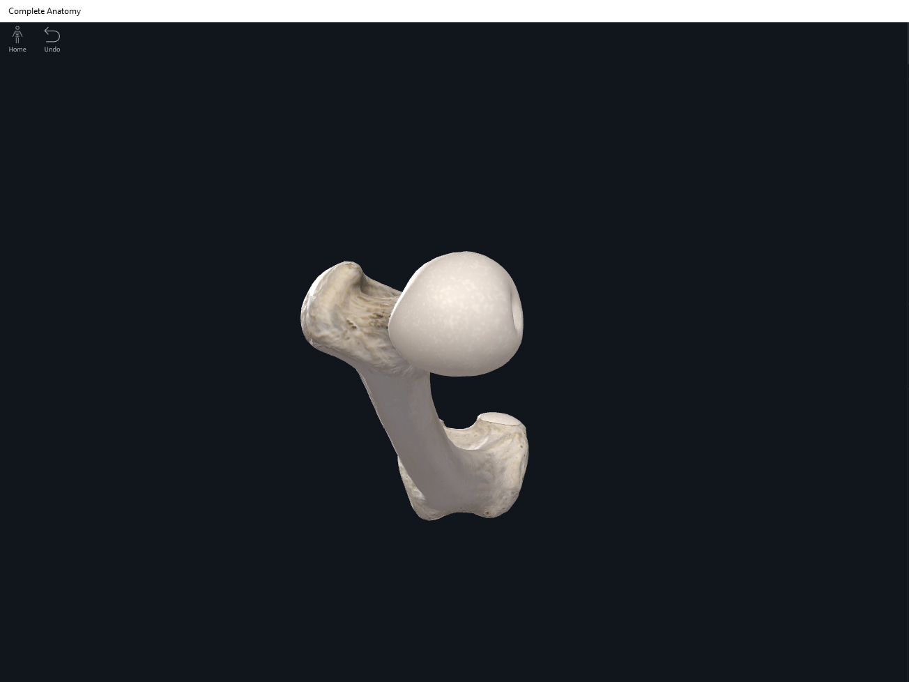
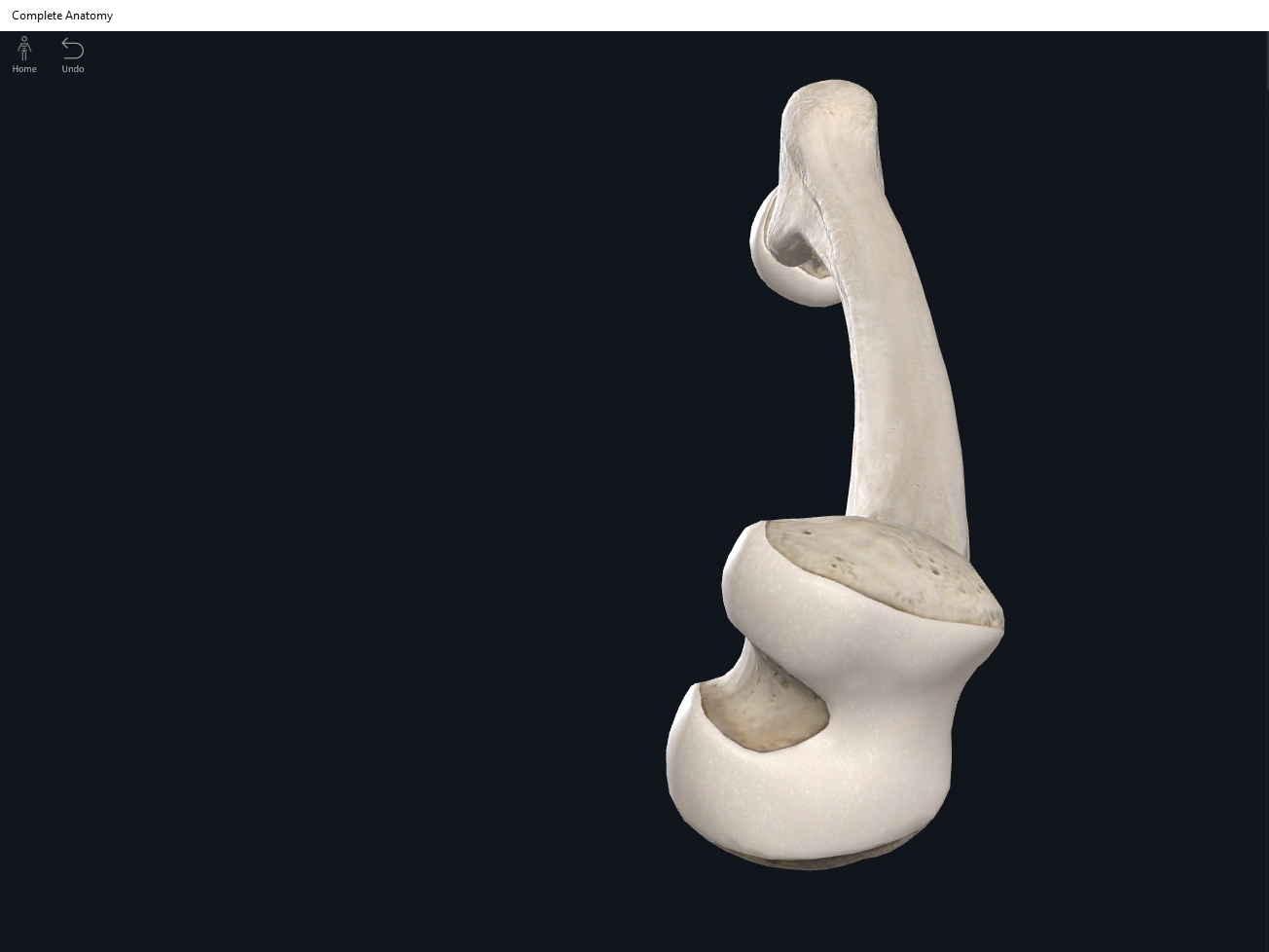
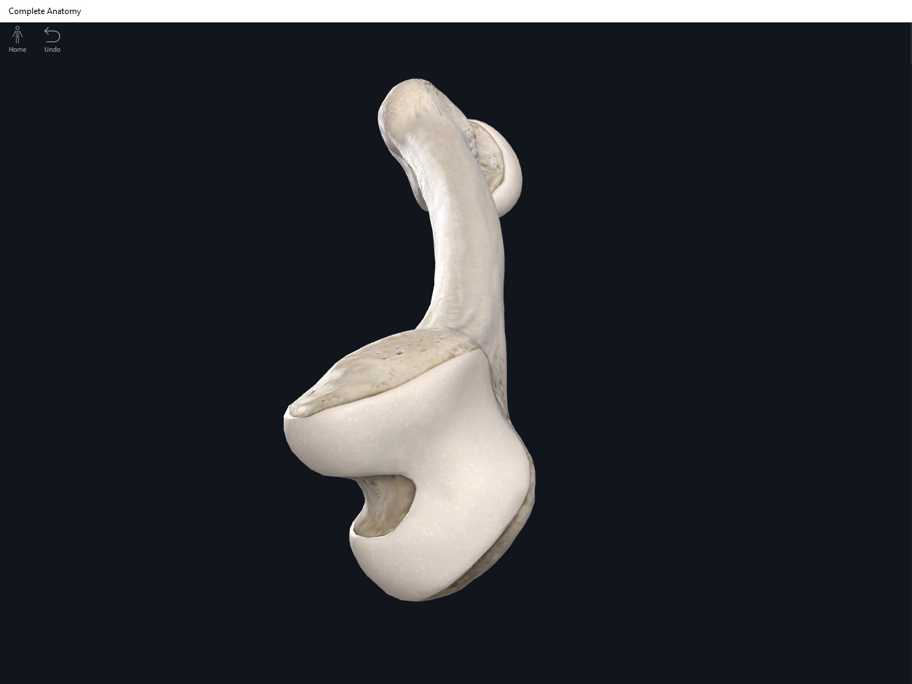
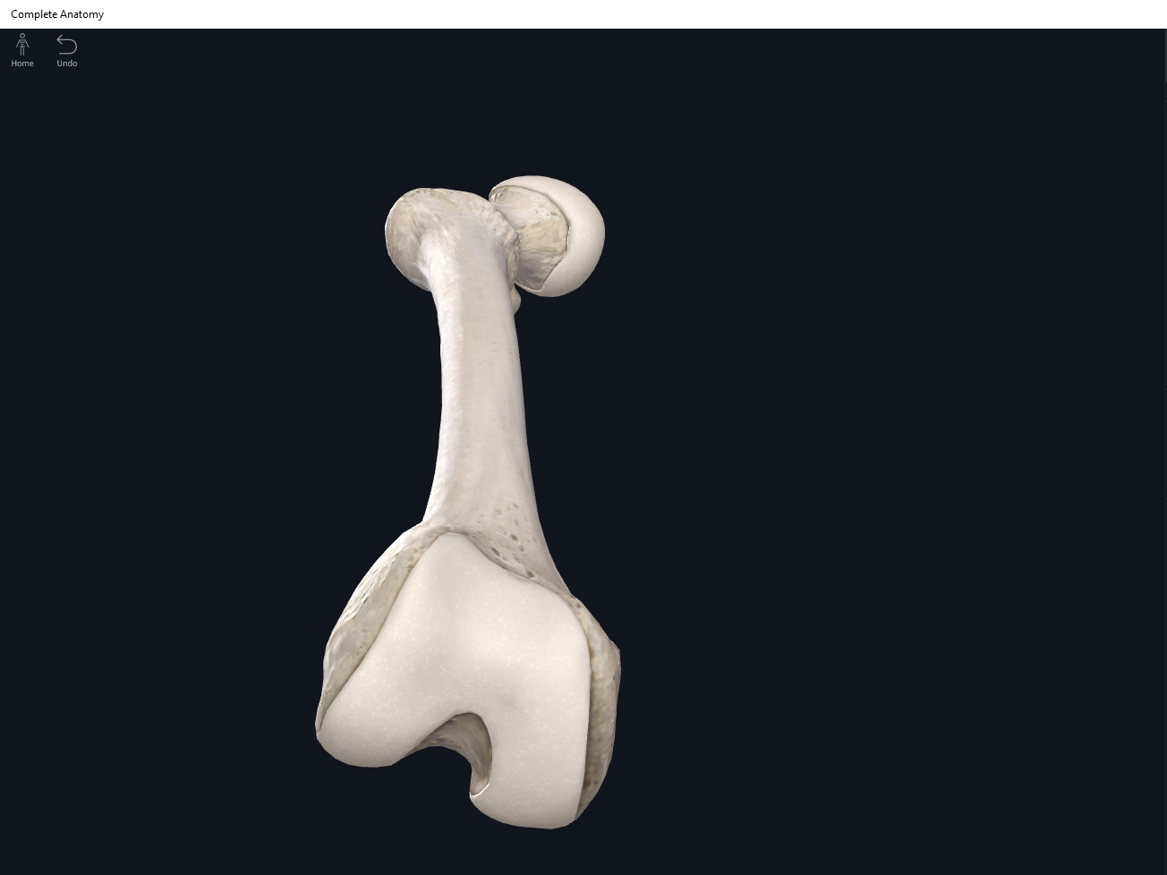
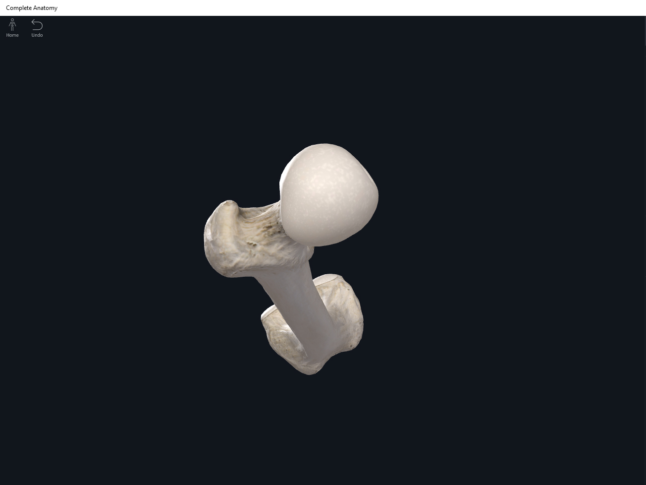
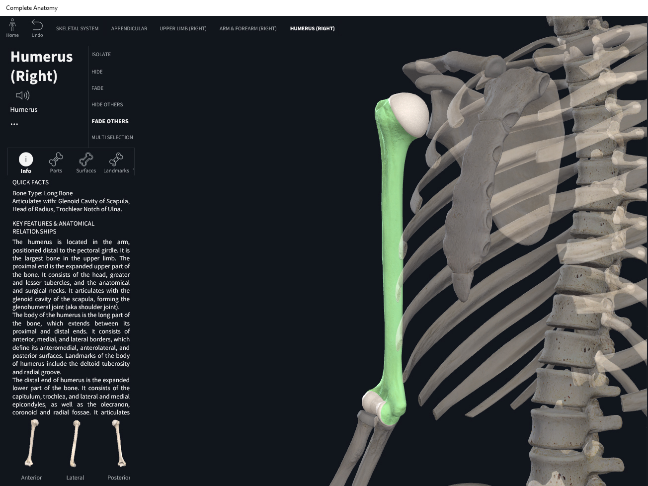
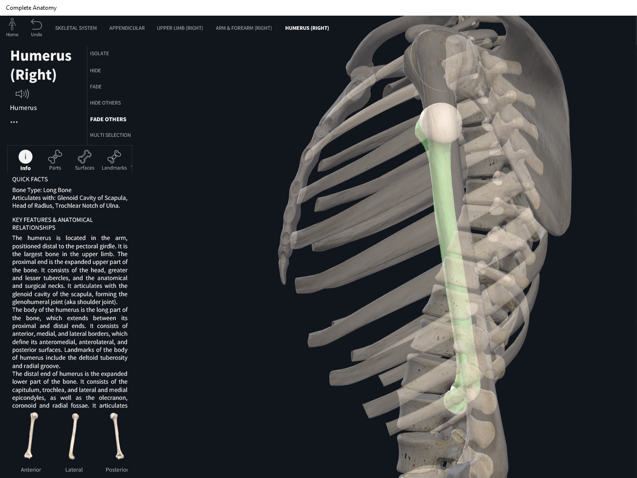
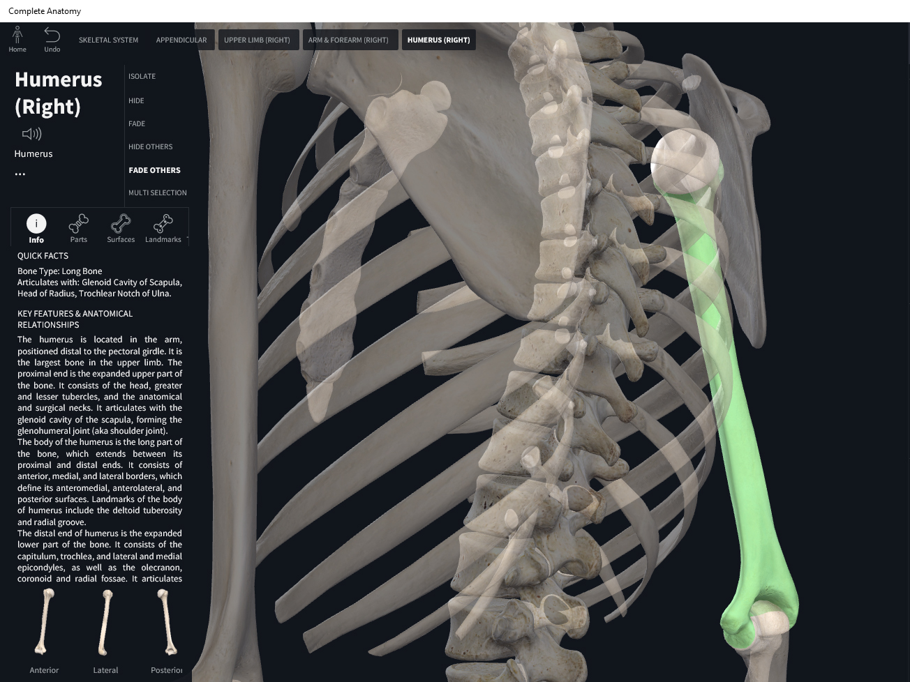
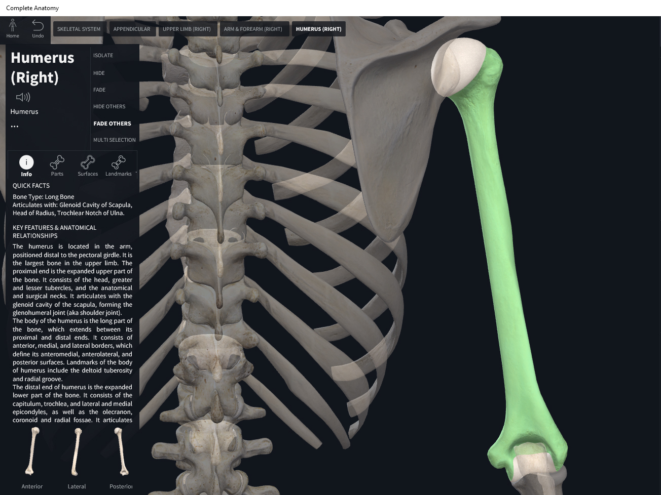
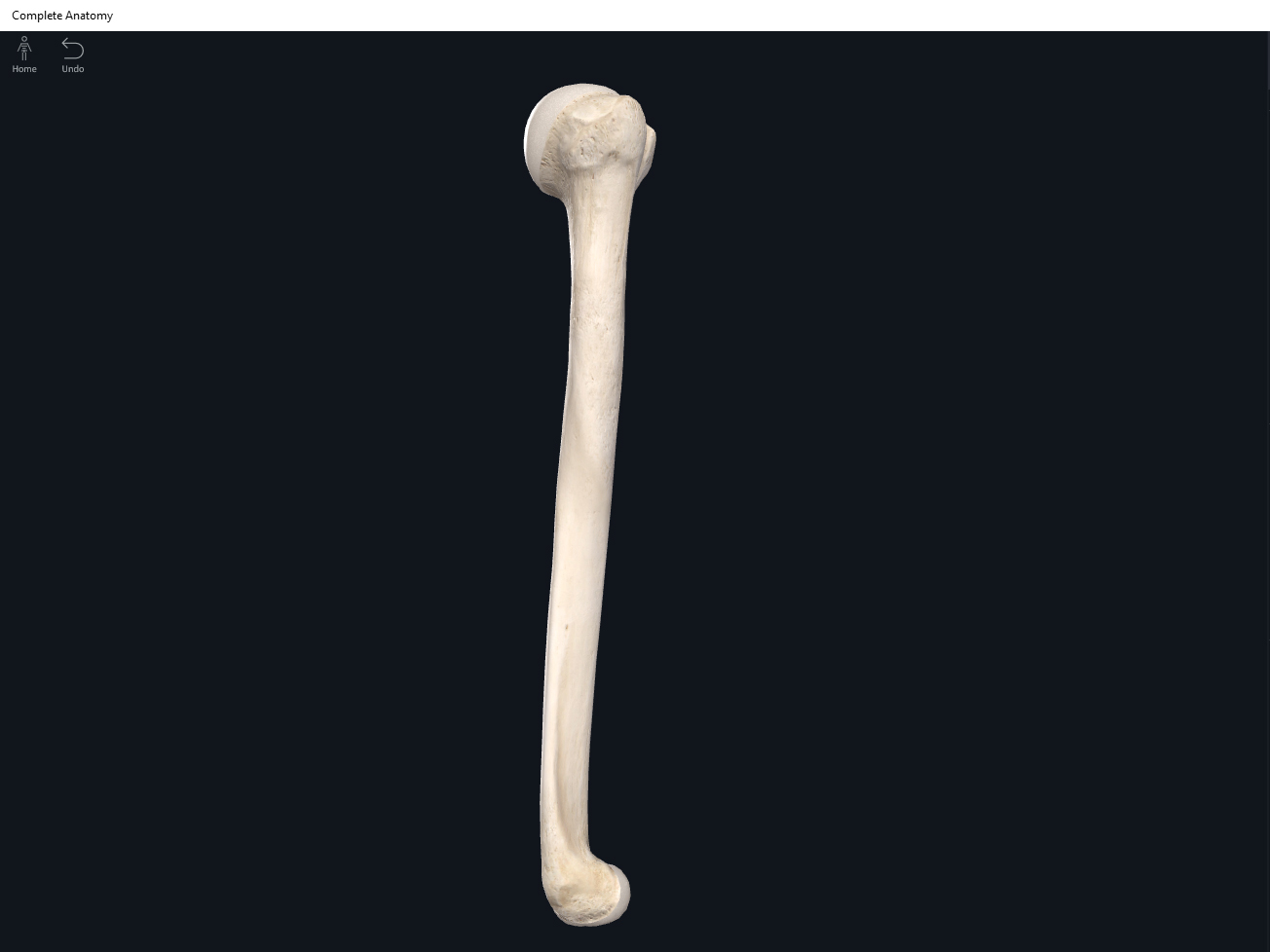
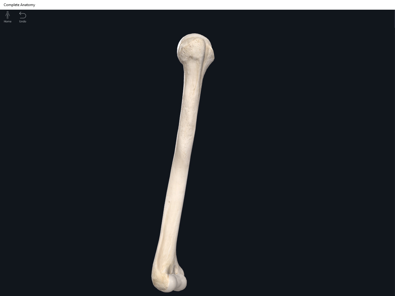
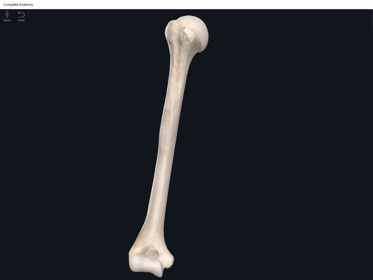
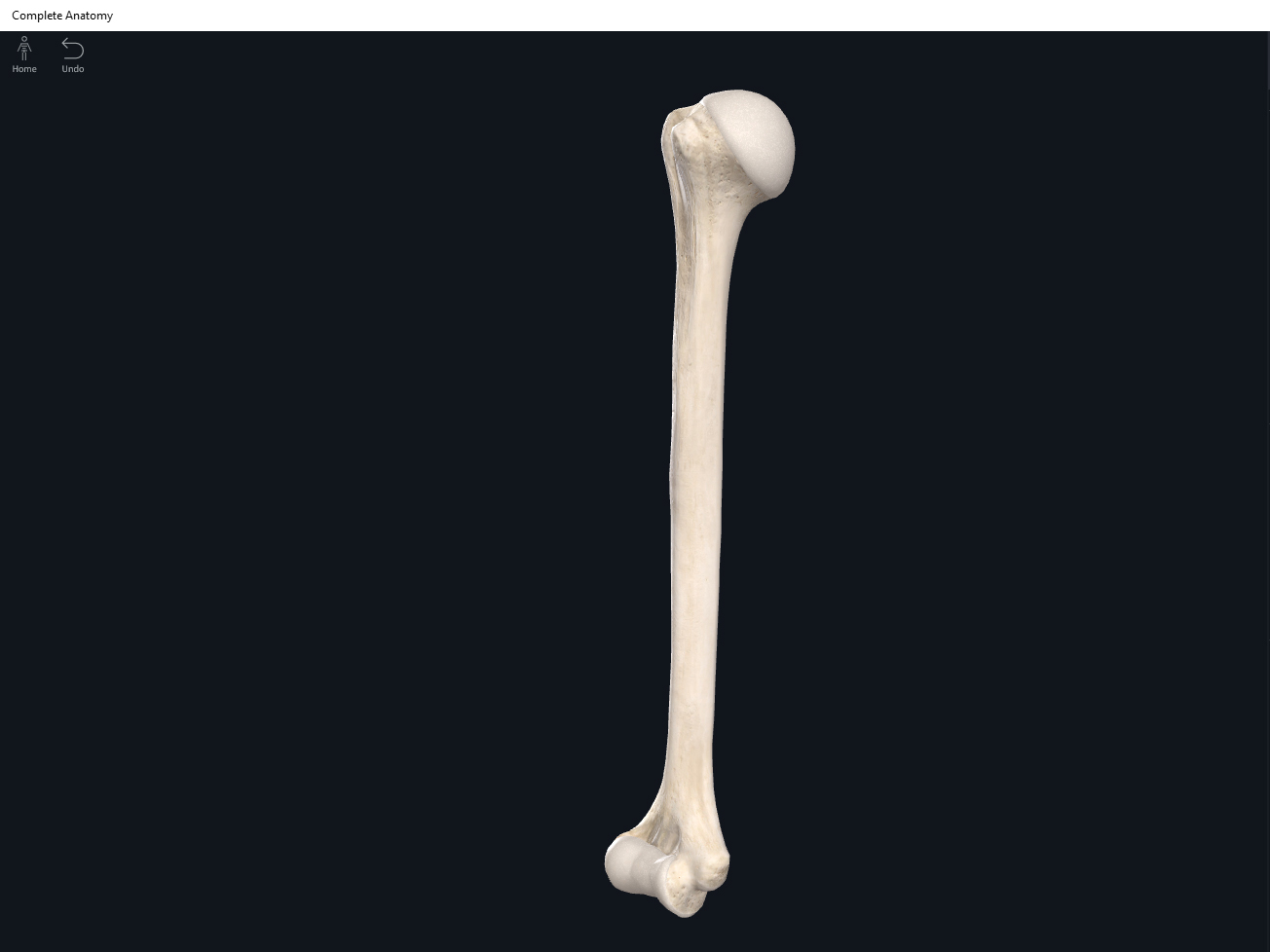
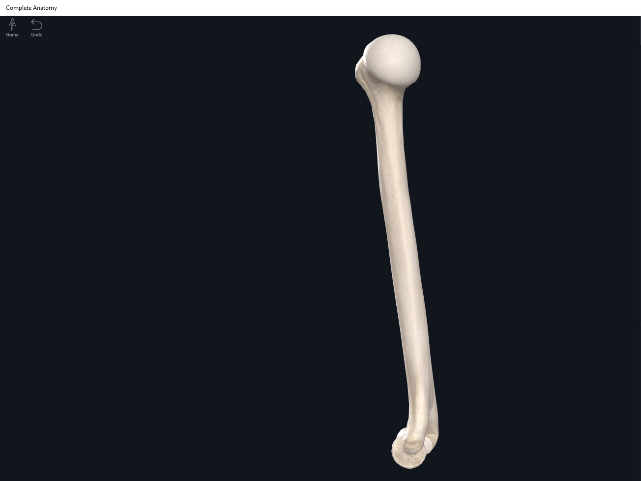
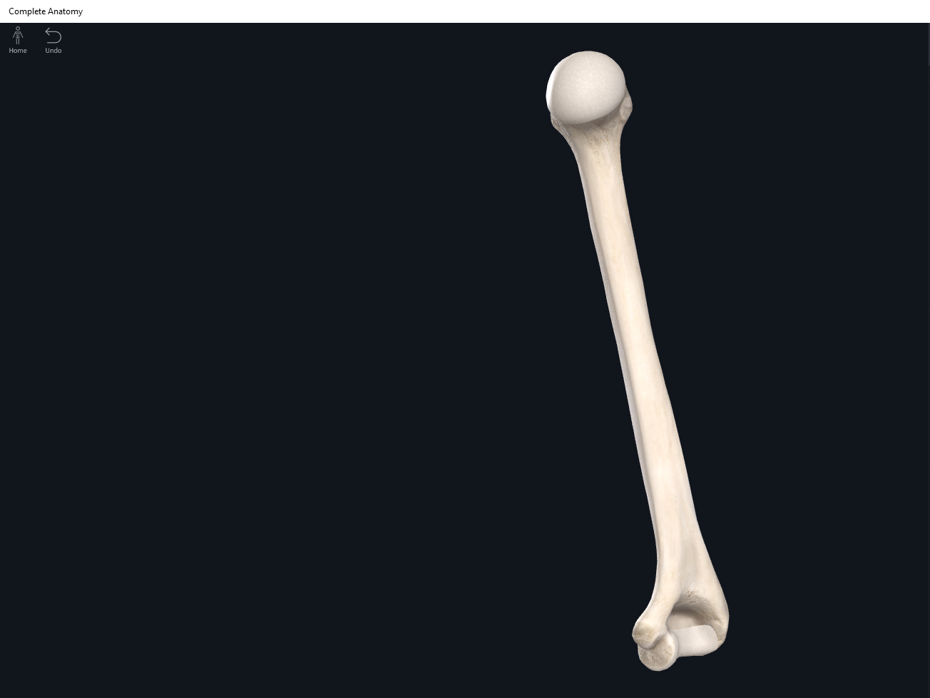
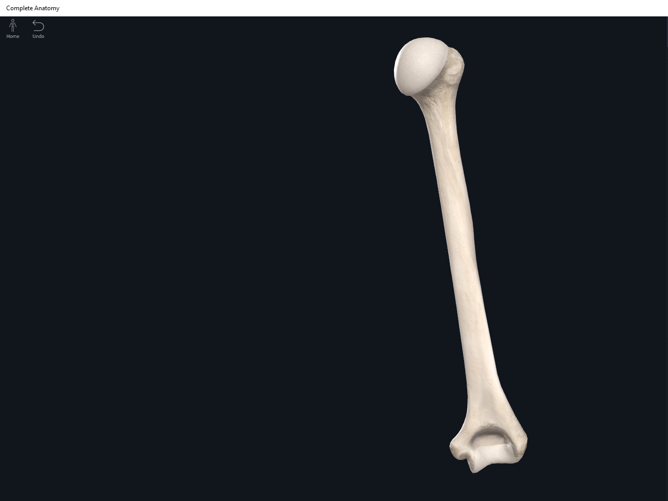
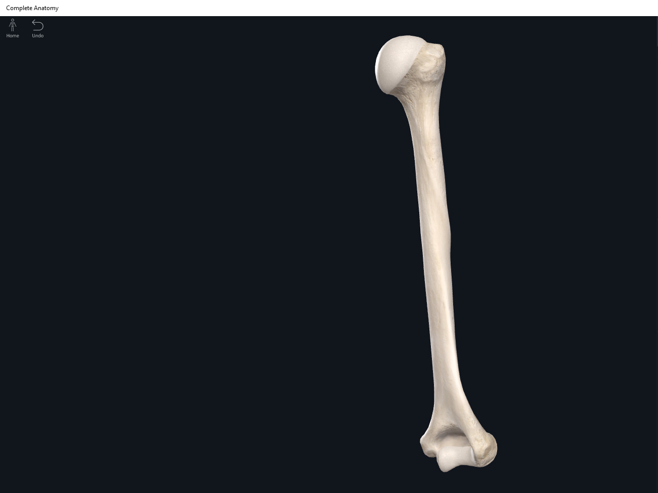
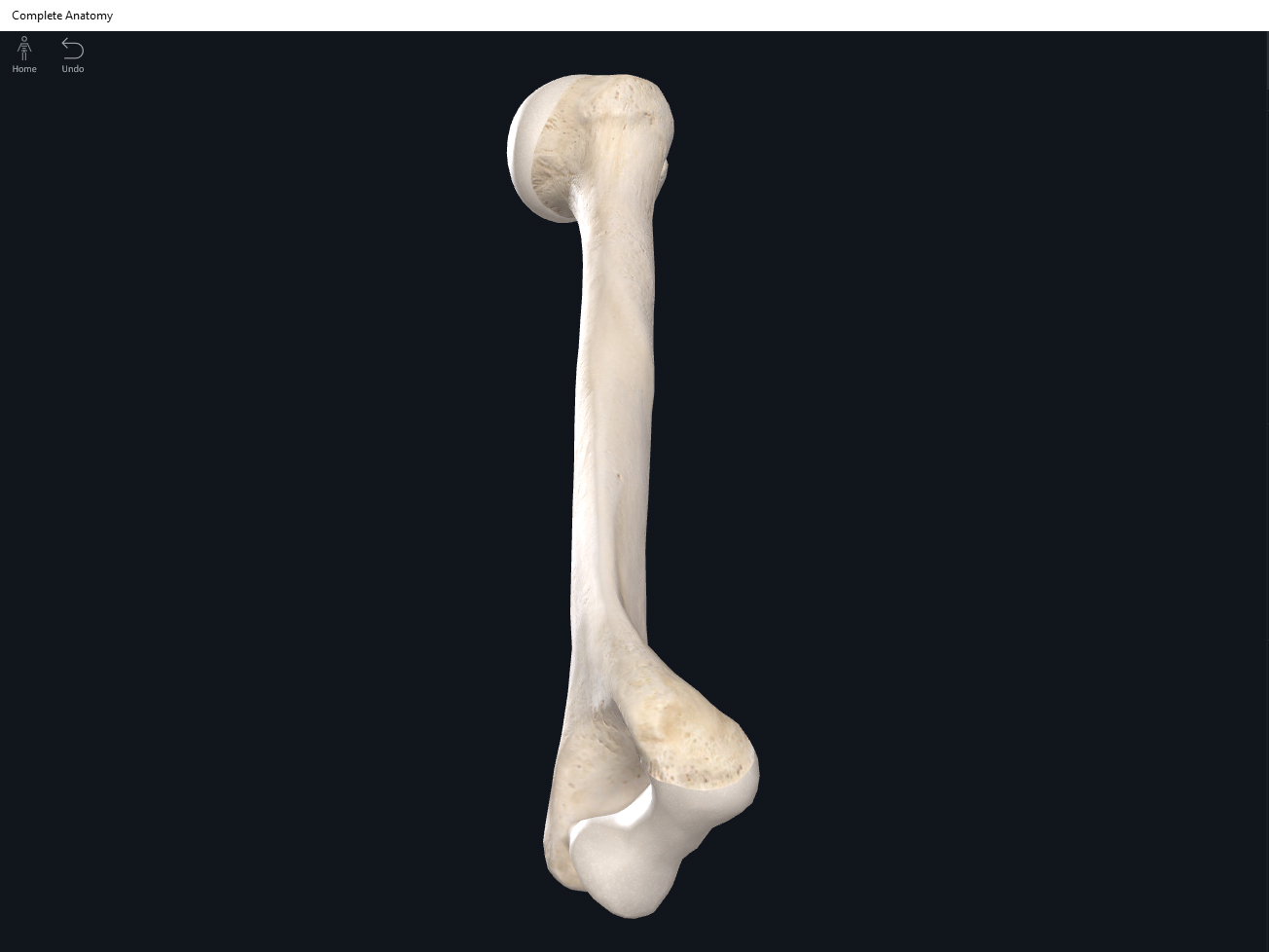
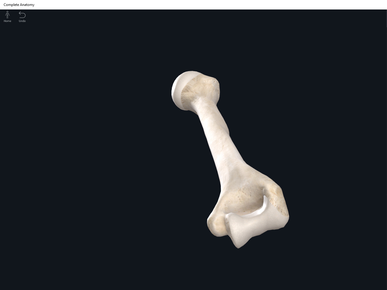
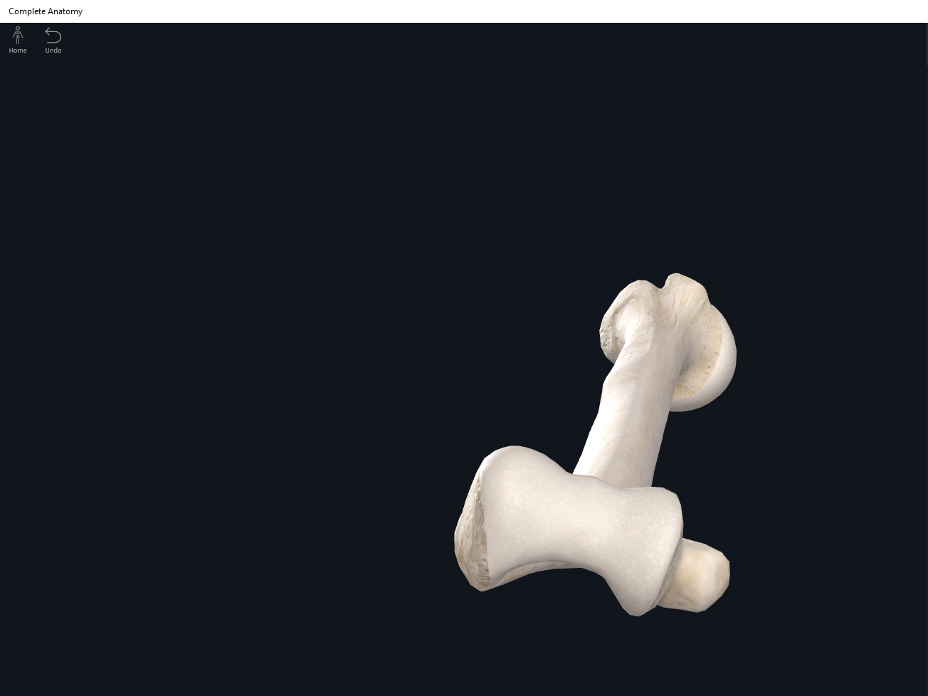
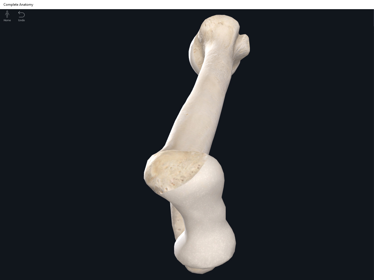
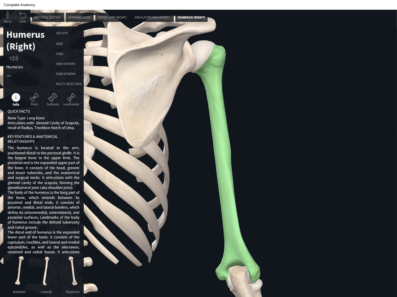
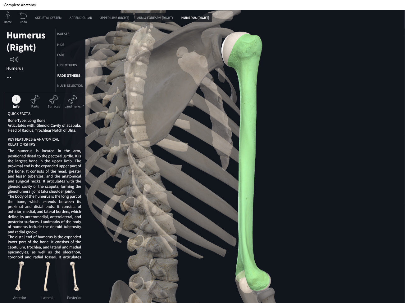
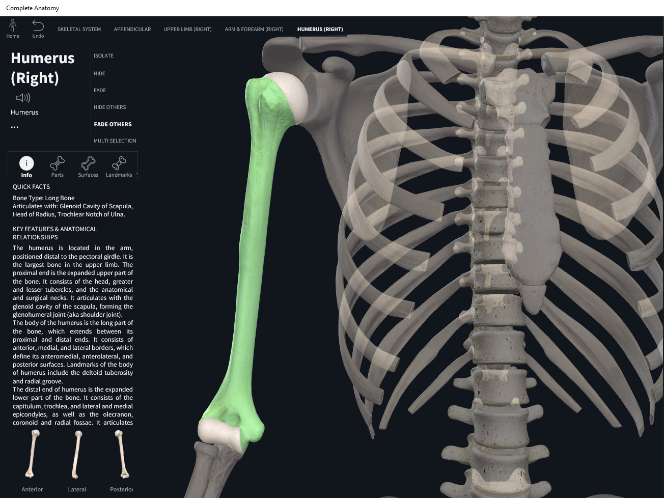
Anatomy & Physiology: Bones—Humerus.
Structure.
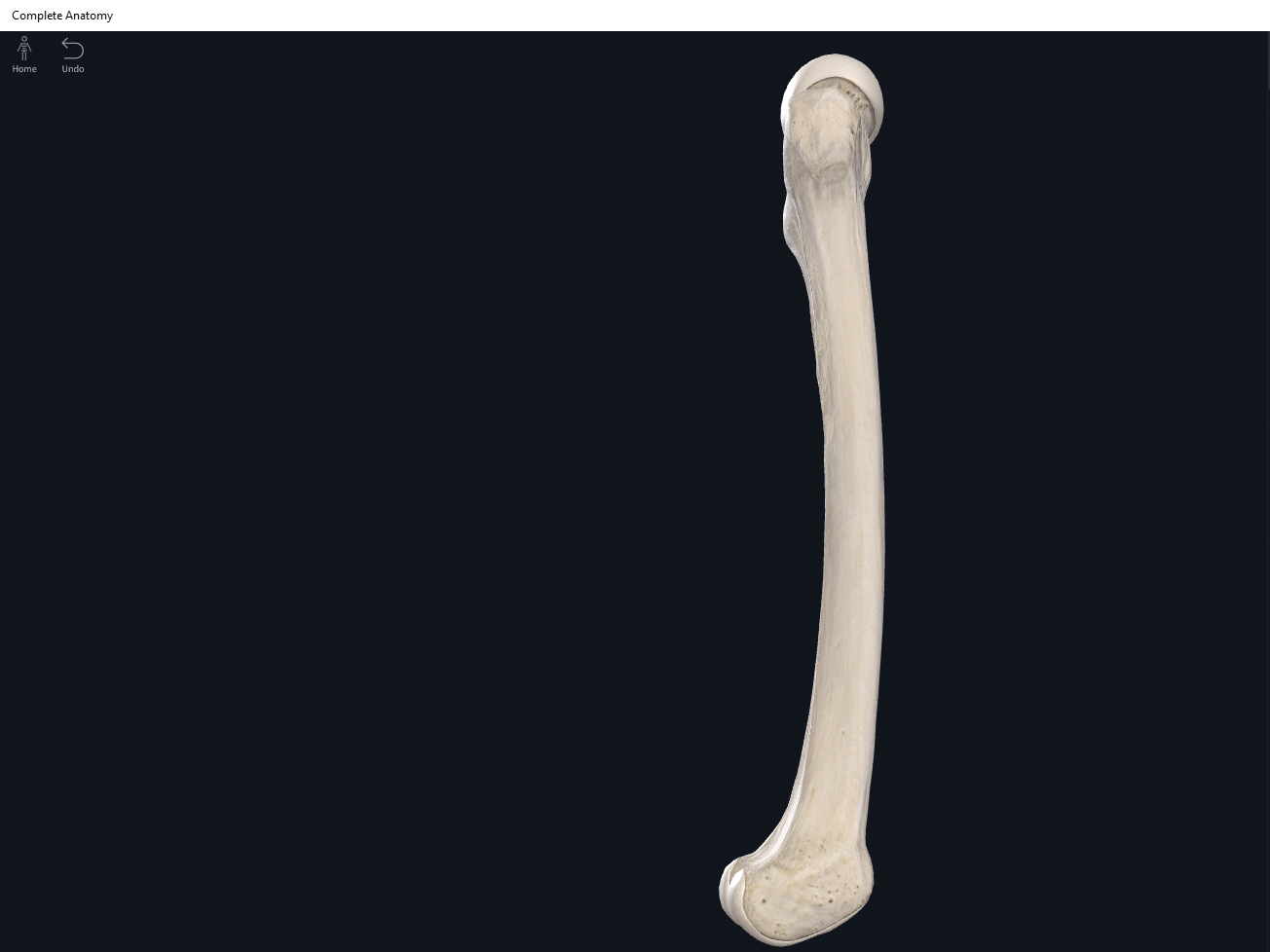
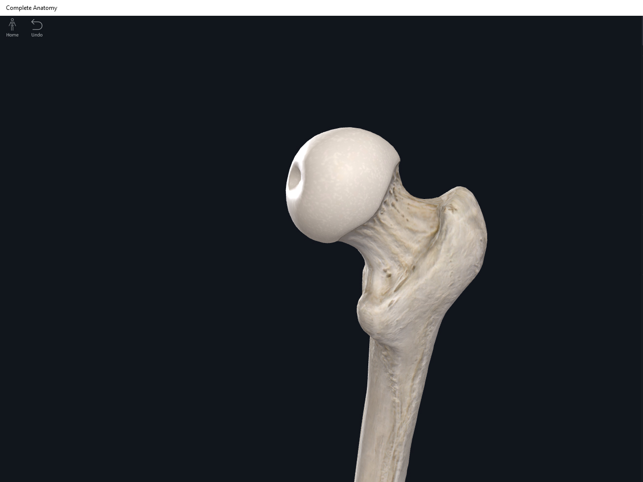
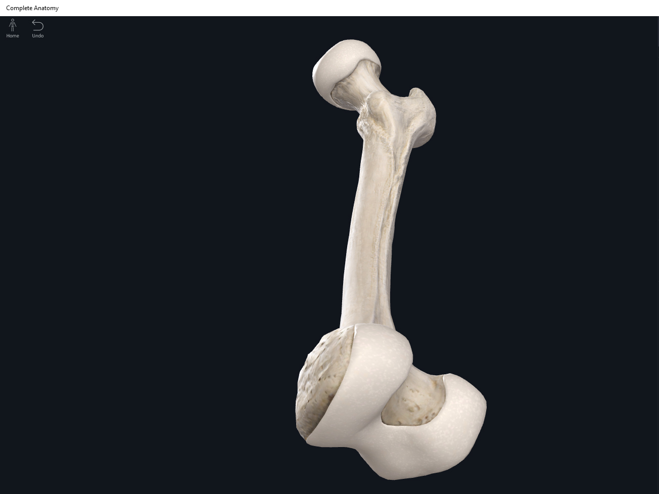
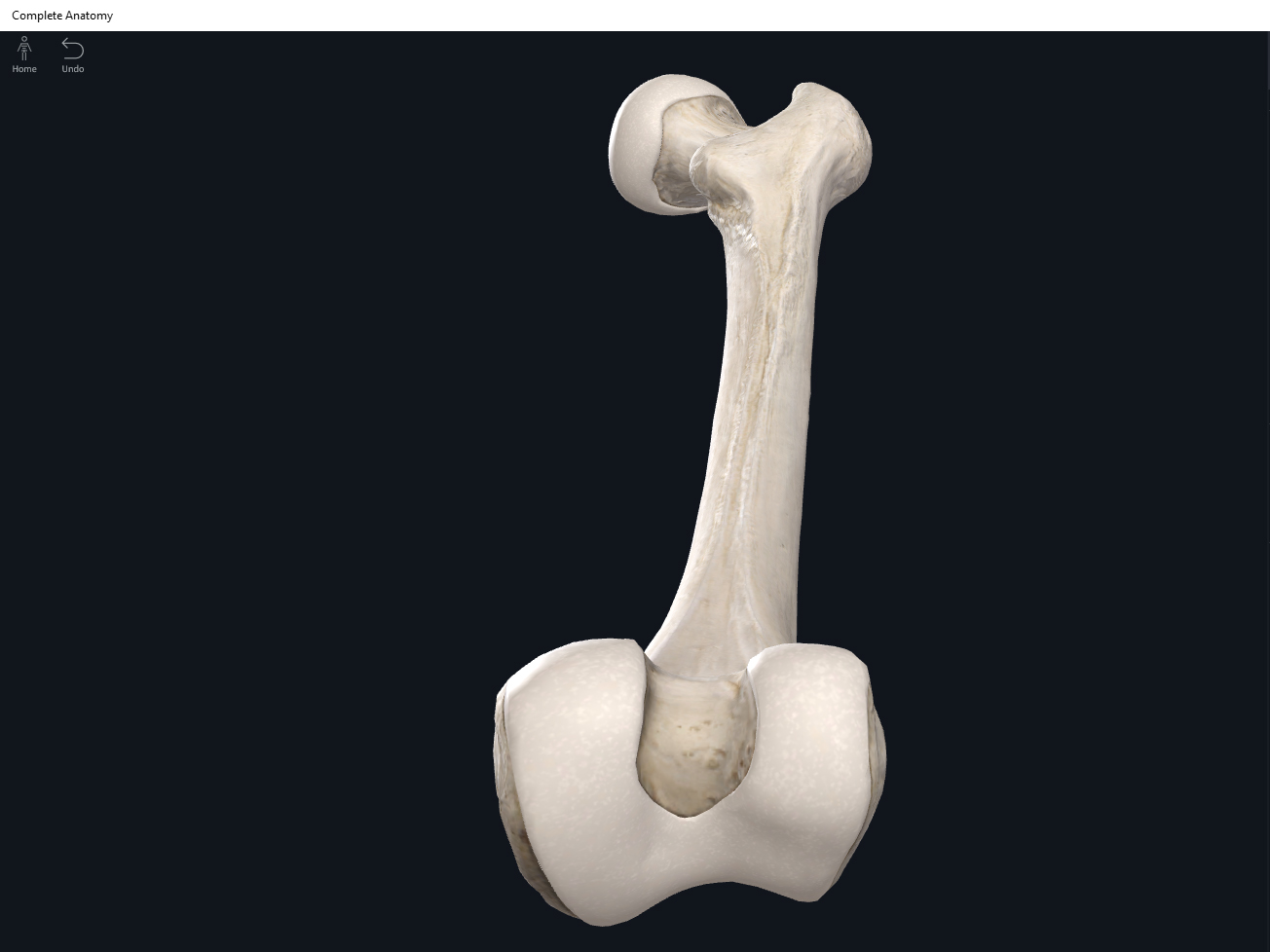
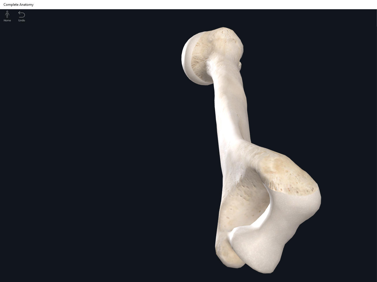
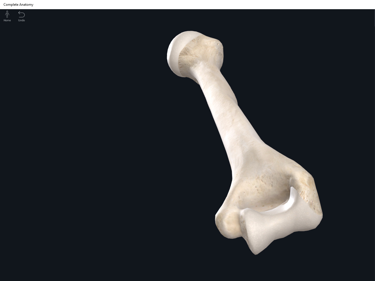
- The largest and longest upper limb bone.
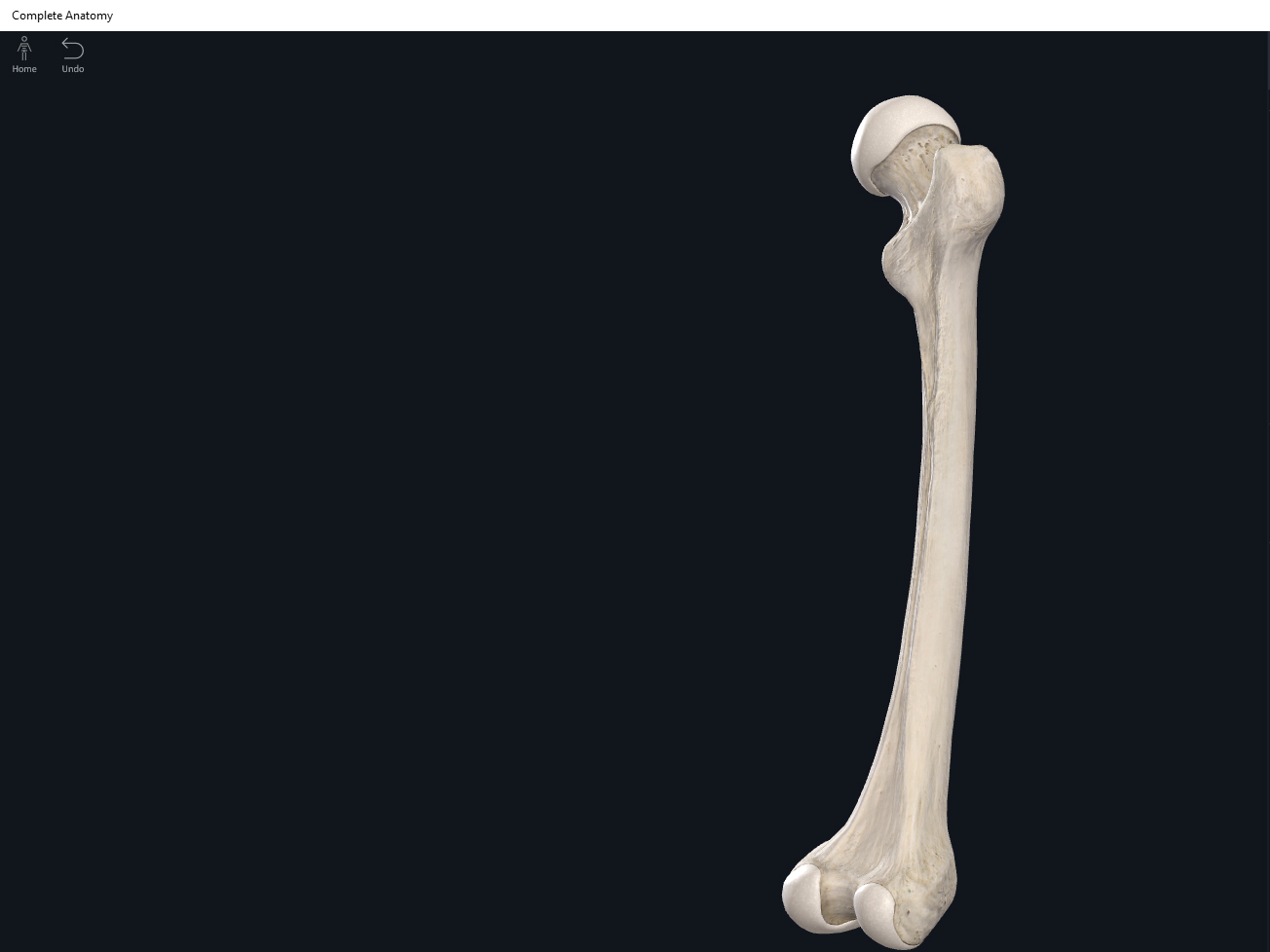
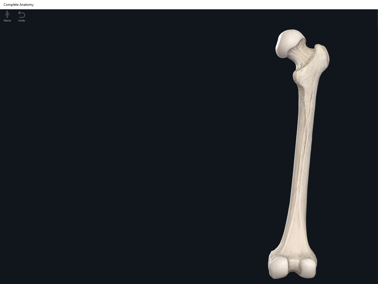
- The humerus articulates with the scapula, radius, and ulna.
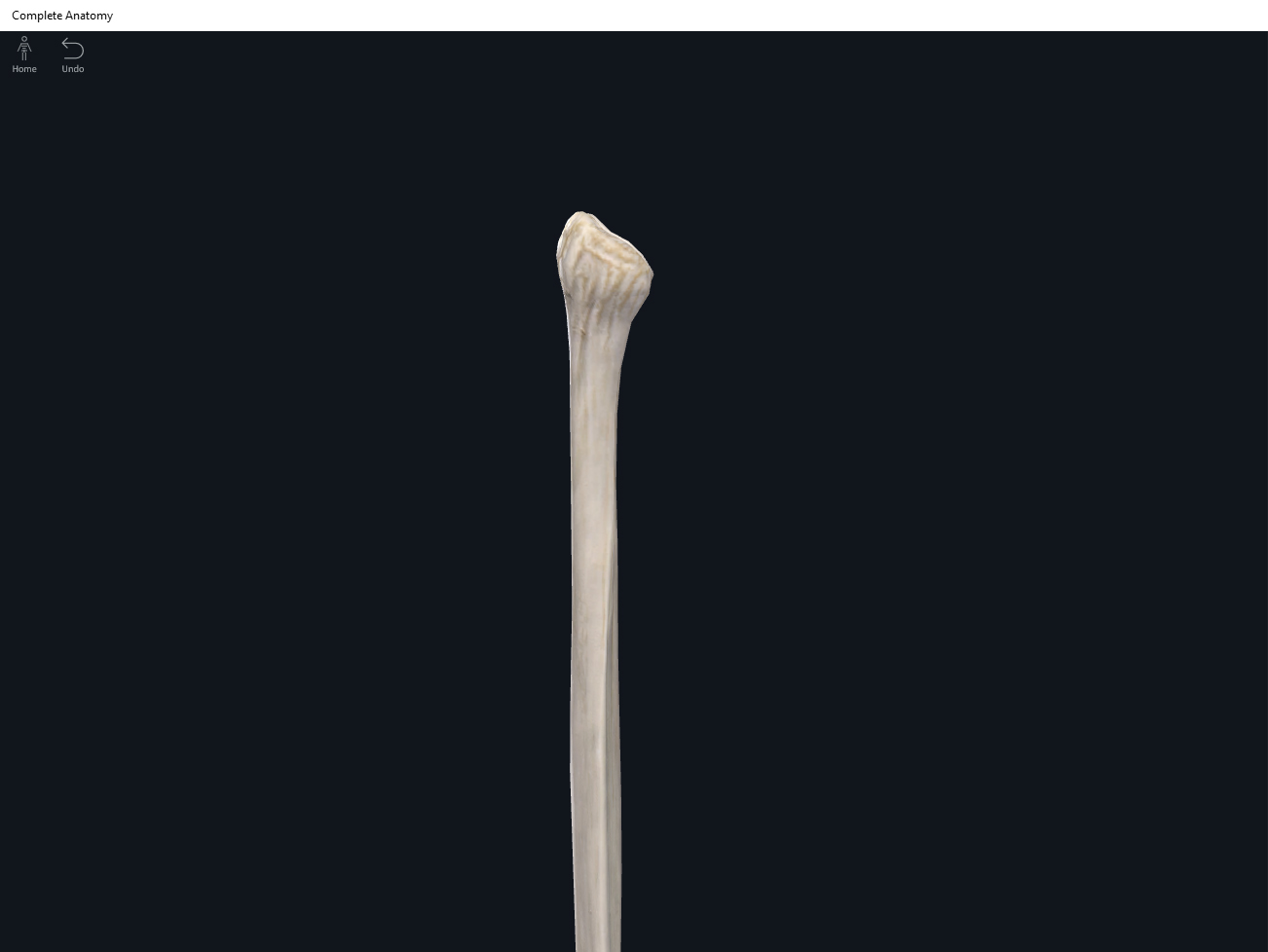
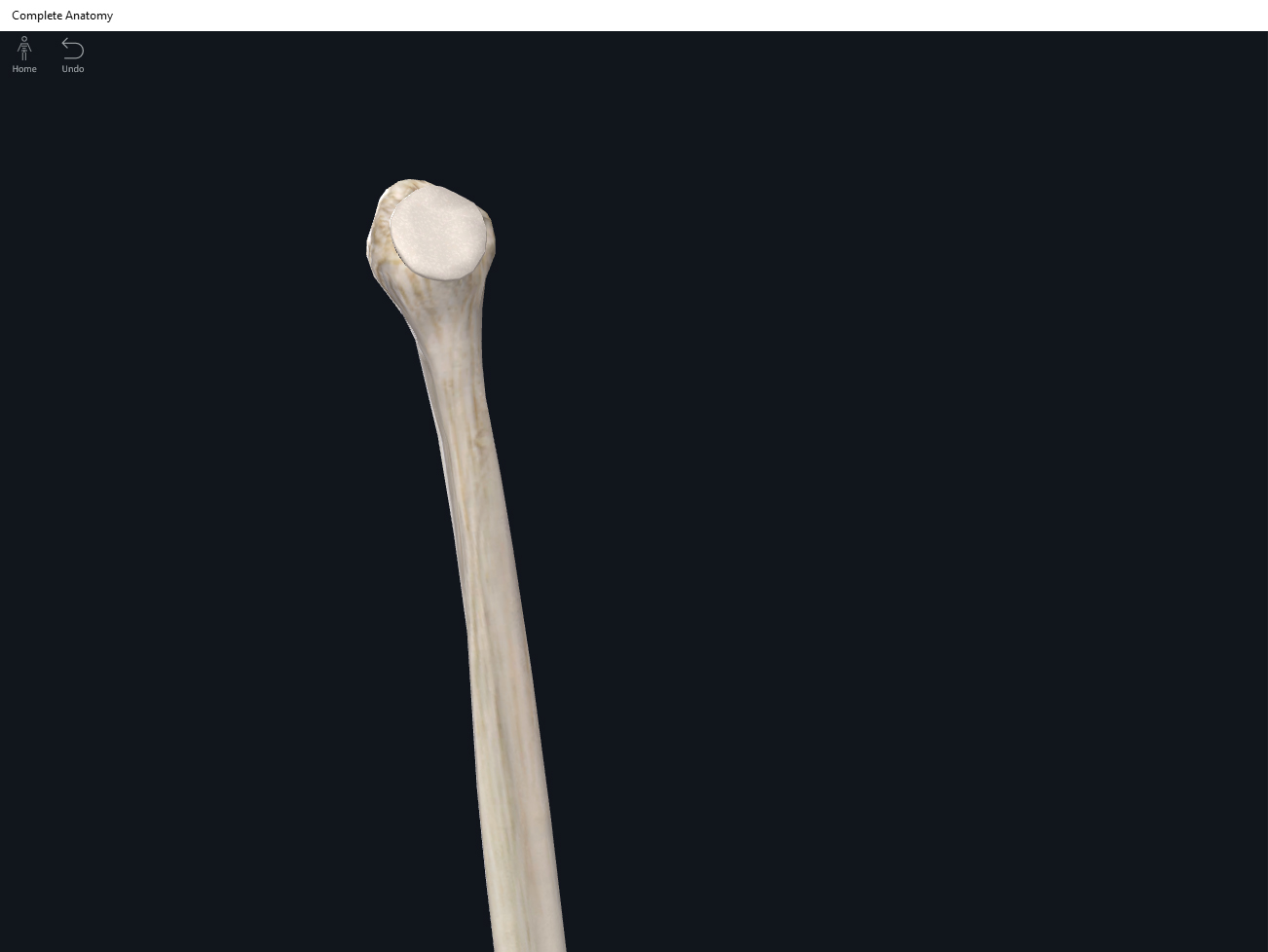
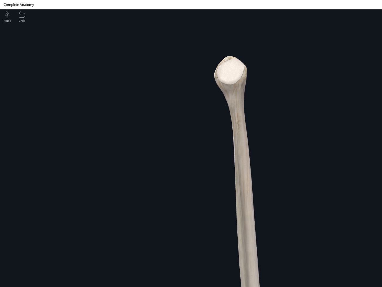
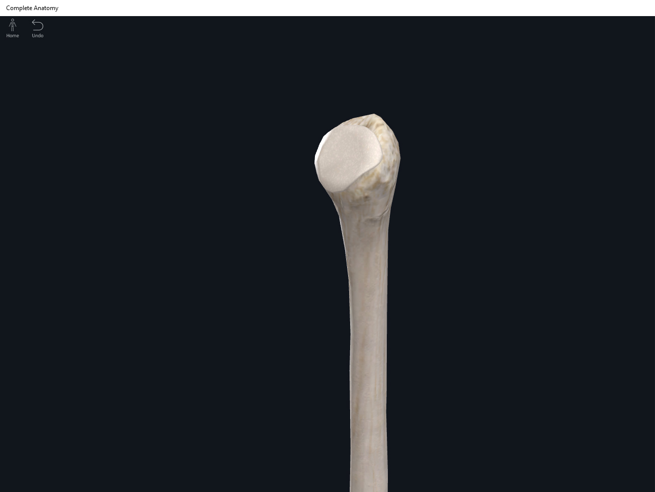
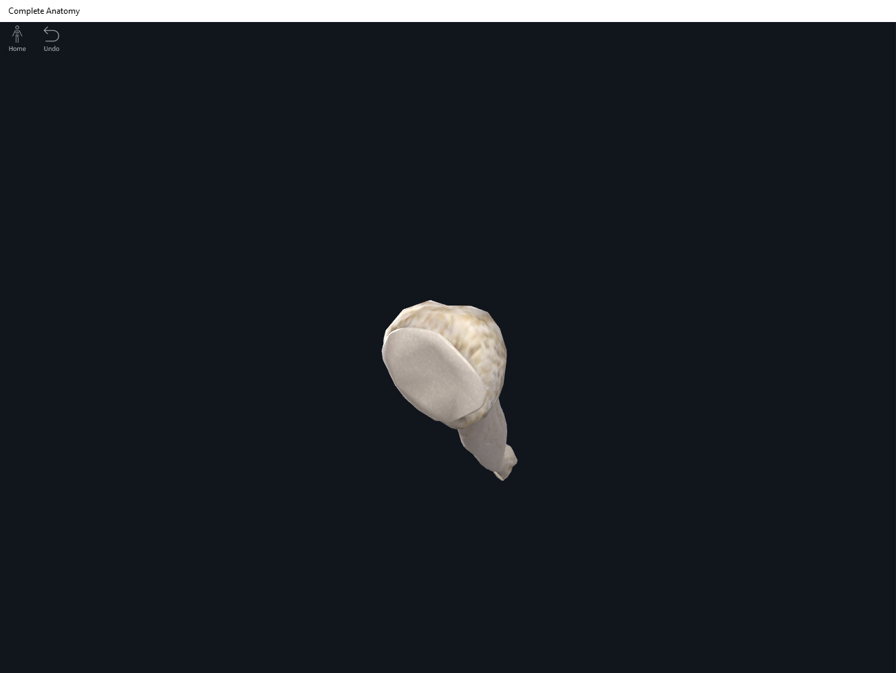
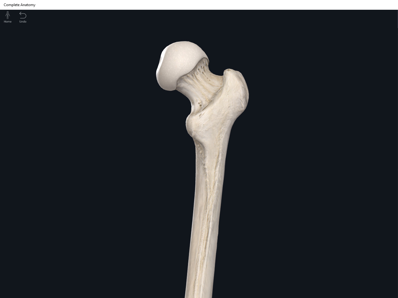
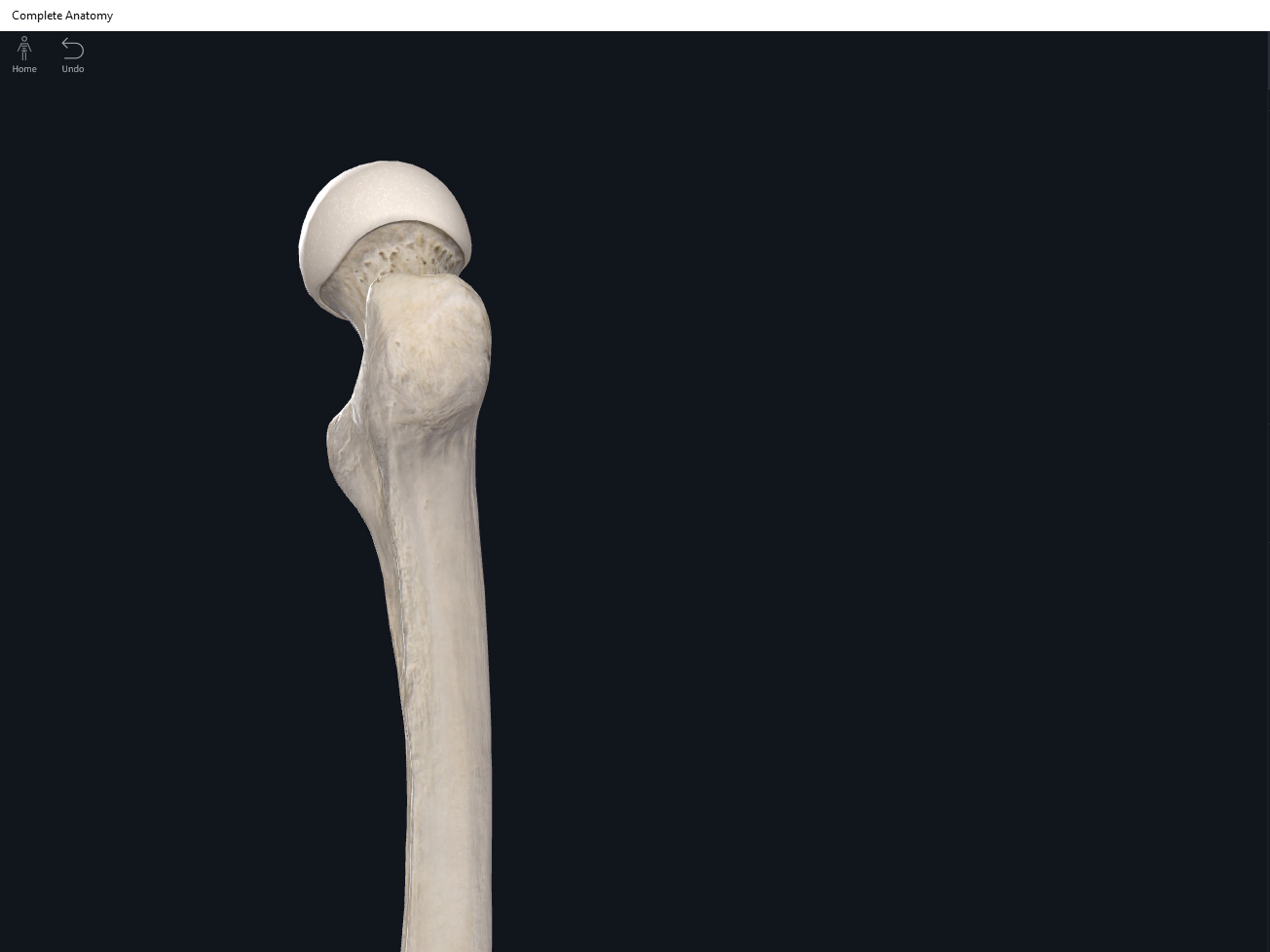
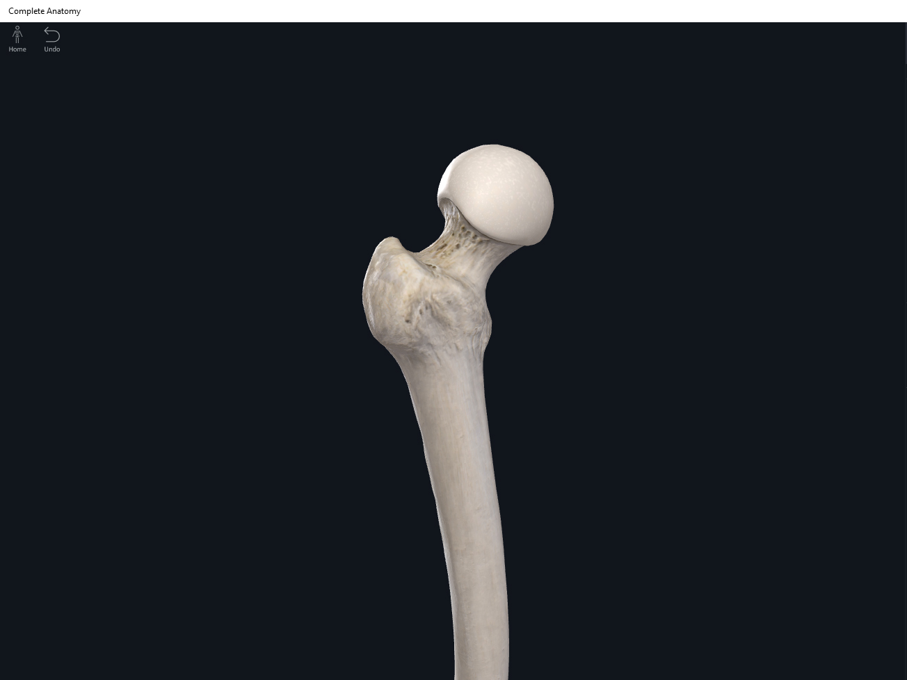
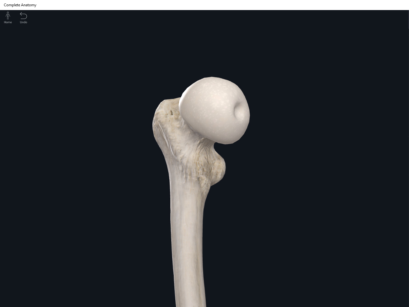
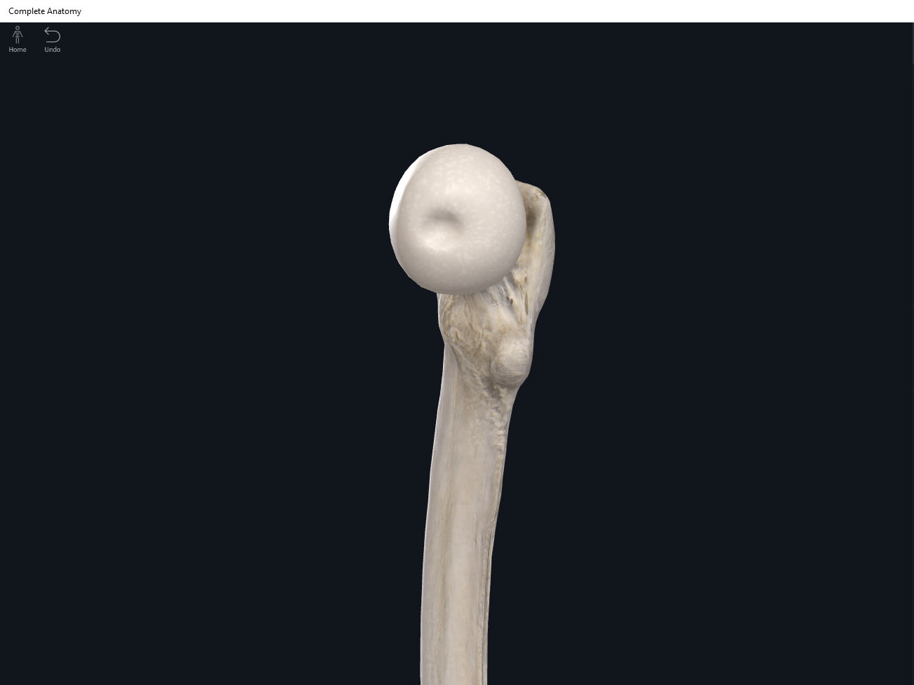
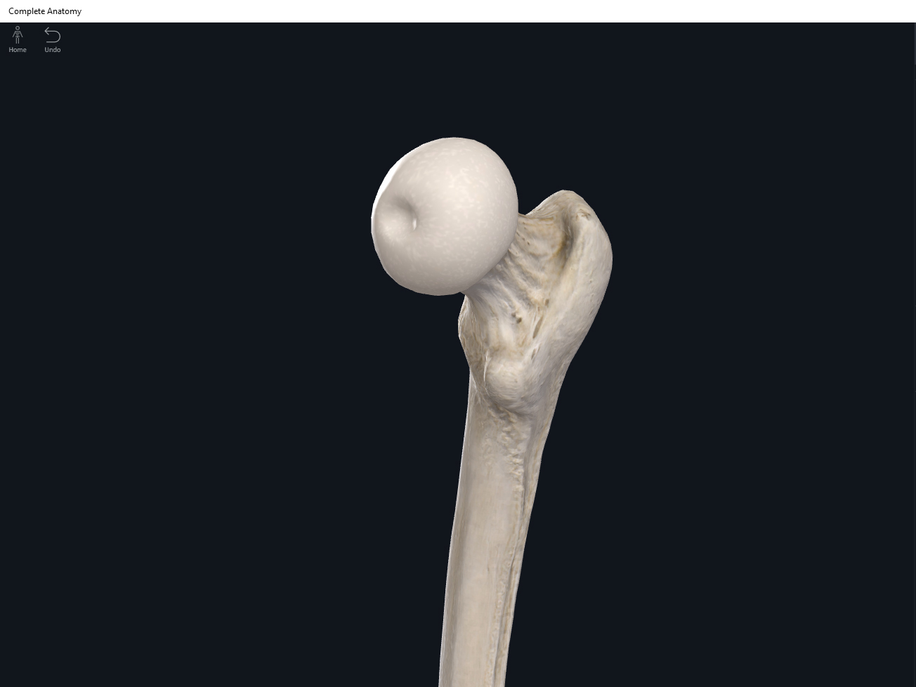
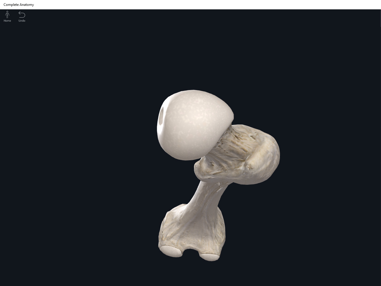
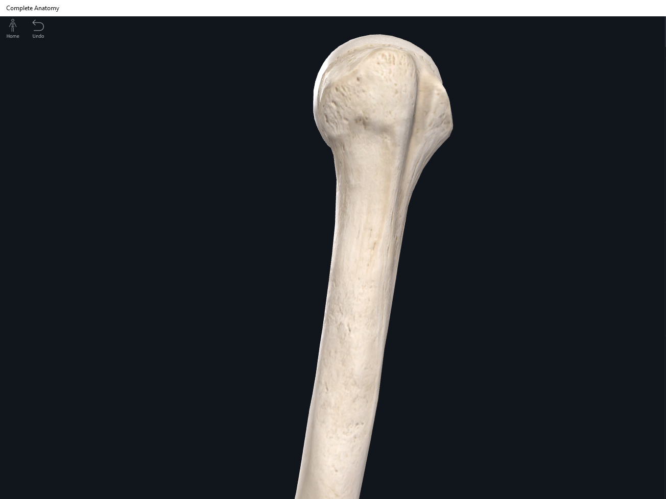
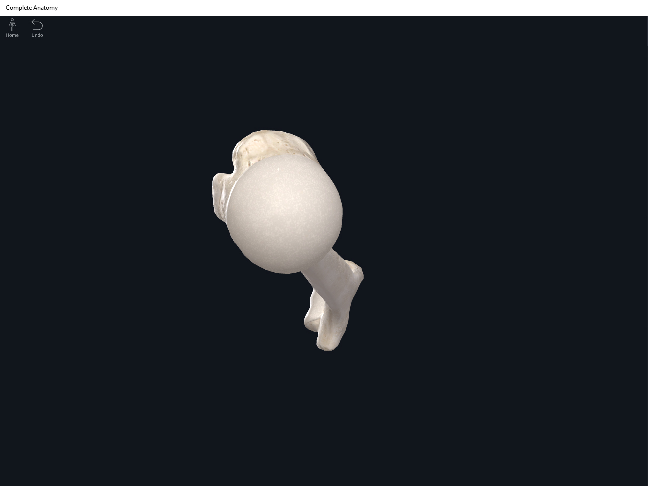
- Head: the proximal end is the head which articulates with the scapula at the glenoid fossa. This forms the shoulder joint.
- Anatomical neck.
- Surgical neck.
- Greater tubercle.
- Lesser tubercle.
- Intertubercular sulcus (groove).
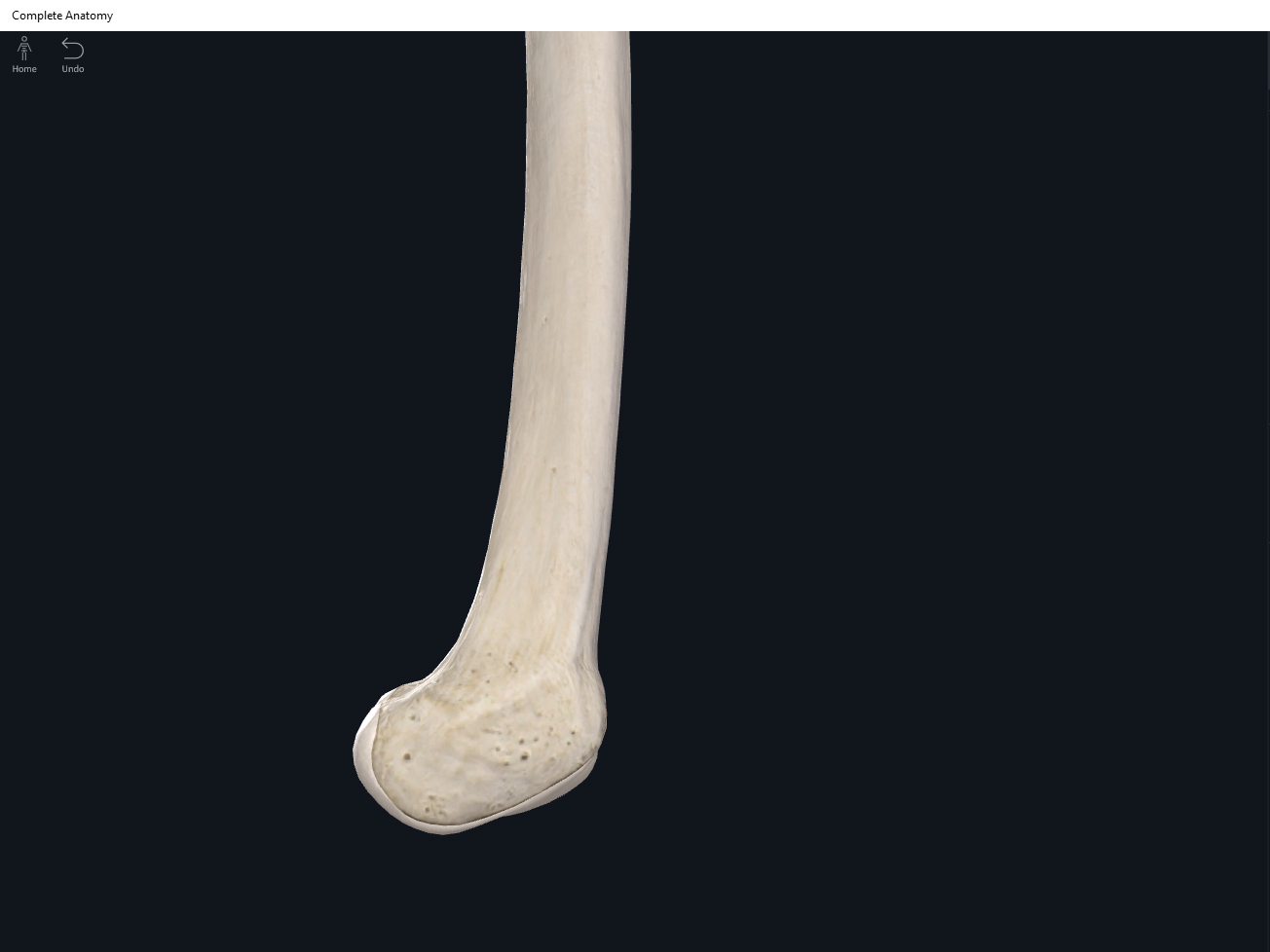
- Body (diaphysis/shaft).
- Deltoid tuberosity: where the tendons of the deltoid attach.
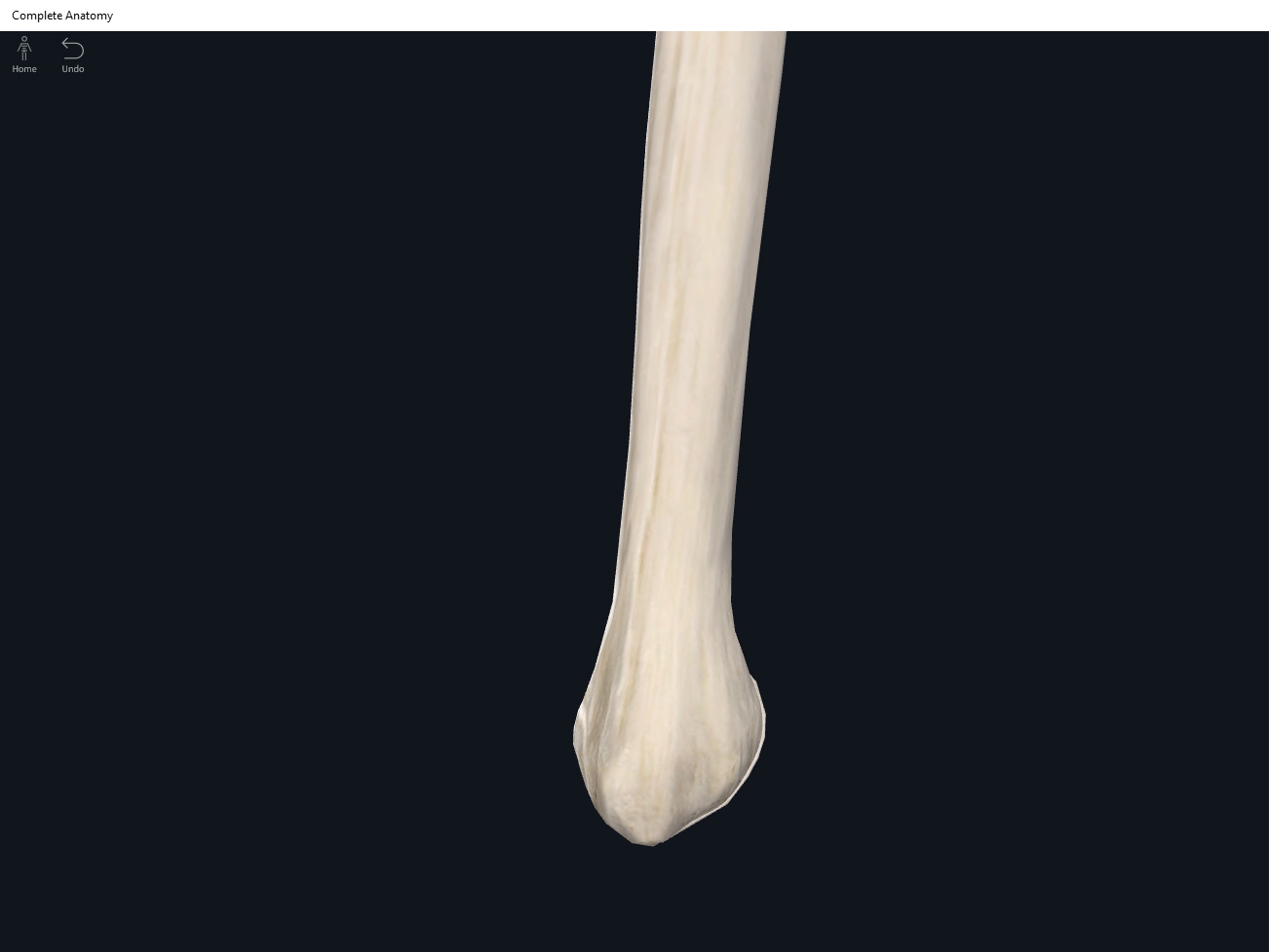
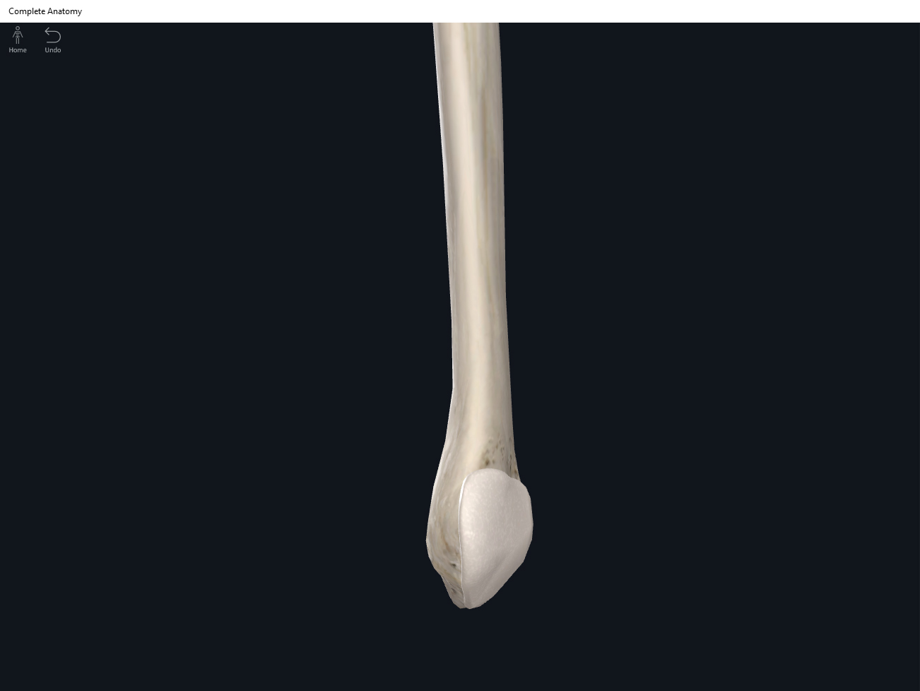
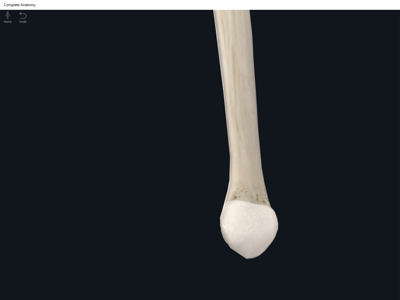
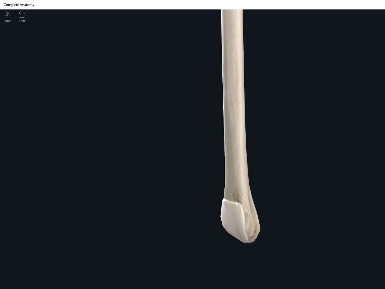
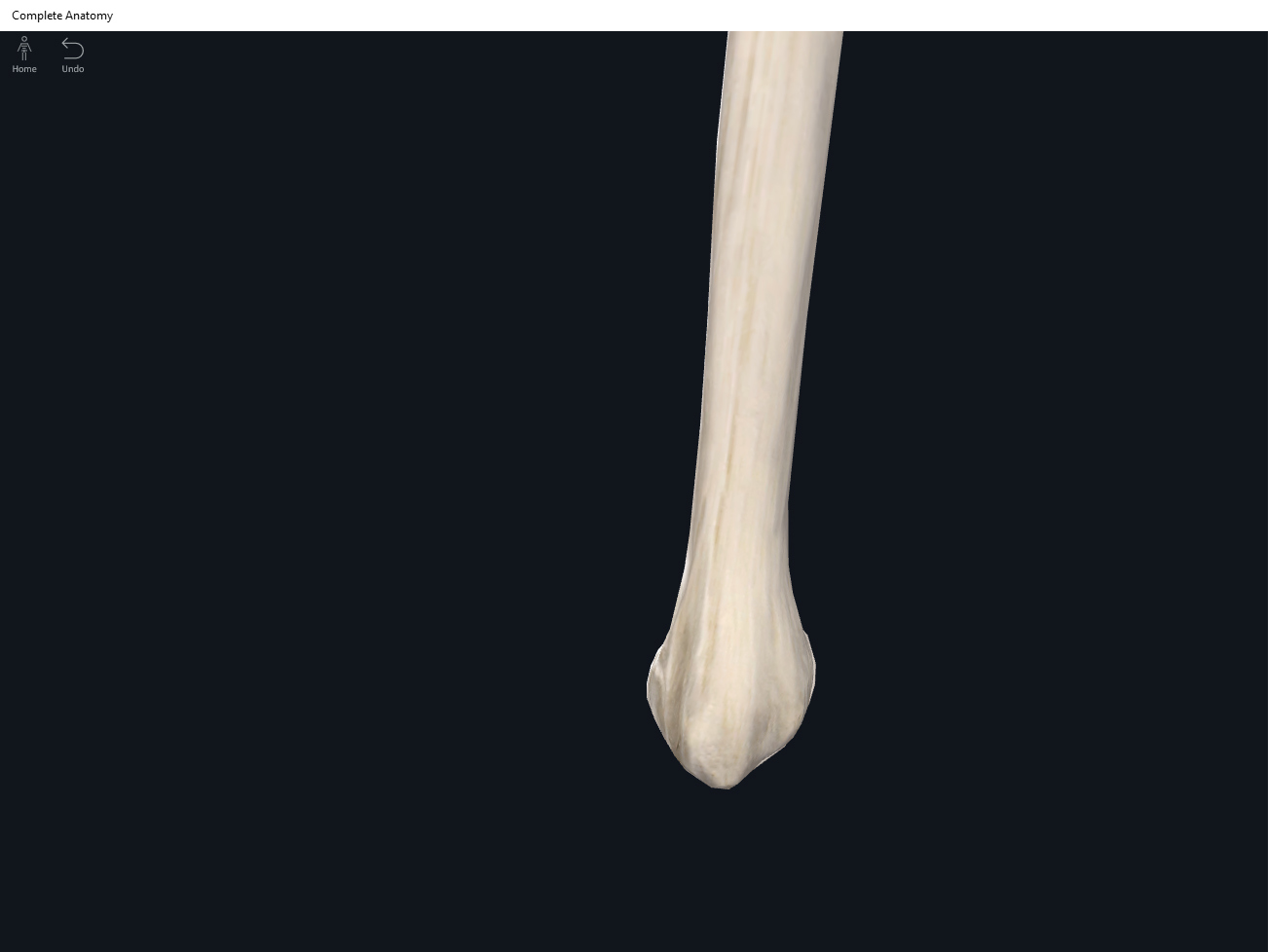
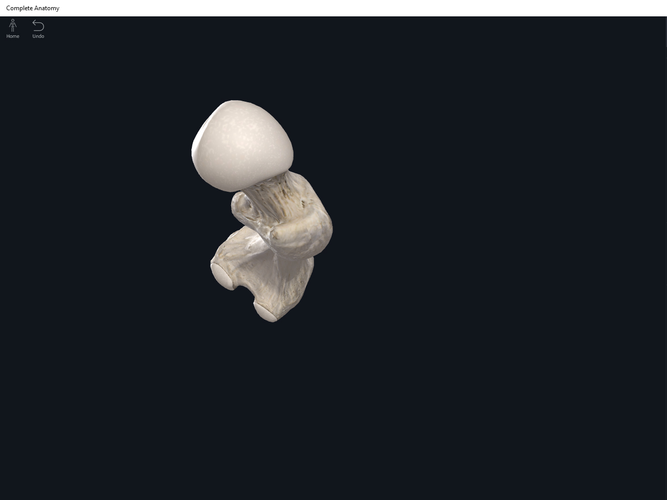
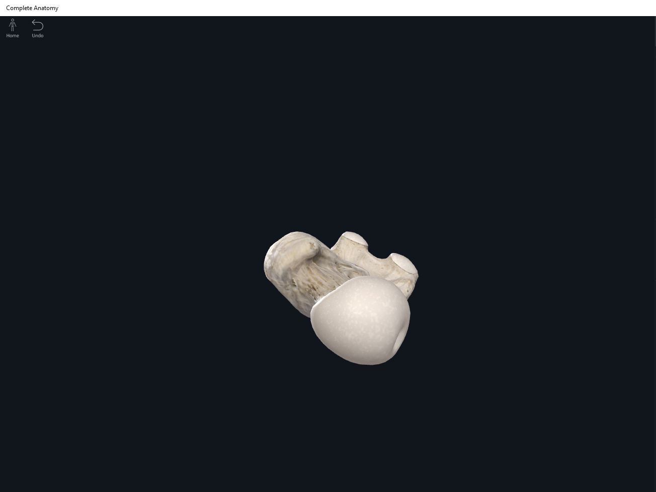
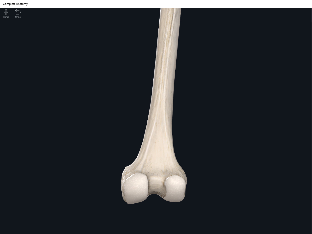
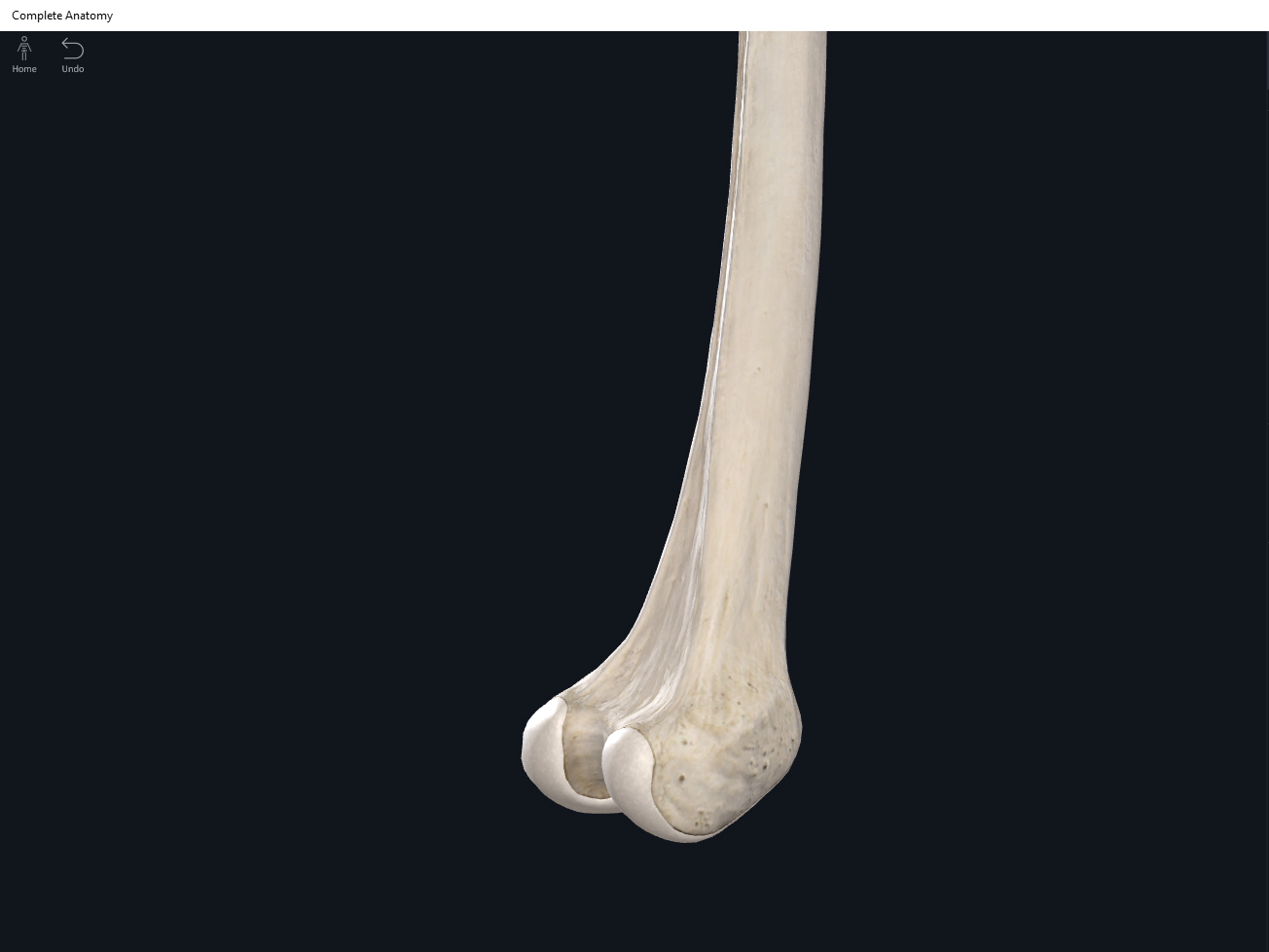
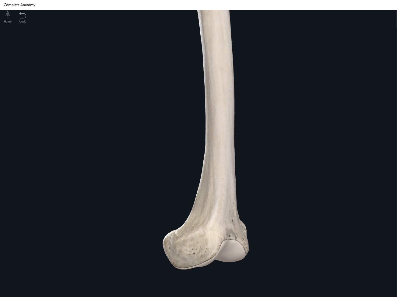
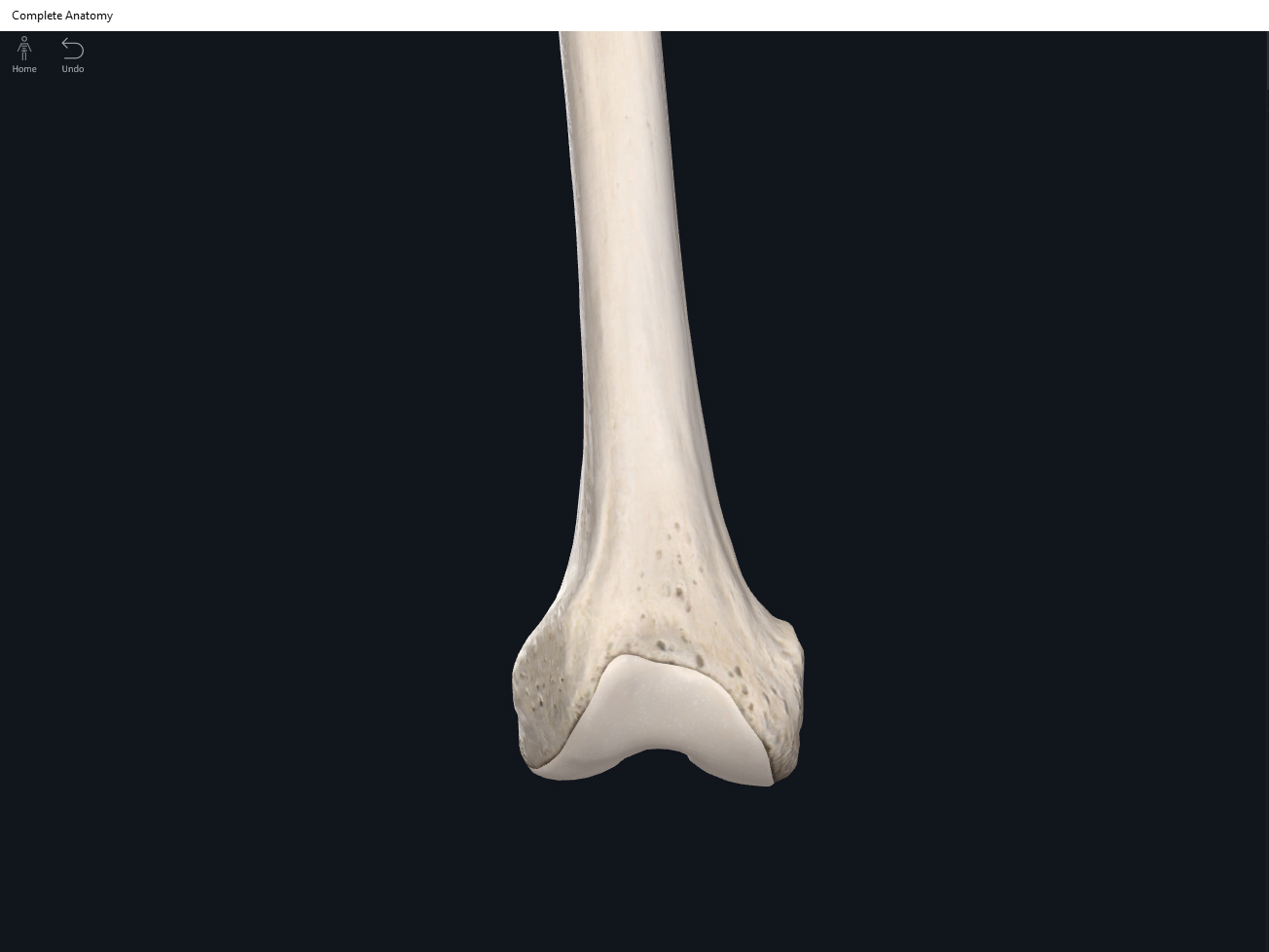
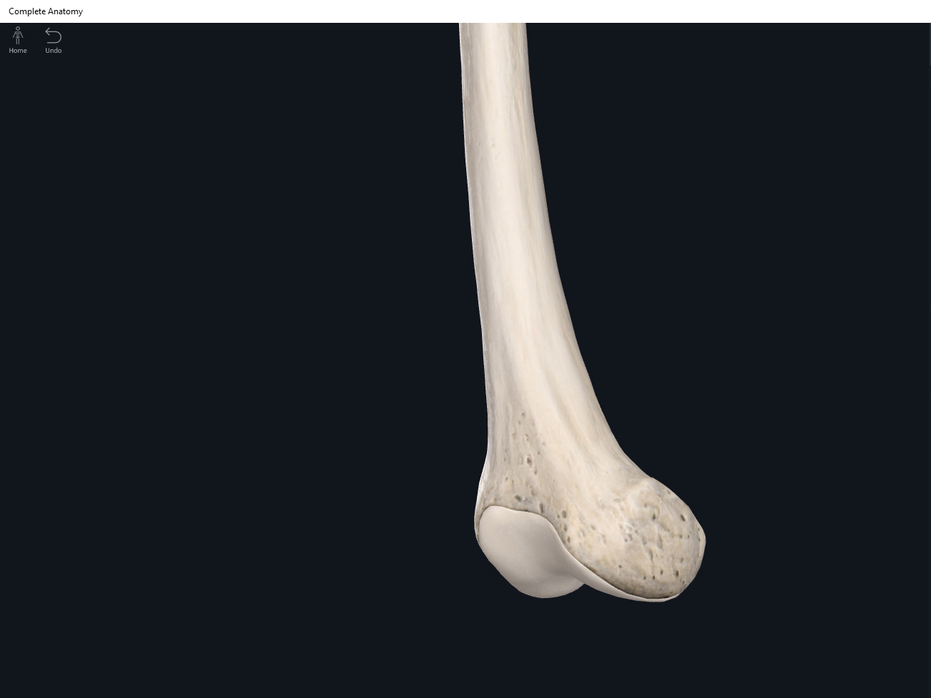
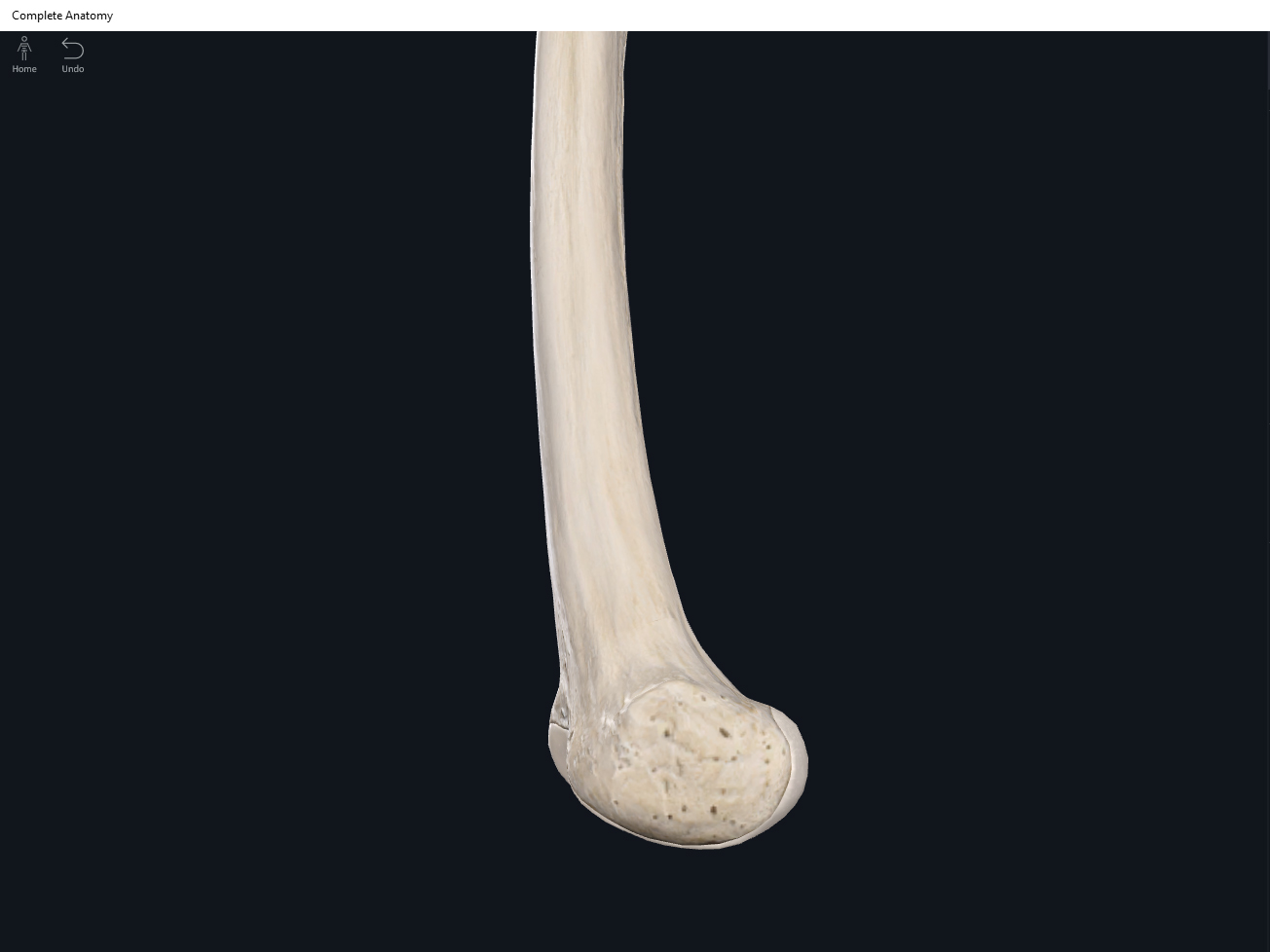
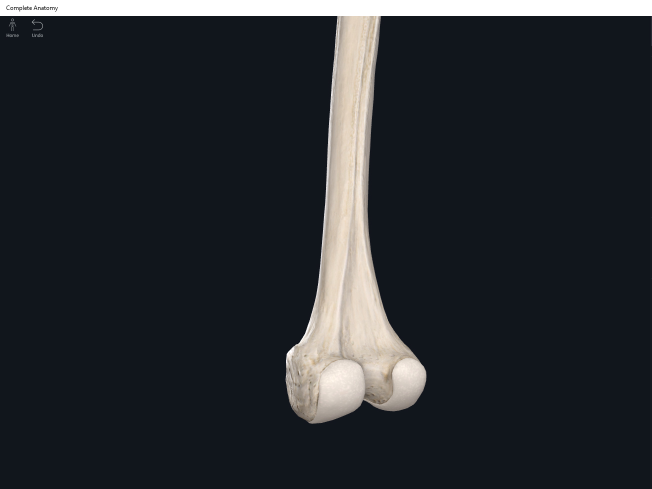
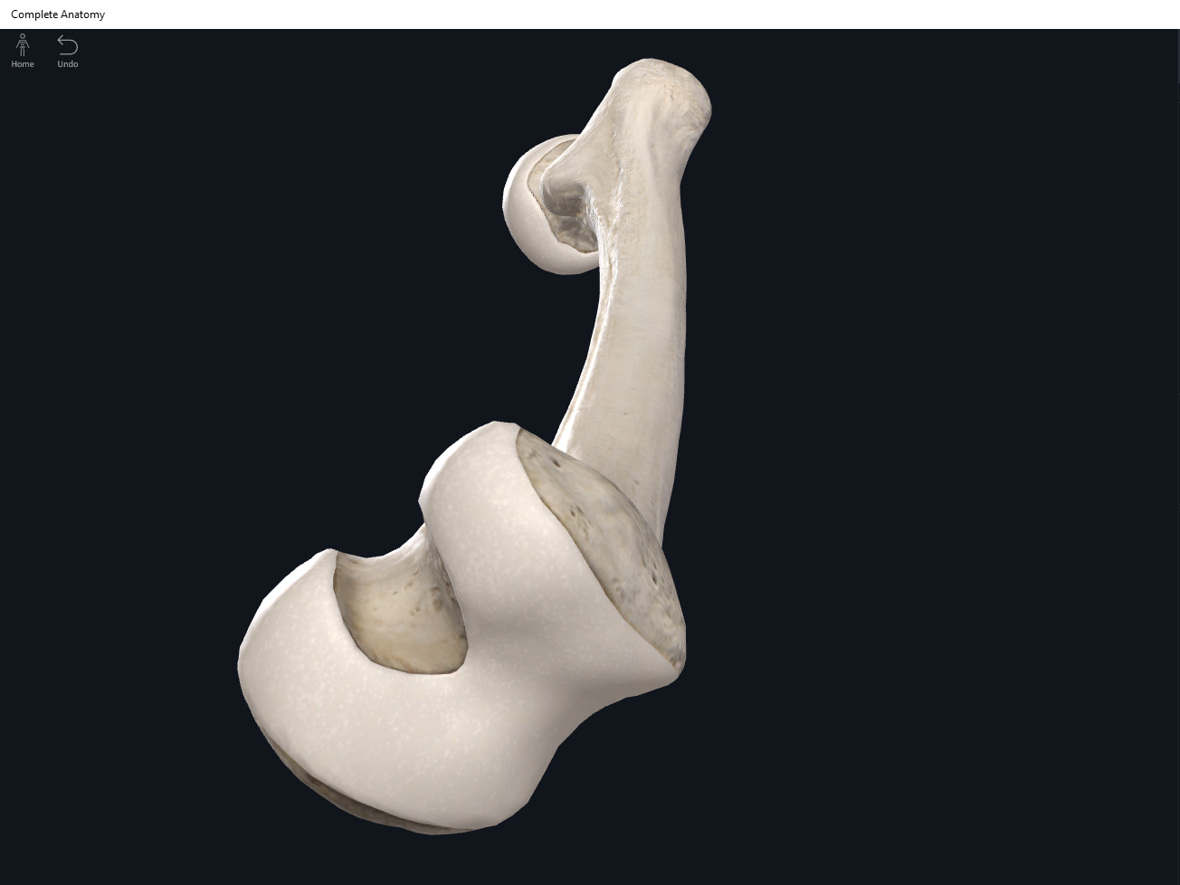
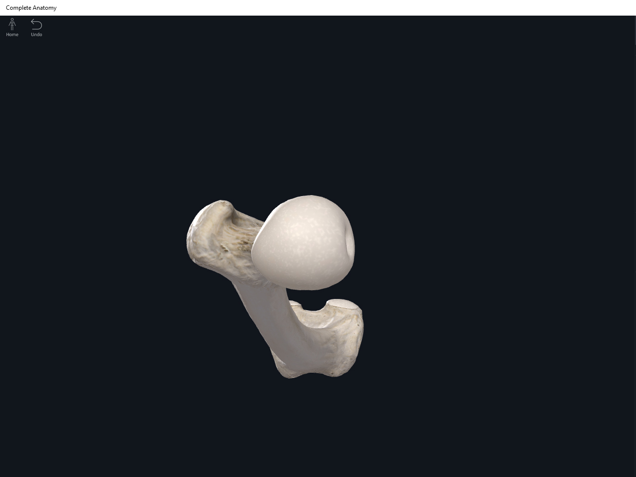
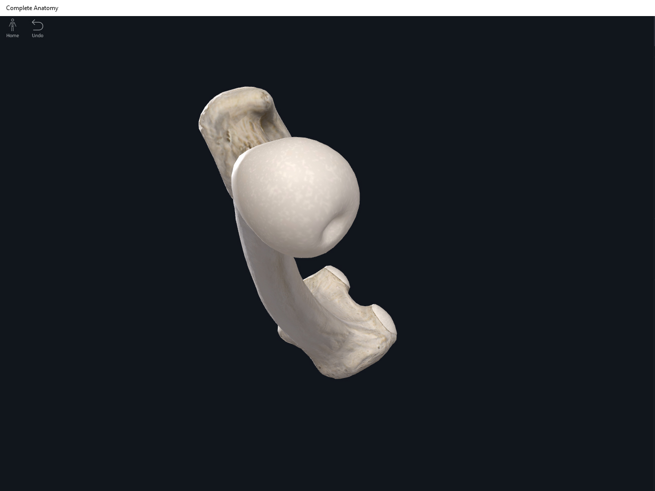
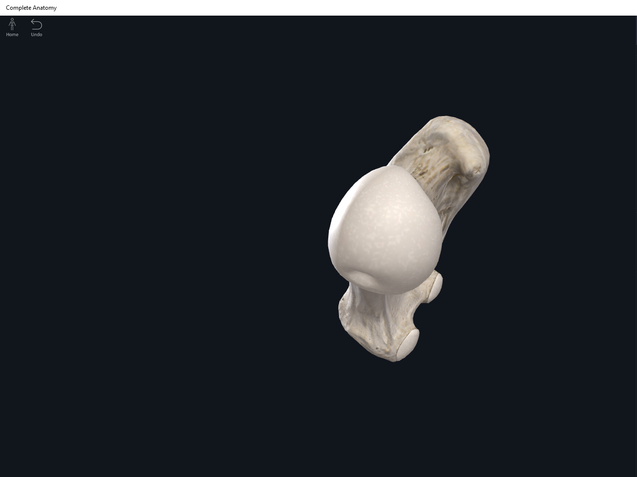
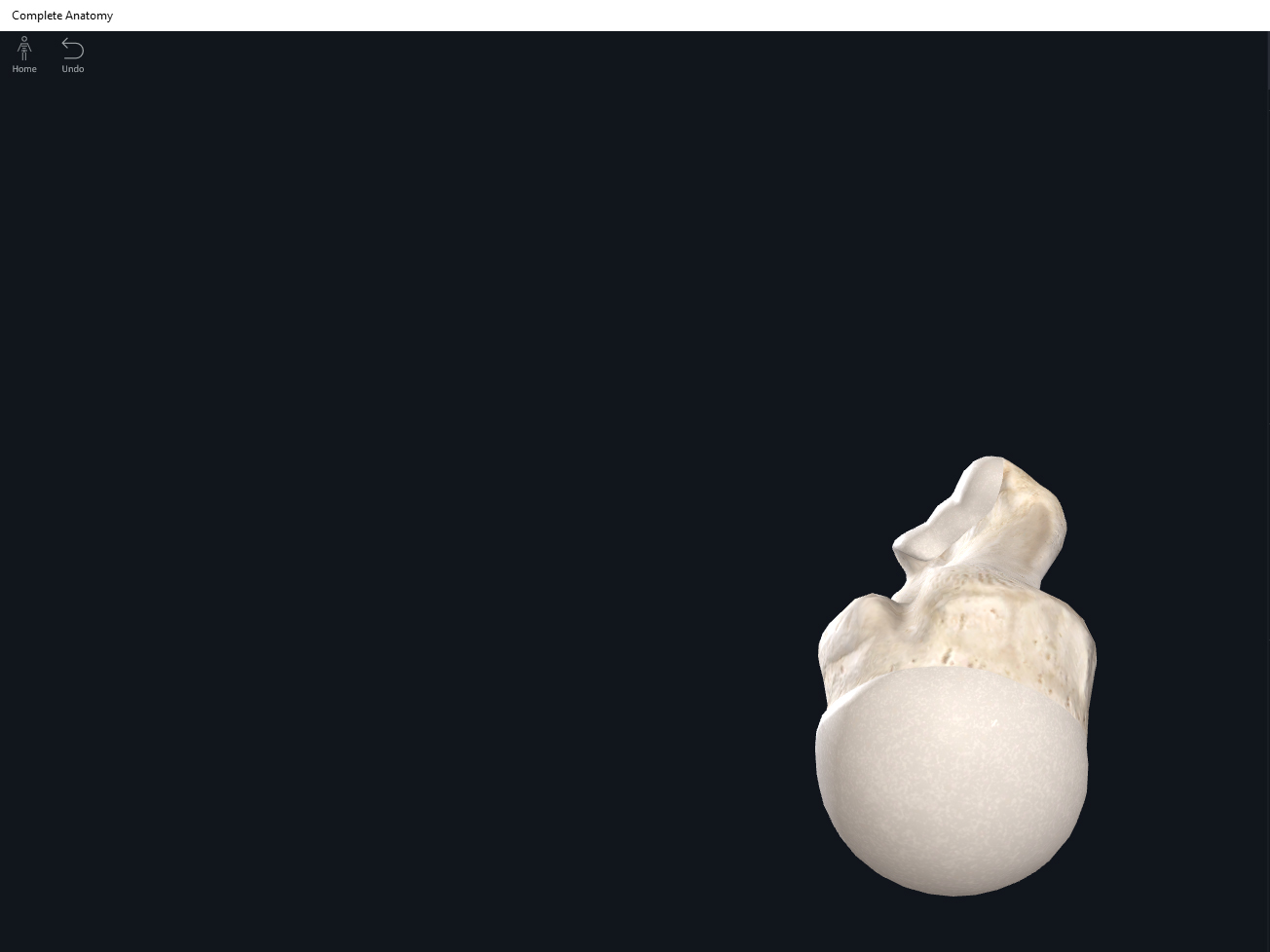
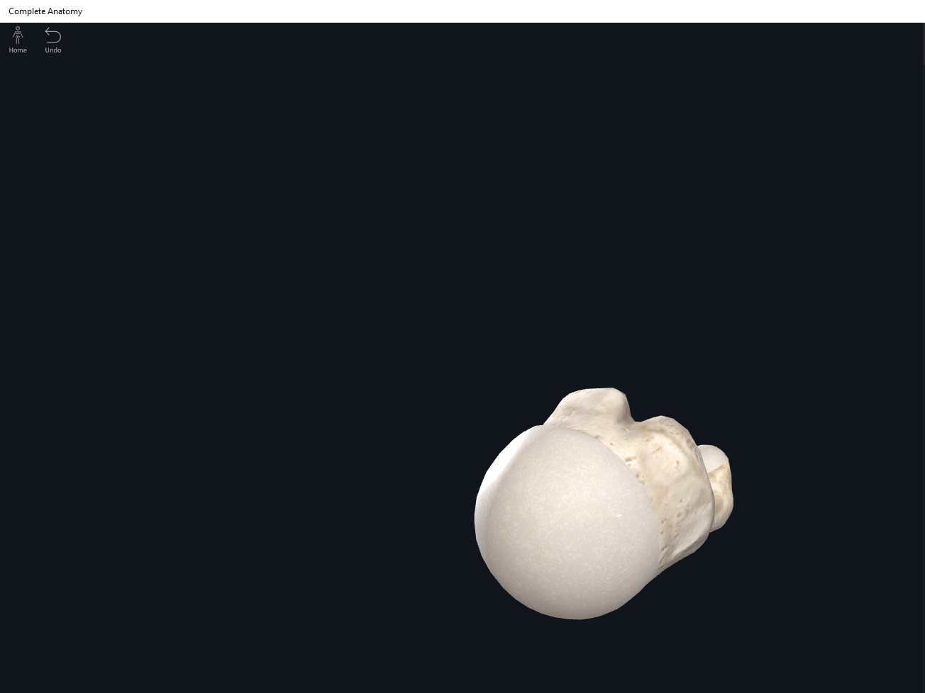
- Capitulum: “capit” means head; a rounded knob-like structure on the lateral side. Articulates with the radial head.
- Radial fossa: anterior depression superior to the capitulum. The radial head gets tucked into this depression when the elbow joint is bent. Most lateral fossa.
- Trochlea: medial to the capitulum. Articulates with the ulnar trochlear notch.
- Coronoid fossa: anterior depression that allows the coronoid process of the ulna to tuck into this depression when the elbow is bent. This fossa is medial to the radial fossa.
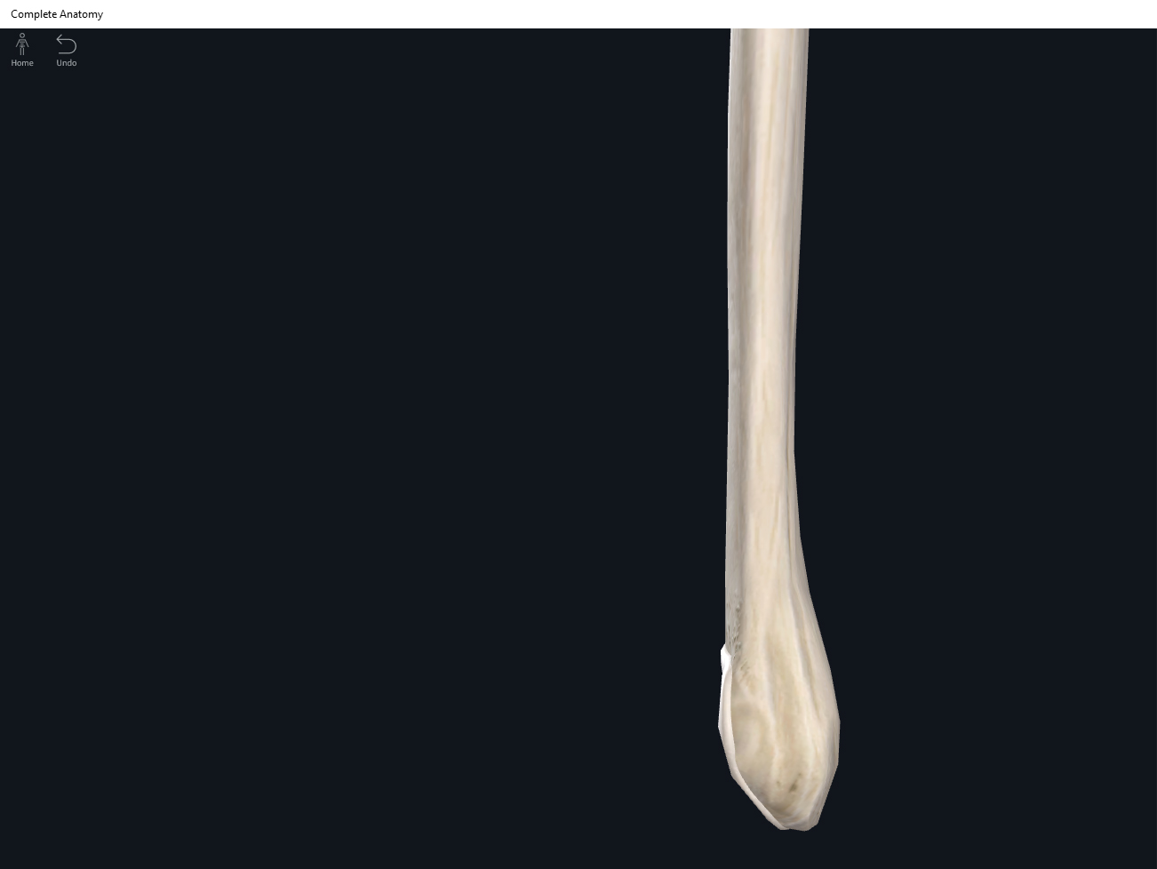
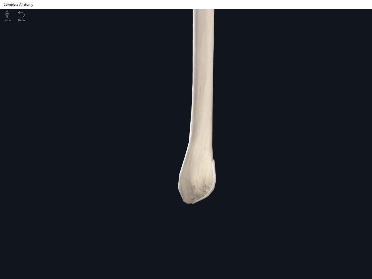
- Olecranon fossa: posterior deep depression that allows the olecranon of the ulna to “tuck” into when the elbow is extended.
- Medial epicondyle.
- Lateral epicondyle.
Function.
Clinical Significance.
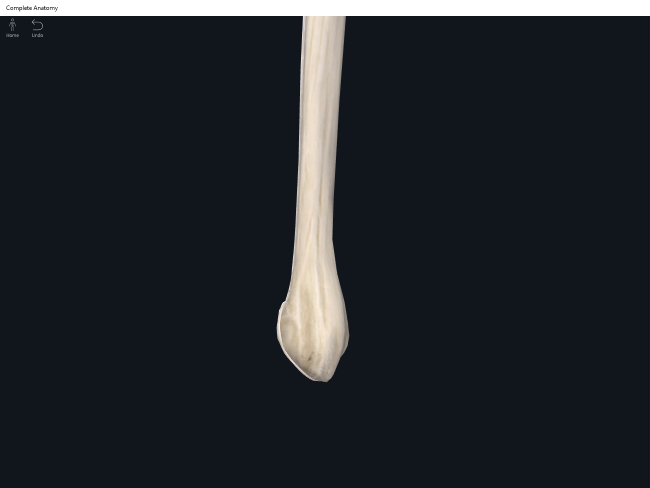
Humerus. Used with permission by 3D4Medical.


References
Biel, A. (2015). Trail guide to the body: A hands-on guide to locating muscles, bones and more.
Cedars-Sinai. (2018). Vertebrae of the spine. Retrieved from https://www.cedars-sinai.org/health-library/diseases-and-conditions/v/vertebrae-of-the-spine.html
Jenkins, G., & Tortora, G. J. (2012). Anatomy and Physiology: From Science to Life, 3rd Edition International Stu. John Wiley & Sons.
Muscolino, J. E. (2017). The muscular system manual: The skeletal muscles of the human body.