Posted on

Muscles: Palmar Interossei.

Download these notes.

Anatomy & Physiology: Muscles—Palmar Interossei.

Structure.

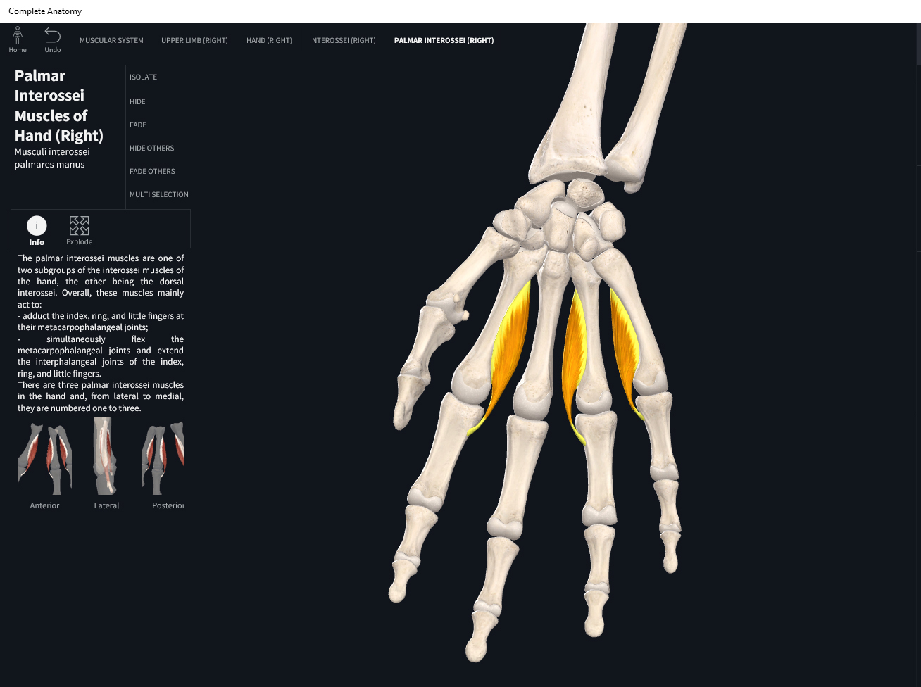
  • Origin: sides of shafts of metacarpals of all fingers (except middle finger).
  • Insertion: sides of bases of proximal phalanges of all fingers (except middle finger).

Function.

  • Concentric action: adduct fingers at metacarpophalangeal joints; flex fingers at metacarpophalangeal joints. Lesser: flexion and extension of fingers 2, 4, and 5 at MCP, and PIP and DIP respectively.
  • Reverse mover action: flex and adduct metacarpals of fingers 2, 4, and 5 at MCP; extend proximal phalanges of fingers 2, 4, and 5 at PIP and DIP.
  • Eccentric action: controls/restrains/slows abduction and extension of fingers 2, 4, and 5 at MCP; and flexion of fingers 2, 4, and 5 at PIP and DIP.
  • Isometric action: stabilizes MCP, PIP, and DIP of fingers 2, 4, and 5.
  • Innervation: ulnar nerve.
  • Arterial supply: branches of radial and ulnar arteries.

Clinical Significance.

More.

References

Biel, A. (2015). Trail guide to the body: A hands-on guide to locating muscles, bones and more.

Cedars-Sinai. (2018). Vertebrae of the spine. Retrieved from https://www.cedars-sinai.org/health-library/diseases-and-conditions/v/vertebrae-of-the-spine.html

Clark, M., Lucett, S., Sutton, B. G., & National Academy of Sports Medicine. (2014). NASM essentials of corrective exercise training. Burlington, MA: Jones & Bartlett Learning.

Jenkins, G., & Tortora, G. J. (2012). Anatomy and Physiology: From Science to Life, 3rd Edition International Stu. John Wiley & Sons.

Muscolino, J. E. (2017). The muscular system manual: The skeletal muscles of the human body.

Posted on

Muscles: Lumbricals Manus.

Download these notes.

Anatomy & Physiology: Muscles—Lumbricals.

Structure.

  • Origin: lateral sides of tendons and flexor digitorum profundus of each finger.
  • Insertion: lateral sides of tendons of extensor digitorum on proximal phalanges of each finger.

Function.

  • Concentric action: flexes each finger at metacarpophalangeal joints and extends fingers at interphalangeal joints. Lesser: abduct and adduct fingers II-V at MCP joints.
  • Reverse mover action: extend proximal phalanges at PIP and DIP; flex metacarpals at MCP; abduct/adduct metacarpals at MCP.
  • Eccentric action: controls/restrains/slows flexion, extension, adduction/abduction of fingers II-V at MCP.
  • Isometric action: stabilizes MCP and interphalangeal joints of fingers II-V.
  • Innervation: median and ulnar nerves.
  • Arterial supply: branches of radial and ulnar arteries.

Clinical Significance.

More.

References

Biel, A. (2015). Trail guide to the body: A hands-on guide to locating muscles, bones and more.

Cedars-Sinai. (2018). Vertebrae of the spine. Retrieved from https://www.cedars-sinai.org/health-library/diseases-and-conditions/v/vertebrae-of-the-spine.html

Clark, M., Lucett, S., Sutton, B. G., & National Academy of Sports Medicine. (2014). NASM essentials of corrective exercise training. Burlington, MA: Jones & Bartlett Learning.

Jenkins, G., & Tortora, G. J. (2012). Anatomy and Physiology: From Science to Life, 3rd Edition International Stu. John Wiley & Sons.

Muscolino, J. E. (2017). The muscular system manual: The skeletal muscles of the human body.

Posted on

Muscles: Adductor Pollicis.

Download these notes.

Anatomy & Physiology: Muscles—Adductor Pollicis.

Structure.

  • Origin: oblique head—capitate and metacarpals II and III; transverse head—metacarpal III.
  • Insertion: medial side of proximal phalanx of thumb by a tendon containing a sesamoid bone.

Function.

  • Concentric action: adducts thumb at carpometacarpal and metacarpophalangeal joints. Lesser: flexion and extension of thumb at MCP and IP joints respectively.
  • Reverse mover action: adducts, flexes trapezium at CMC; flex and extend metacarpal of thumb at MCP and IP respectively.
  • Eccentric action: controls/restrains/slows thumb abduction at CMC, extension of thumb at CMC and MCP; flexion at IP of thumb.
  • Isometric action: stabilizes CMC, MCP, and IP of thumb.
  • Innervation: ulnar nerves.
  • Arterial supply: branches of radial artery.

Clinical Significance.

More.

References

Biel, A. (2015). Trail guide to the body: A hands-on guide to locating muscles, bones and more.

Cedars-Sinai. (2018). Vertebrae of the spine. Retrieved from https://www.cedars-sinai.org/health-library/diseases-and-conditions/v/vertebrae-of-the-spine.html

Clark, M., Lucett, S., Sutton, B. G., & National Academy of Sports Medicine. (2014). NASM essentials of corrective exercise training. Burlington, MA: Jones & Bartlett Learning.

Jenkins, G., & Tortora, G. J. (2012). Anatomy and Physiology: From Science to Life, 3rd Edition International Stu. John Wiley & Sons.

Muscolino, J. E. (2017). The muscular system manual: The skeletal muscles of the human body.

Posted on

Muscles: Opponens Digiti Minimi.

Download these notes.

Anatomy & Physiology: Muscles—Opponens Digiti Minimi.

Structure.

  • Origin: flexor retinaculum and hamate.
  • Insertion: medial side of 5th metacarpal.

Function.

  • Concentric action: moves little finger across palm to touch the thumb. Lesser: flex little finger at CMC; lateral rotation of little finger at CMC; adduction of little finger at CMC.
  • Reverse mover action: flexion, medial rotation and adduction of hamate at 5th CMC.
  • Eccentric action: controls/restrains/slows little finger movements at CMC.
  • Isometric action: stabilize CMC of little finger.
  • Innervation: ulnar nerve.
  • Arterial supply: branches of ulnar artery.

Clinical Significance.

More.

References

Biel, A. (2015). Trail guide to the body: A hands-on guide to locating muscles, bones and more.

Cedars-Sinai. (2018). Vertebrae of the spine. Retrieved from https://www.cedars-sinai.org/health-library/diseases-and-conditions/v/vertebrae-of-the-spine.html

Clark, M., Lucett, S., Sutton, B. G., & National Academy of Sports Medicine. (2014). NASM essentials of corrective exercise training. Burlington, MA: Jones & Bartlett Learning.

Jenkins, G., & Tortora, G. J. (2012). Anatomy and Physiology: From Science to Life, 3rd Edition International Stu. John Wiley & Sons.

Muscolino, J. E. (2017). The muscular system manual: The skeletal muscles of the human body.

Posted on

Muscles: Flexor Digiti Minimi.

Download these notes.

Anatomy & Physiology: Muscles—Flexor Digiti Minimi (fingers).

Structure.

  • Origin: flexor retinaculum and hamate.
  • Insertion: medial side of proximal phalanx of little finger.

Function.

  • Concentric action: flexes pinky finger at carpometacarpal and metacarpophalangeal joints.
  • Reverse mover action: flexion of 5th metacarpal at MCP; distally glide the hamate at 5th CMC joint.
  • Eccentric action: controls/restrains/slows the CMC and MCP joints of little finger.
  • Isometric action: stabilize CMC and MCP of little finger.
  • Innervation: ulnar nerve.
  • Arterial supply: branches of ulnar artery.

Clinical Significance.

More.

References

Biel, A. (2015). Trail guide to the body: A hands-on guide to locating muscles, bones and more.

Cedars-Sinai. (2018). Vertebrae of the spine. Retrieved from https://www.cedars-sinai.org/health-library/diseases-and-conditions/v/vertebrae-of-the-spine.html

Clark, M., Lucett, S., Sutton, B. G., & National Academy of Sports Medicine. (2014). NASM essentials of corrective exercise training. Burlington, MA: Jones & Bartlett Learning.

Jenkins, G., & Tortora, G. J. (2012). Anatomy and Physiology: From Science to Life, 3rd Edition International Stu. John Wiley & Sons.

Muscolino, J. E. (2017). The muscular system manual: The skeletal muscles of the human body.

Posted on

Muscles: Abductor Digiti Minimi Brevis.

Download these notes.

Anatomy & Physiology: Muscles—Abductor Digiti Minimi (fingers).

Structure.

  • Origin: pisiform and tendon of flexor carpi ulnaris.
  • Insertion: medial side of proximal phalanx of littler finger.

Function.

  • Concentric action: abducts and flexes little finger at metacarpophalangeal joint.
  • Reverse mover action: abduct 5th metacarpal at MCP; abduct hamate at CMC of little finger; flex 5th metacarpal at MCO; distally glide the pisiform at CMC; extend proximal phalanx of little finger at PIP; extend middle phalanx of little finger at DIP.
  • Eccentric action: controls/restrains/slows little finger at MCP, CMC, DIP, and PIP joints.
  • Isometric action: stabilize little finger at MCP, CMC, DIP, PIP.
  • Innervation: ulnar nerve.
  • Arterial supply: branches of ulnar artery.

Clinical Significance.

More.

References

Biel, A. (2015). Trail guide to the body: A hands-on guide to locating muscles, bones and more.

Cedars-Sinai. (2018). Vertebrae of the spine. Retrieved from https://www.cedars-sinai.org/health-library/diseases-and-conditions/v/vertebrae-of-the-spine.html

Clark, M., Lucett, S., Sutton, B. G., & National Academy of Sports Medicine. (2014). NASM essentials of corrective exercise training. Burlington, MA: Jones & Bartlett Learning.

Jenkins, G., & Tortora, G. J. (2012). Anatomy and Physiology: From Science to Life, 3rd Edition International Stu. John Wiley & Sons.

Muscolino, J. E. (2017). The muscular system manual: The skeletal muscles of the human body.

Posted on

Muscles: Opponens Pollicis.

Download these notes.

Anatomy & Physiology: Muscles—Opponens Pollicis.

Structure.

  • Origin: flexor retinaculum, scaphoid, and trapezium.
  • Insertion: lateral side of first metacarpal.

Function.

  • Concentric action: move thumb across palm to little finger (bring thumb and little finger together).
  • Reverse mover action: flexion, lateral rotation, and abduction of trapezium.
  • Eccentric action: controls/restrains/slows thumb movements.
  • Isometric action: stabilizes thumb at CMC.
  • Innervation: median nerve.
  • Arterial supply: branches of radial artery.

Clinical Significance.

More.

References

Biel, A. (2015). Trail guide to the body: A hands-on guide to locating muscles, bones and more.

Cedars-Sinai. (2018). Vertebrae of the spine. Retrieved from https://www.cedars-sinai.org/health-library/diseases-and-conditions/v/vertebrae-of-the-spine.html

Clark, M., Lucett, S., Sutton, B. G., & National Academy of Sports Medicine. (2014). NASM essentials of corrective exercise training. Burlington, MA: Jones & Bartlett Learning.

Jenkins, G., & Tortora, G. J. (2012). Anatomy and Physiology: From Science to Life, 3rd Edition International Stu. John Wiley & Sons.

Muscolino, J. E. (2017). The muscular system manual: The skeletal muscles of the human body.

Posted on

Muscles: Flexor Pollicis Brevis.

Download these notes.

Anatomy & Physiology: Muscles—Flexor Pollicis Brevis.

Structure.

  • Origin: flexor retinaculum, trapezium, capitate, and trapezoid.
  • Insertion: lateral side of proximal phalanx of thumb.

Function.

  • Concentric action: flexes thumb at carpometacarpal and metacarpophalangeal joints; thumb abduction at CMC.
  • Reverse mover action: flexion of trapezium at CMC; flexion of thumb at MCP; abduction of trapezium at CMC.
  • Eccentric action: controls/restrains/slows thumb extension at CMC and MCP; adduction of thumb at CMC.
  • Isometric action: stabilizes thumb at CMC and MCP.
  • Innervation: median and ulnar nerves.
  • Arterial supply: branches of radial artery.

Clinical Significance.

More.

References

Biel, A. (2015). Trail guide to the body: A hands-on guide to locating muscles, bones and more.

Cedars-Sinai. (2018). Vertebrae of the spine. Retrieved from https://www.cedars-sinai.org/health-library/diseases-and-conditions/v/vertebrae-of-the-spine.html

Clark, M., Lucett, S., Sutton, B. G., & National Academy of Sports Medicine. (2014). NASM essentials of corrective exercise training. Burlington, MA: Jones & Bartlett Learning.

Jenkins, G., & Tortora, G. J. (2012). Anatomy and Physiology: From Science to Life, 3rd Edition International Stu. John Wiley & Sons.

Muscolino, J. E. (2017). The muscular system manual: The skeletal muscles of the human body.

Posted on

Muscles: Abductor Pollicis Brevis.

Download these notes.

Anatomy & Physiology: Muscles—Abductor Pollicis Brevis.

Structure.

  • Origin: flexor retinaculum, scaphoid, and trapezium.
  • Insertion: lateral side of proximal phalanx of thumb.

Function.

  • Concentric action: abducts thumb at carpometacarpal joint. Lesser: thumb flexion at MCO; thumb extension at CMC and IP.
  • Reverse mover action: abudction of trapezium at CMC; flexion of thumb at MCP; trapezium extension at CMC; extension of proximal phalanx of thumb at IP.
  • Eccentric action: controls/restrains/slows thumb adduction at CMC; thumb extension at MCP; flexion of thumb at CMC and IP.
  • Isometric action: stabilizes CMC, MCP, and IP joints of thumb.
  • Innervation: median nerve.
  • Arterial supply: radial artery.

Clinical Significance.

More.

References

Biel, A. (2015). Trail guide to the body: A hands-on guide to locating muscles, bones and more.

Cedars-Sinai. (2018). Vertebrae of the spine. Retrieved from https://www.cedars-sinai.org/health-library/diseases-and-conditions/v/vertebrae-of-the-spine.html

Clark, M., Lucett, S., Sutton, B. G., & National Academy of Sports Medicine. (2014). NASM essentials of corrective exercise training. Burlington, MA: Jones & Bartlett Learning.

Jenkins, G., & Tortora, G. J. (2012). Anatomy and Physiology: From Science to Life, 3rd Edition International Stu. John Wiley & Sons.

Muscolino, J. E. (2017). The muscular system manual: The skeletal muscles of the human body.

Posted on

Muscles: Extensor Indicis.

Download these notes.

Anatomy & Physiology: Muscles—Extensor Indicis.

Structure.

  • Origin: posterior surface of ulna.
  • Insertion: tendon of extensor digitorum of index finger.

Function.

  • One of the deep distal four group.
  • Concentric action: extends distal and middle phalanges of index finger at interphalangeal joints, proximal phalanx of index finger at metacarpophalangeal joint and hand at wrist joint. Lesser: adduction index finger at MCP; supination.
  • Reverse mover action: extension of 2nd metacarpal at MCP; extension of proximal phalanx at PIP; middle phalanx extension at DIP.
  • Eccentric action: controls/restrains/slows index finger flexion at MCP, DIP, PIP; wrist flexion; abduction of index finger; pronation.
  • Isometric action: stabilize MCP, DIP, PIP of index finger; wrist; radioulnar joints.
  • Innervation: deep radial nerve.
  • Arterial supply: posterior interosseus artery; perforating branches of anterior interosseus artery.

Clinical Significance.


References

Biel, A. (2015). Trail guide to the body: A hands-on guide to locating muscles, bones and more.

Cedars-Sinai. (2018). Vertebrae of the spine. Retrieved from https://www.cedars-sinai.org/health-library/diseases-and-conditions/v/vertebrae-of-the-spine.html

Clark, M., Lucett, S., Sutton, B. G., & National Academy of Sports Medicine. (2014). NASM essentials of corrective exercise training. Burlington, MA: Jones & Bartlett Learning.

Jenkins, G., & Tortora, G. J. (2012). Anatomy and Physiology: From Science to Life, 3rd Edition International Stu. John Wiley & Sons.

Muscolino, J. E. (2017). The muscular system manual: The skeletal muscles of the human body.