Posted on

Muscles: Flexor Carpi Ulnaris.

Download these notes.

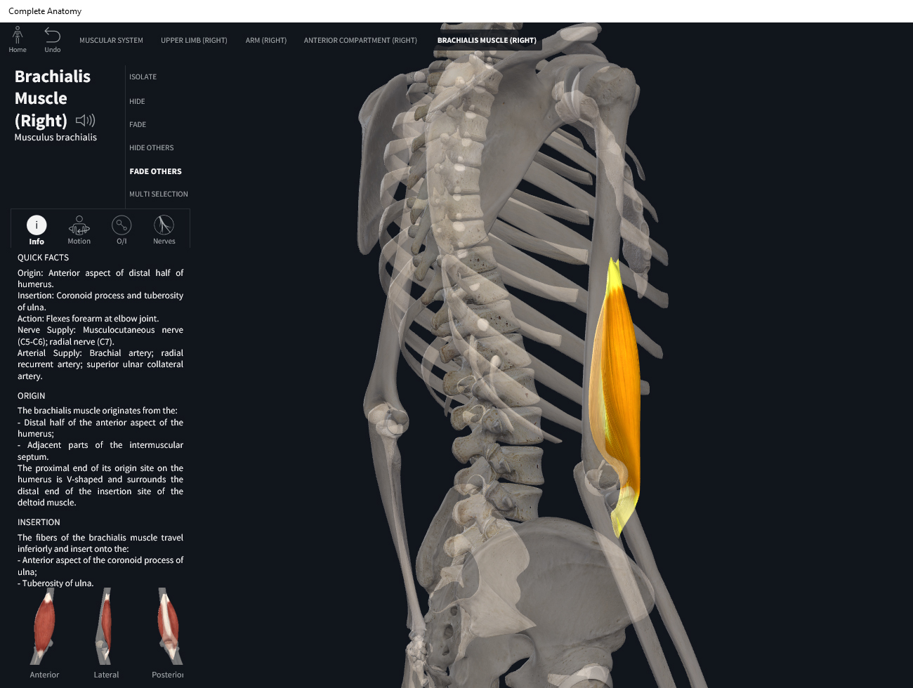
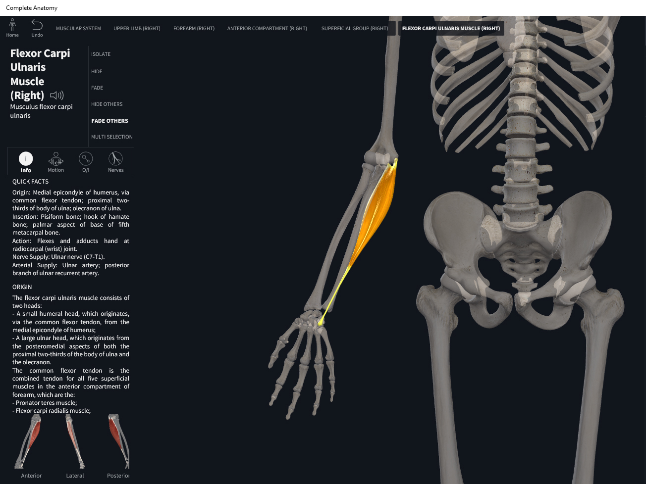
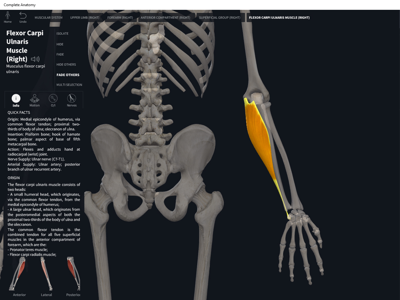
Anatomy & Physiology: Muscles—Flexor Carpi Ulnaris.

Structure.

  • Origin: medial epicondyle of humerus and superior posterior border of ulna.
  • Insertion: pisiform, hamate, and base of fifth metacarpal.

Function.

  • Concentric action: flexes and adducts hand (ulnar deviation) at wrist. Lesser: elbow flexion.
  • Reverse mover action: flexion and deviations at wrist joint; elbow flexion.
  • Eccentric action: controls/restrains/slows wrist extension and radial deviation, elbow extension.
  • Isometric action: stabilize wrist and elbow.
  • Innervation: ulnar nerve.
  • Arterial supply: ulnar artery.

Clinical Significance.


References

Biel, A. (2015). Trail guide to the body: A hands-on guide to locating muscles, bones and more.

Cedars-Sinai. (2018). Vertebrae of the spine. Retrieved from https://www.cedars-sinai.org/health-library/diseases-and-conditions/v/vertebrae-of-the-spine.html

Clark, M., Lucett, S., Sutton, B. G., & National Academy of Sports Medicine. (2014). NASM essentials of corrective exercise training. Burlington, MA: Jones & Bartlett Learning.

Jenkins, G., & Tortora, G. J. (2012). Anatomy and Physiology: From Science to Life, 3rd Edition International Stu. John Wiley & Sons.

Muscolino, J. E. (2017). The muscular system manual: The skeletal muscles of the human body.

Posted on

Muscles: Palmaris Longus.

Download these notes.

Anatomy & Physiology: Muscles—Palmaris Longus.

Structure.

  • Origin: medial epicondyle of humerus.
  • Insertion: flexor retinaculum and palmar aponeurosis (fascia in center of palm).

Function.

  • Concentric action: weakly flexes hand at wrist joint. Lesser: wrinkles skin of palm; elbow flexion; pronation; radial deviation of hand; ulnar deviation of hand.
  • Reverse mover action: forearm flexion at wrist; elbow flexion; pronation; radial and ulnar deviation at wrist.
  • Eccentric action: controls/restrains/slows wrist extension, wrist ulnar and radial deviation, elbow extension, supination.
  • Isometric action: stabilize wrist and elbow joints.
  • Innervation: median nerve.
  • Arterial supply: ulnar artery.

Clinical Significance.

References

Biel, A. (2015). Trail guide to the body: A hands-on guide to locating muscles, bones and more.

Cedars-Sinai. (2018). Vertebrae of the spine. Retrieved from https://www.cedars-sinai.org/health-library/diseases-and-conditions/v/vertebrae-of-the-spine.html

Clark, M., Lucett, S., Sutton, B. G., & National Academy of Sports Medicine. (2014). NASM essentials of corrective exercise training. Burlington, MA: Jones & Bartlett Learning.

Jenkins, G., & Tortora, G. J. (2012). Anatomy and Physiology: From Science to Life, 3rd Edition International Stu. John Wiley & Sons.

Muscolino, J. E. (2017). The muscular system manual: The skeletal muscles of the human body.

Posted on

Muscles: Flexor Carpi Radialis.

Download these notes.

Anatomy & Physiology: Muscles—Flexor Carpi Radialis.

Structure.

  • Origin: medial epicondyle of humerus.
  • Insertion: second and third metacarpals.

Function.

  • Concentric action: flexes and abducts hand (radial deviation) at wrist joint. Lesser: elbow flexion, forearm pronation.
  • Reverse mover action: forearm flexion, radial deviation at wrist joint, elbow flexion, pronation, lateral rotation at shoulder joint.
  • Eccentric action: controls/restrains/slows wrist extension, ulnar deviation, elbow extension, supination.
  • Isometric action: stabilize wrist, elbow, and radioulnar joint.
  • Innervation: median nerve.
  • Arterial supply: ulnar and radial arteries (branch of brachial artery).

Clinical Significance.


References

Biel, A. (2015). Trail guide to the body: A hands-on guide to locating muscles, bones and more.

Cedars-Sinai. (2018). Vertebrae of the spine. Retrieved from https://www.cedars-sinai.org/health-library/diseases-and-conditions/v/vertebrae-of-the-spine.html

Clark, M., Lucett, S., Sutton, B. G., & National Academy of Sports Medicine. (2014). NASM essentials of corrective exercise training. Burlington, MA: Jones & Bartlett Learning.

Jenkins, G., & Tortora, G. J. (2012). Anatomy and Physiology: From Science to Life, 3rd Edition International Stu. John Wiley & Sons.

Muscolino, J. E. (2017). The muscular system manual: The skeletal muscles of the human body.

Posted on

Muscles: Supinator.

Download these notes.

Anatomy & Physiology: Muscles—Supinator.

Structure.

  • Origin: lateral epicondyle of humerus, and ridge near radial notch of ulna (supinator crest).
  • Insertion: lateral surface of proximal 1/3 of radius.

Function.

  • Concentric action: forearm supination. Lesser degree: elbow flexion.
  • Reverse muscle action: supination; arm flexion; medial rotation of arm at glenohumeral joint.
  • Eccentric action: forearm pronation (lesser degree: elbow extension).
  • Isometric action: stabilization of the proximal radioulnar joint and elbow.
  • Innervation: deep radial nerve.
  • Arterial supply: branches off radial artery; interosseus recurrent and posterior interosseus arteries.

Clinical Significance.


References

Biel, A. (2015). Trail guide to the body: A hands-on guide to locating muscles, bones and more.

Cedars-Sinai. (2018). Vertebrae of the spine. Retrieved from https://www.cedars-sinai.org/health-library/diseases-and-conditions/v/vertebrae-of-the-spine.html

Clark, M., Lucett, S., Sutton, B. G., & National Academy of Sports Medicine. (2014). NASM essentials of corrective exercise training. Burlington, MA: Jones & Bartlett Learning.

Jenkins, G., & Tortora, G. J. (2012). Anatomy and Physiology: From Science to Life, 3rd Edition International Stu. John Wiley & Sons.

Muscolino, J. E. (2017). The muscular system manual: The skeletal muscles of the human body.

Posted on

Muscles: Pronator Quadratus.

Download these notes.

Anatomy & Physiology: Muscles—Pronator Quadratus.

Structure.

  • Origin: distal portion of ulnar shaft.
  • Insertion: distal portion of radial shaft.

Function.

  • Concentric action: pronates forearm.
  • Reverse mover action: pronates forearm; lateral rotation of arm at glenohumeral joint.
  • Eccentric action: supination.
  • Isometric action: stabilization of the distal radioulnar joint.
  • Innervation: median nerve.
  • Arterial supply: anterior interosseus artery (branch of ulnar artery).

Clinical Significance.


References

Biel, A. (2015). Trail guide to the body: A hands-on guide to locating muscles, bones and more.

Cedars-Sinai. (2018). Vertebrae of the spine. Retrieved from https://www.cedars-sinai.org/health-library/diseases-and-conditions/v/vertebrae-of-the-spine.html

Clark, M., Lucett, S., Sutton, B. G., & National Academy of Sports Medicine. (2014). NASM essentials of corrective exercise training. Burlington, MA: Jones & Bartlett Learning.

Jenkins, G., & Tortora, G. J. (2012). Anatomy and Physiology: From Science to Life, 3rd Edition International Stu. John Wiley & Sons.

Muscolino, J. E. (2017). The muscular system manual: The skeletal muscles of the human body.

Posted on

Muscles: Pronator Teres.

Download these notes.

Anatomy & Physiology: Muscles—Pronator Teres.

Structure.

  • Origin: medial epicondyle of humerus, and coronoid process of ulna.
  • Insertion: midlateral surface of radius.

Function.

  • Concentric action: pronates forearm at radioulnar joints and weakly flexes forearm at elbow joint.
  • Reverse mover action: pronation of forearm; elbow flexion.
  • Eccentric action: controls/restrains/slows elbow supination and elbow extension.
  • Isometric action: stabilization of the elbow.
  • Innervation: median nerve.
  • Arterial supply: ulnar artery, radial artery.

Clinical Significance.


References

Biel, A. (2015). Trail guide to the body: A hands-on guide to locating muscles, bones and more.

Cedars-Sinai. (2018). Vertebrae of the spine. Retrieved from https://www.cedars-sinai.org/health-library/diseases-and-conditions/v/vertebrae-of-the-spine.html

Clark, M., Lucett, S., Sutton, B. G., & National Academy of Sports Medicine. (2014). NASM essentials of corrective exercise training. Burlington, MA: Jones & Bartlett Learning.

Jenkins, G., & Tortora, G. J. (2012). Anatomy and Physiology: From Science to Life, 3rd Edition International Stu. John Wiley & Sons.

Muscolino, J. E. (2017). The muscular system manual: The skeletal muscles of the human body.

Posted on

Muscles: Anconeus.

Download these notes.

Anatomy & Physiology: Muscles—Aconeus.

Structure.

  • Origin: lateral epicondyle of humerus.
  • Insertion: olecranon and superior portion of ulnar shaft.

Function.

  • Concentric action: elbow extension.
  • Reverse mover action: elbow extension.
  • Eccentric action: elbow flexion.
  • Isometric action: stabilization of the elbow.
  • Innervation: radial nerve.
  • Arterial supply: deep brachial artery.

Clinical Significance.

References

Biel, A. (2015). Trail guide to the body: A hands-on guide to locating muscles, bones and more.

Cedars-Sinai. (2018). Vertebrae of the spine. Retrieved from https://www.cedars-sinai.org/health-library/diseases-and-conditions/v/vertebrae-of-the-spine.html

Clark, M., Lucett, S., Sutton, B. G., & National Academy of Sports Medicine. (2014). NASM essentials of corrective exercise training. Burlington, MA: Jones & Bartlett Learning.

Jenkins, G., & Tortora, G. J. (2012). Anatomy and Physiology: From Science to Life, 3rd Edition International Stu. John Wiley & Sons.

Muscolino, J. E. (2017). The muscular system manual: The skeletal muscles of the human body.

Posted on

Muscles: Triceps Brachii.

Download these notes.

Anatomy & Physiology: Muscles—Triceps Brachii.

Structure.

  • Origin: long head—infraglenoid tubercle of scapula, a projection inferior to glenoid cavity; lateral head—lateral and posterior surfaces of humerus superior to radial groove; medial head—posterior surface of humerus inferior to groove for radial nerve.
  • Insertion: olecranon process of ulna.

Function.

  • Concentric action: elbow extension; shoulder extension. Adduction of shoulder (long head); shoulder extension (glenohumeral joint).
  • Reverse mover action: arm extension at elbow; scapular downward rotation; protraction and lateral tilt of scapula.
  • Eccentric action: controls/restrains/slows elbow flexion, shoulder flexion, shoulder abduction, horizontal flexion, scapular upward rotation, scapular retraction, scpular medial tilt.
  • Isometric action: stabilization of the elbow and shoulder girdle.
  • Innervation: radial nerve.
  • Arterial supply: deep brachial artery; circumflex scapular artery.

Clinical Significance.


References

Biel, A. (2015). Trail guide to the body: A hands-on guide to locating muscles, bones and more.

Cedars-Sinai. (2018). Vertebrae of the spine. Retrieved from https://www.cedars-sinai.org/health-library/diseases-and-conditions/v/vertebrae-of-the-spine.html

Clark, M., Lucett, S., Sutton, B. G., & National Academy of Sports Medicine. (2014). NASM essentials of corrective exercise training. Burlington, MA: Jones & Bartlett Learning.

Jenkins, G., & Tortora, G. J. (2012). Anatomy and Physiology: From Science to Life, 3rd Edition International Stu. John Wiley & Sons.

Muscolino, J. E. (2017). The muscular system manual: The skeletal muscles of the human body.

Posted on

Muscles: Brachioradialis.

Download these notes.

Anatomy & Physiology: Muscles—Brachioradialis.

Structure.

  • Origin: lateral border of distal end of humerus.
  • Insertion: superior to styloid process of radius.

Function.

  • Concentric action: elbow flexion; supinates and pronates forearm at radioulnar joints to neutral position.
  • Reverse mover action: arm flexion at elbow; supination; pronation; medial rotation of arm at glenohumeral joint.
  • Eccentric action: elbow extension.
  • Isometric action: stabilization of the elbow.
  • Innervation: radial nerve.
  • Arterial supply: branches of brachial artery; radial artery.

Clinical Significance.


References

Biel, A. (2015). Trail guide to the body: A hands-on guide to locating muscles, bones and more.

Cedars-Sinai. (2018). Vertebrae of the spine. Retrieved from https://www.cedars-sinai.org/health-library/diseases-and-conditions/v/vertebrae-of-the-spine.html

Clark, M., Lucett, S., Sutton, B. G., & National Academy of Sports Medicine. (2014). NASM essentials of corrective exercise training. Burlington, MA: Jones & Bartlett Learning.

Jenkins, G., & Tortora, G. J. (2012). Anatomy and Physiology: From Science to Life, 3rd Edition International Stu. John Wiley & Sons.

Muscolino, J. E. (2017). The muscular system manual: The skeletal muscles of the human body.

Posted on

Muscles: Brachialis.

Download these notes.

Anatomy & Physiology: Muscles—Brachialis.

Structure.

  • Origin: distal, anterior surface of humerus.
  • Insertion: ulnar tuberosity and coronoid process of ulna.

Function.

  • Concentric action: elbow flexion.
  • Reverse mover action: elbow flexion.
  • Eccentric action: elbow extension.
  • Isometric action: stabilization of the elbow.
  • Innervation: musculocutaneous and radial nerve.
  • Arterial supply: brachial artery.

Clinical Significance.


References

Biel, A. (2015). Trail guide to the body: A hands-on guide to locating muscles, bones and more.

Cedars-Sinai. (2018). Vertebrae of the spine. Retrieved from https://www.cedars-sinai.org/health-library/diseases-and-conditions/v/vertebrae-of-the-spine.html

Clark, M., Lucett, S., Sutton, B. G., & National Academy of Sports Medicine. (2014). NASM essentials of corrective exercise training. Burlington, MA: Jones & Bartlett Learning.

Jenkins, G., & Tortora, G. J. (2012). Anatomy and Physiology: From Science to Life, 3rd Edition International Stu. John Wiley & Sons.

Muscolino, J. E. (2017). The muscular system manual: The skeletal muscles of the human body.