Anatomy & Physiology: Muscles—Trapezius, Upper.
Structure.
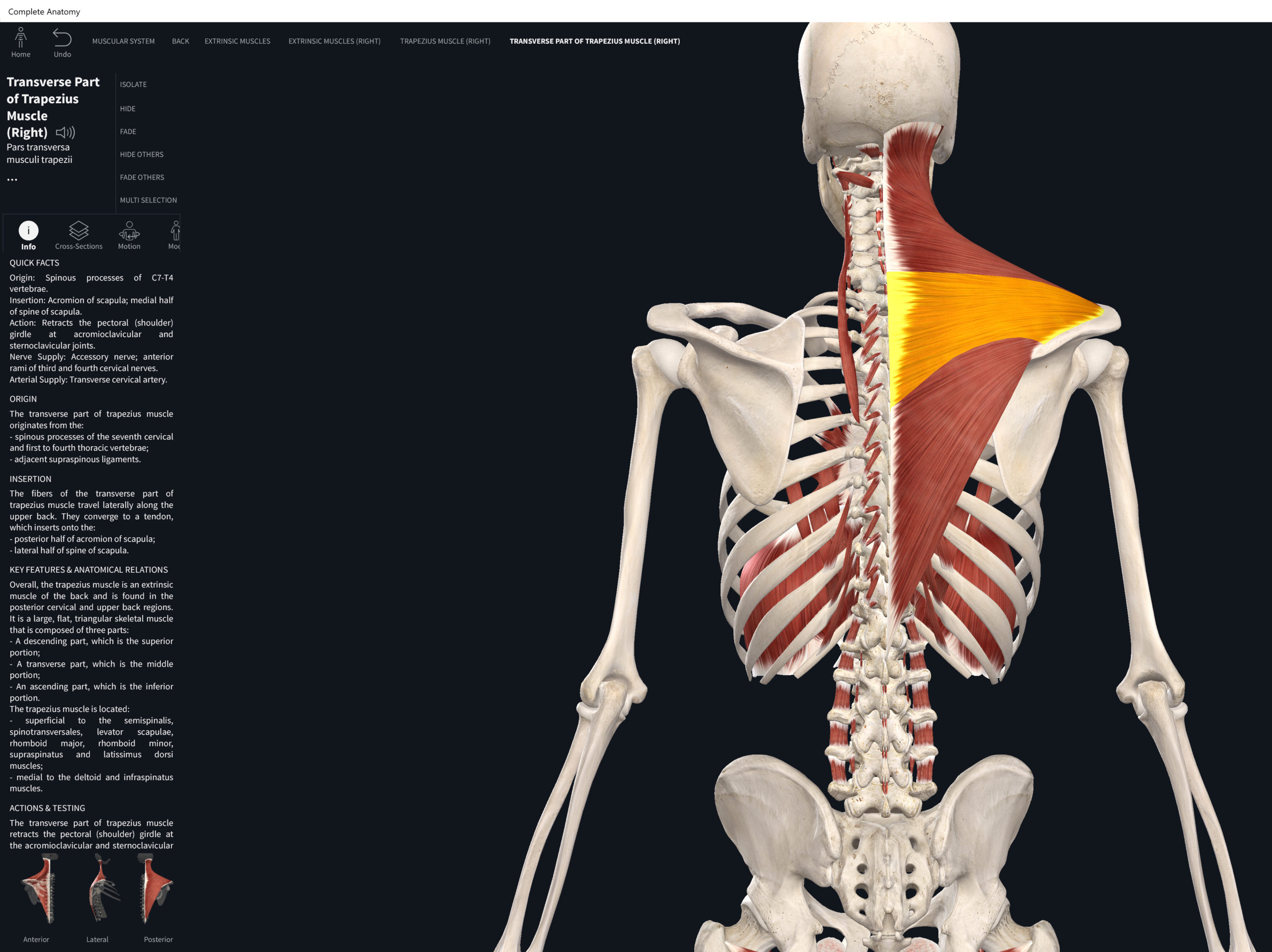
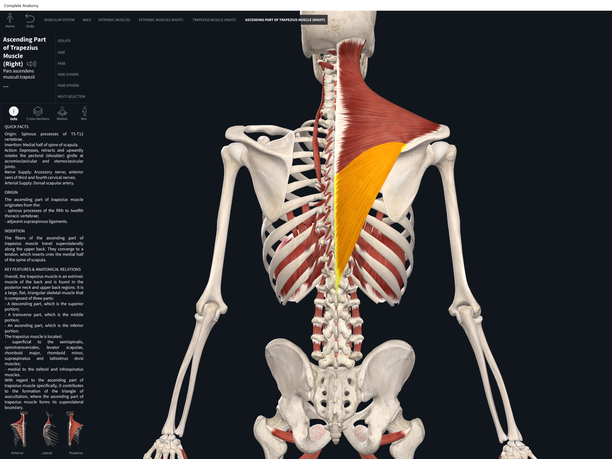
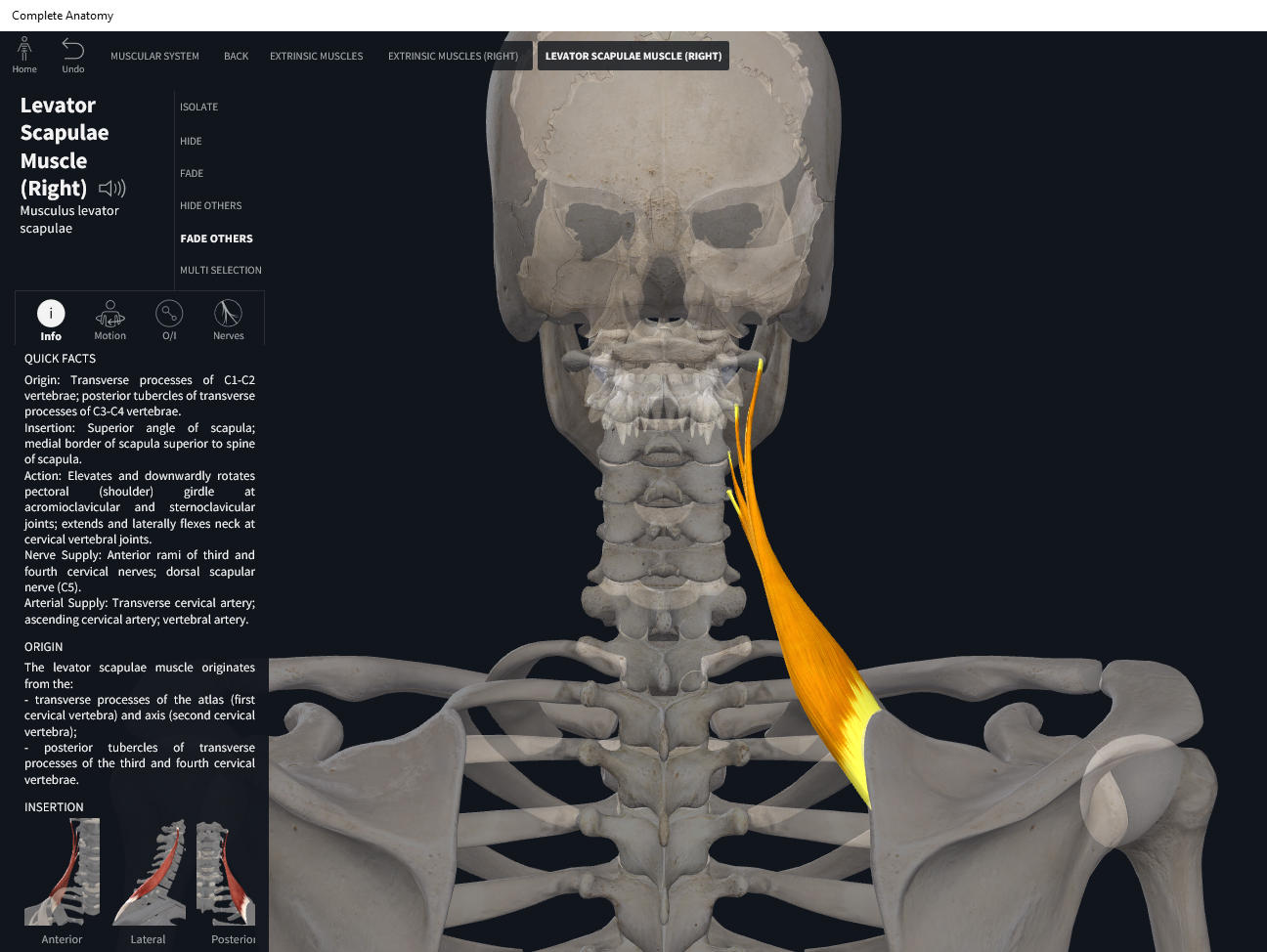
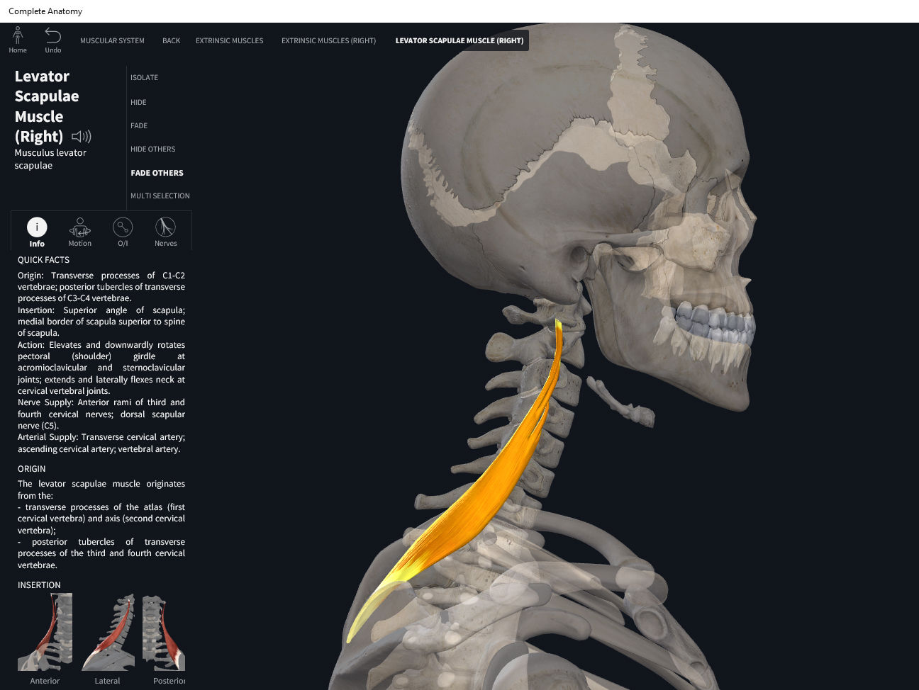
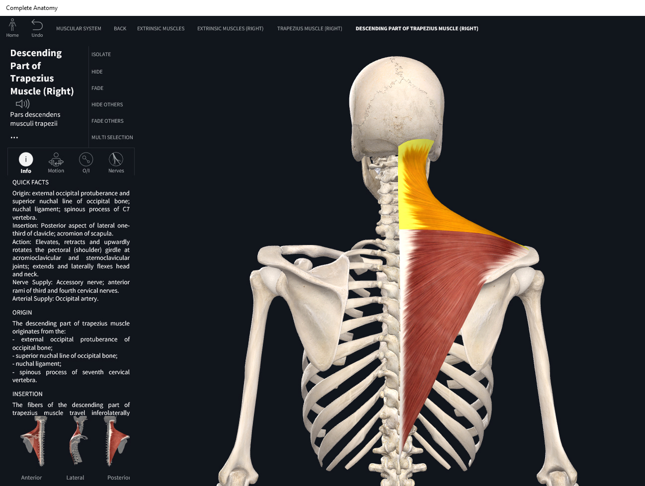
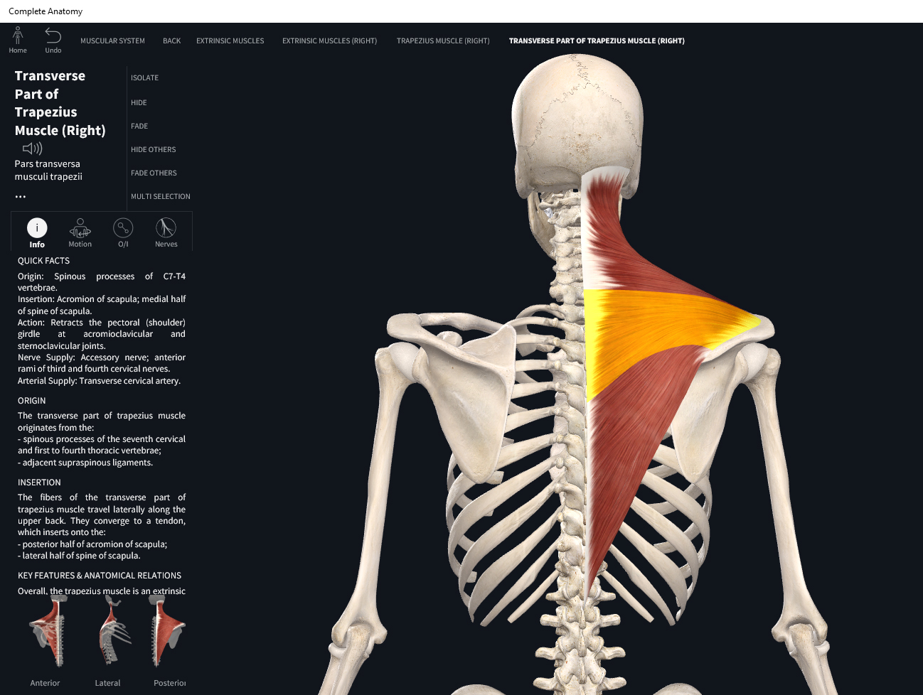
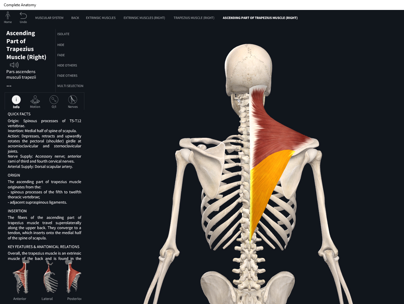
- Origin: superior nuchal line, ligamentum nuchae; spinous process of C7, T1-T12.
- Insertion: lateral 1/3 of clavicle; acromion process of scapula.
Function.
- Concentric action: cervical extension, lateral flexion and rotation; scapular elevation at scapulocostal joint (ScC); scapular retraction at ScC; upward rotation of the scapula at the ScC.
- Reverse mover actions: head and neck extension at spinal joints; contralateral rotation of the head and neck at spinal joints; lateral flexion of the head and neck at spinal joints.
- Eccentric action: cervical flexion, lateral flexion, and rotation; scapular depression. Controls/restrains/slows scapular protraction at ScC and ipsilateral rotation of the trunk at the spinal joints.
- Isometric action: stabilization of the medial border of scapula and cervical spine.
- Innervation: accessory XI nerve; cervical spinal nerves C3-C5.
- Arterial supply: transverse cervical artery (branch from thyrocervical trunk), dorsal scapular artery (branch from subclavian artery).
Clinical Significance.

References
Biel, A. (2015). Trail guide to the body: A hands-on guide to locating muscles, bones and more.
Cedars-Sinai. (2018). Vertebrae of the spine. Retrieved from https://www.cedars-sinai.org/health-library/diseases-and-conditions/v/vertebrae-of-the-spine.html
Clark, M., Lucett, S., Sutton, B. G., & National Academy of Sports Medicine. (2014). NASM essentials of corrective exercise training. Burlington, MA: Jones & Bartlett Learning.
Jenkins, G., & Tortora, G. J. (2012). Anatomy and Physiology: From Science to Life, 3rd Edition International Stu. John Wiley & Sons.
Muscolino, J. E. (2017). The muscular system manual: The skeletal muscles of the human body.