Posted on

Muscles: Triceps Brachii.

Download these notes.

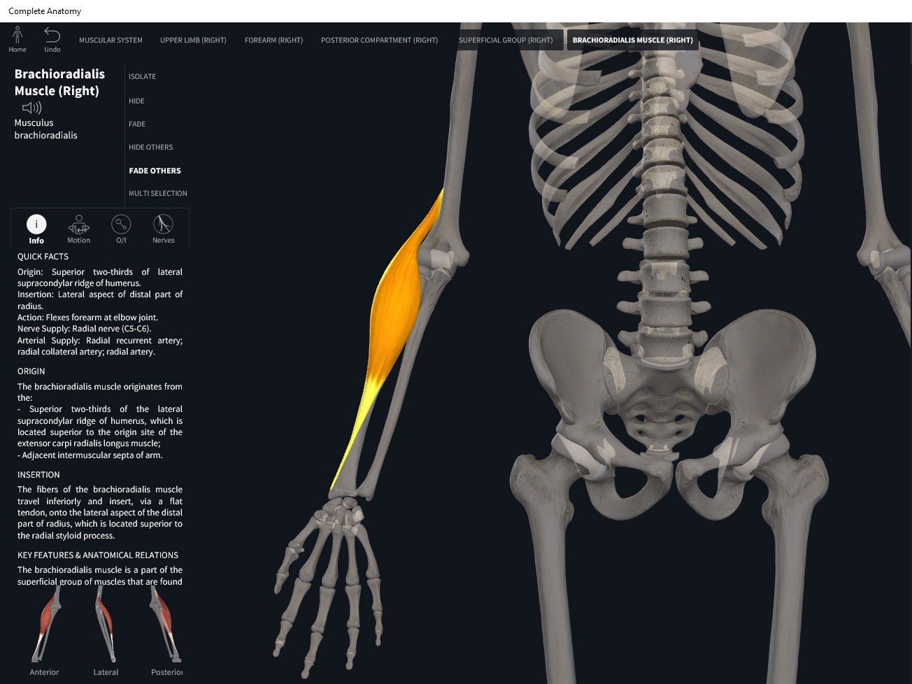
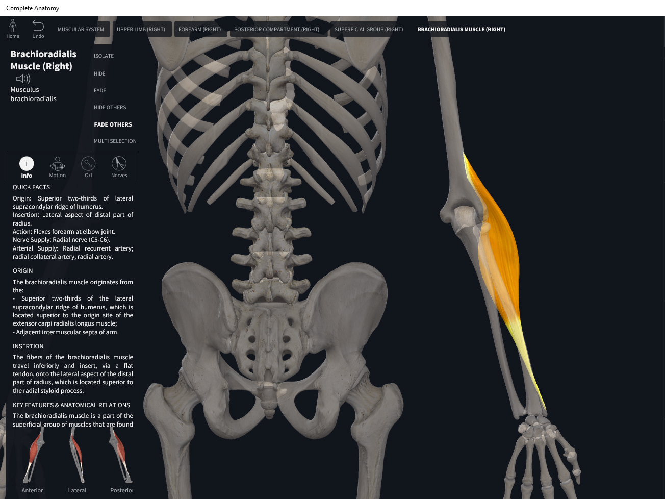
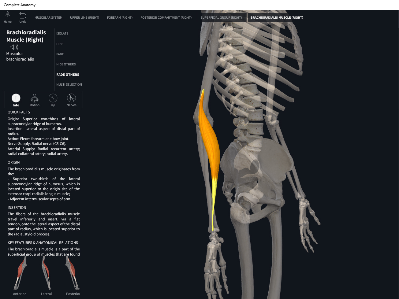
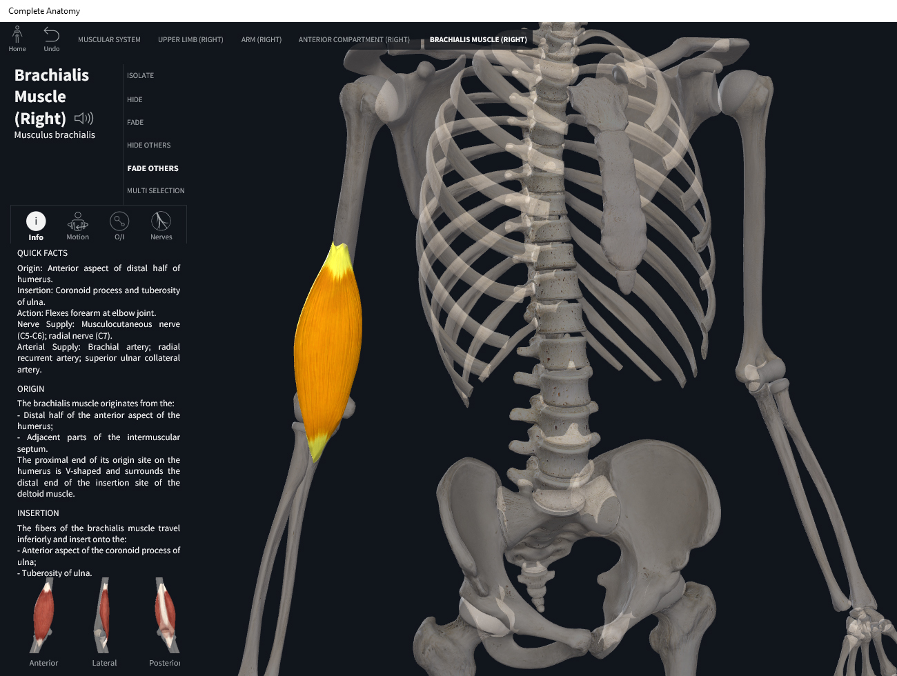
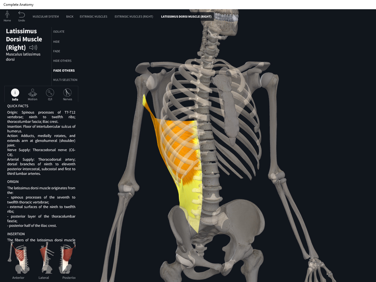
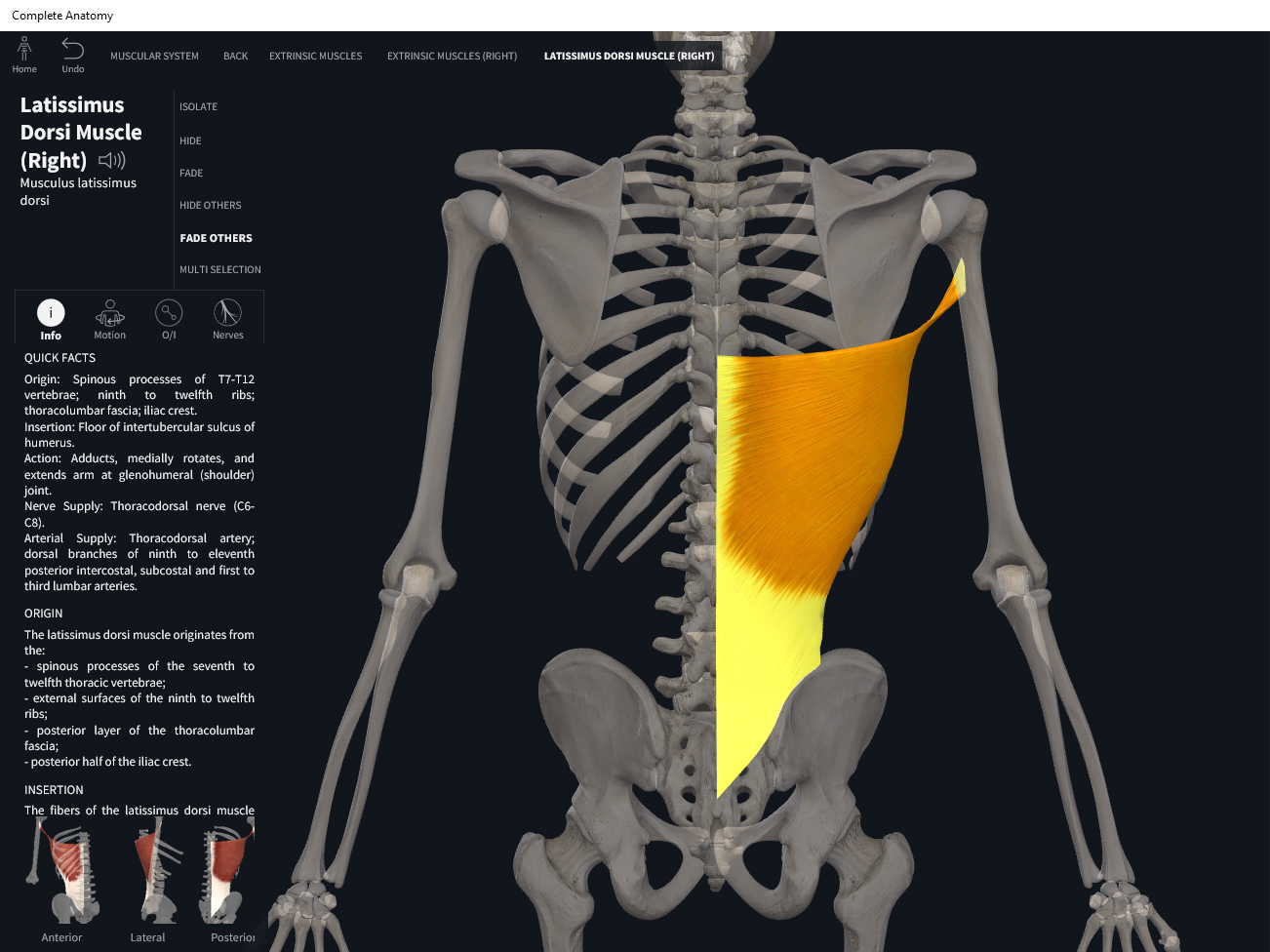
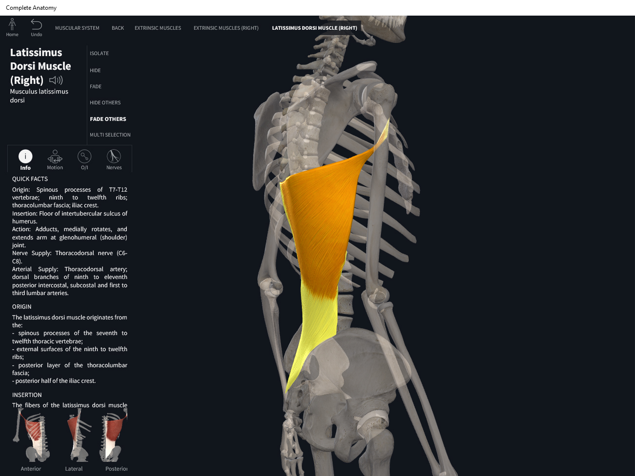
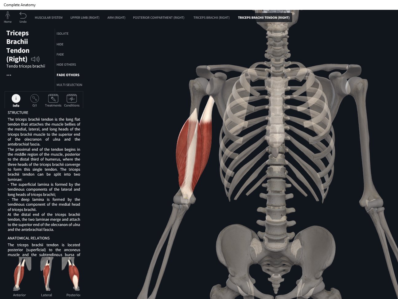
Anatomy & Physiology: Muscles—Triceps Brachii.

Structure.

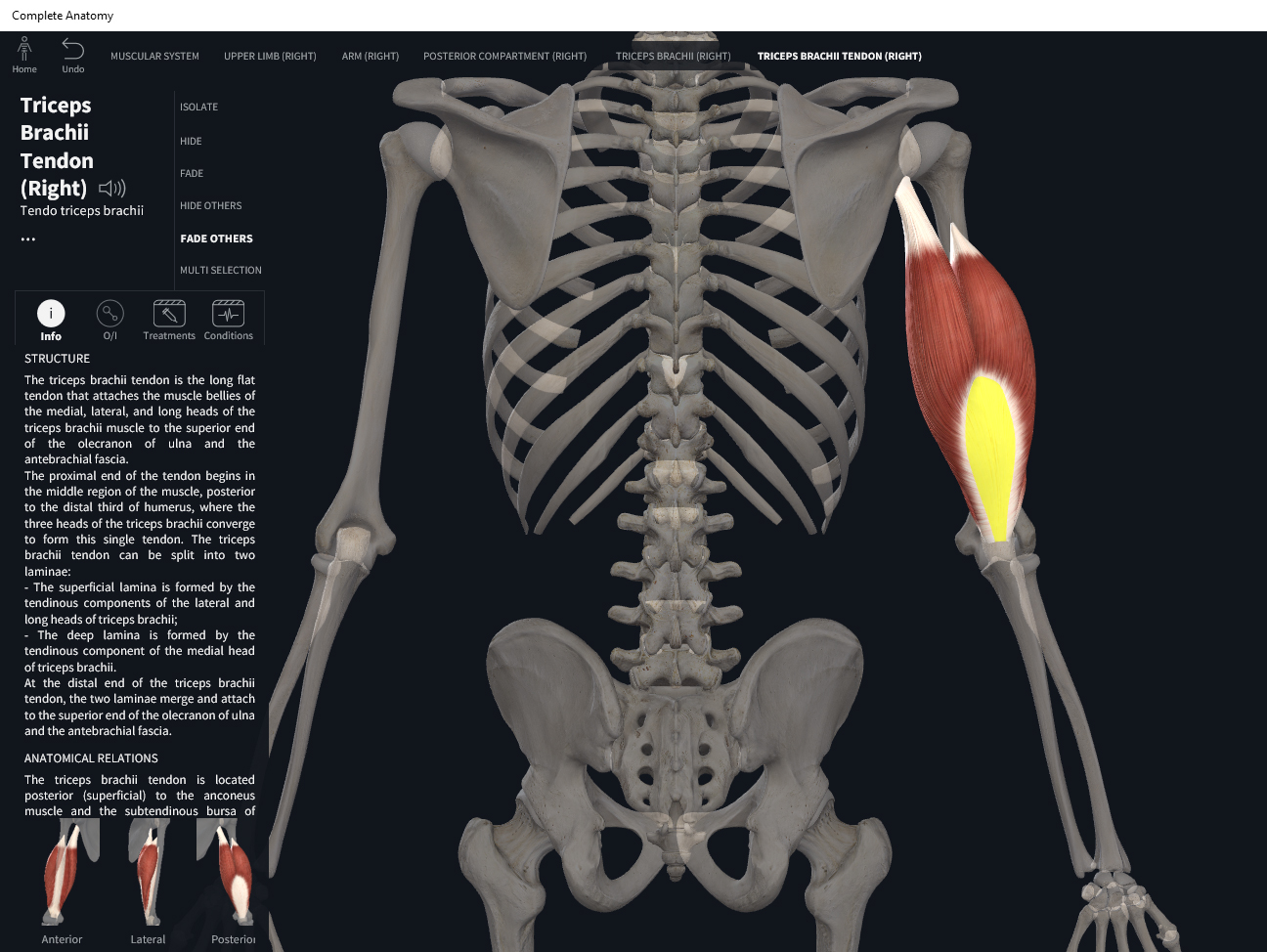
  • Origin: long head—infraglenoid tubercle of scapula, a projection inferior to glenoid cavity; lateral head—lateral and posterior surfaces of humerus superior to radial groove; medial head—posterior surface of humerus inferior to groove for radial nerve.
  • Insertion: olecranon process of ulna.

Function.

  • Concentric action: elbow extension; shoulder extension. Adduction of shoulder (long head); shoulder extension (glenohumeral joint).
  • Reverse mover action: arm extension at elbow; scapular downward rotation; protraction and lateral tilt of scapula.
  • Eccentric action: controls/restrains/slows elbow flexion, shoulder flexion, shoulder abduction, horizontal flexion, scapular upward rotation, scapular retraction, scpular medial tilt.
  • Isometric action: stabilization of the elbow and shoulder girdle.
  • Innervation: radial nerve.
  • Arterial supply: deep brachial artery; circumflex scapular artery.

Clinical Significance.


References

Biel, A. (2015). Trail guide to the body: A hands-on guide to locating muscles, bones and more.

Cedars-Sinai. (2018). Vertebrae of the spine. Retrieved from https://www.cedars-sinai.org/health-library/diseases-and-conditions/v/vertebrae-of-the-spine.html

Clark, M., Lucett, S., Sutton, B. G., & National Academy of Sports Medicine. (2014). NASM essentials of corrective exercise training. Burlington, MA: Jones & Bartlett Learning.

Jenkins, G., & Tortora, G. J. (2012). Anatomy and Physiology: From Science to Life, 3rd Edition International Stu. John Wiley & Sons.

Muscolino, J. E. (2017). The muscular system manual: The skeletal muscles of the human body.

Posted on

Muscles: Brachioradialis.

Download these notes.

Anatomy & Physiology: Muscles—Brachioradialis.

Structure.

  • Origin: lateral border of distal end of humerus.
  • Insertion: superior to styloid process of radius.

Function.

  • Concentric action: elbow flexion; supinates and pronates forearm at radioulnar joints to neutral position.
  • Reverse mover action: arm flexion at elbow; supination; pronation; medial rotation of arm at glenohumeral joint.
  • Eccentric action: elbow extension.
  • Isometric action: stabilization of the elbow.
  • Innervation: radial nerve.
  • Arterial supply: branches of brachial artery; radial artery.

Clinical Significance.


References

Biel, A. (2015). Trail guide to the body: A hands-on guide to locating muscles, bones and more.

Cedars-Sinai. (2018). Vertebrae of the spine. Retrieved from https://www.cedars-sinai.org/health-library/diseases-and-conditions/v/vertebrae-of-the-spine.html

Clark, M., Lucett, S., Sutton, B. G., & National Academy of Sports Medicine. (2014). NASM essentials of corrective exercise training. Burlington, MA: Jones & Bartlett Learning.

Jenkins, G., & Tortora, G. J. (2012). Anatomy and Physiology: From Science to Life, 3rd Edition International Stu. John Wiley & Sons.

Muscolino, J. E. (2017). The muscular system manual: The skeletal muscles of the human body.

Posted on

Muscles: Brachialis.

Download these notes.

Anatomy & Physiology: Muscles—Brachialis.

Structure.

  • Origin: distal, anterior surface of humerus.
  • Insertion: ulnar tuberosity and coronoid process of ulna.

Function.

  • Concentric action: elbow flexion.
  • Reverse mover action: elbow flexion.
  • Eccentric action: elbow extension.
  • Isometric action: stabilization of the elbow.
  • Innervation: musculocutaneous and radial nerve.
  • Arterial supply: brachial artery.

Clinical Significance.


References

Biel, A. (2015). Trail guide to the body: A hands-on guide to locating muscles, bones and more.

Cedars-Sinai. (2018). Vertebrae of the spine. Retrieved from https://www.cedars-sinai.org/health-library/diseases-and-conditions/v/vertebrae-of-the-spine.html

Clark, M., Lucett, S., Sutton, B. G., & National Academy of Sports Medicine. (2014). NASM essentials of corrective exercise training. Burlington, MA: Jones & Bartlett Learning.

Jenkins, G., & Tortora, G. J. (2012). Anatomy and Physiology: From Science to Life, 3rd Edition International Stu. John Wiley & Sons.

Muscolino, J. E. (2017). The muscular system manual: The skeletal muscles of the human body.

Posted on

Muscles: Biceps Brachii.

Download these notes.

Anatomy & Physiology: Muscles—Biceps Brachii.

Structure.

  • Origin: short head—corocoid process; long head—tubercle above glenoid cavity on scapula (supraglenoid tubercle).
  • Insertion: radial tuberosity of radius, and bicipital aponeurosis.

Function.

  • Concentric action: elbow flexion, supination of radioulnar joint, shoulder flexion.
  • Reverse mover action: arm flexion at elbow; supination; medial rotation at glenohumeral joint; upward tilt of scapula; scapular downward rotation; lateral tilt of scapula; scapular retraction.
  • Eccentric action: elbow extension, pronation of radioulnar joint, shoulder extension.
  • Isometric action: stabilization of the elbow and shoulder girdle.
  • Innervation: musculocutaneous nerve.
  • Arterial supply: branches of the brachial artery; anterior circumflex humeral artery.

Clinical Significance.


References

Biel, A. (2015). Trail guide to the body: A hands-on guide to locating muscles, bones and more.

Cedars-Sinai. (2018). Vertebrae of the spine. Retrieved from https://www.cedars-sinai.org/health-library/diseases-and-conditions/v/vertebrae-of-the-spine.html

Clark, M., Lucett, S., Sutton, B. G., & National Academy of Sports Medicine. (2014). NASM essentials of corrective exercise training. Burlington, MA: Jones & Bartlett Learning.

Jenkins, G., & Tortora, G. J. (2012). Anatomy and Physiology: From Science to Life, 3rd Edition International Stu. John Wiley & Sons.

Muscolino, J. E. (2017). The muscular system manual: The skeletal muscles of the human body.

Posted on

Muscles: Subscapularis.

Download these notes.

Anatomy & Physiology: Muscles—Subscapularis.

Structure.

  • Origin: subscapular fossa of scapula.
  • Insertion: lesser tubercle of humerus.

Function.

  • Concentric action: shoulder internal/medial rotation.
  • Reverse mover action: medial tilt of scapula.
  • Eccentric action: controls/restrains/slows shoulder external/lateral rotation, and scapular lateral tilt.
  • Isometric action: stabilization of the shoulder girdle.
  • Innervation: upper and lower subscapular nerve.
  • Arterial supply: circumflex scapular artery (branch of subscapular artery); dorsal scapular and suprascapular arteries; lateral thoracic artery.

Clinical Significance.

More.

References

Biel, A. (2015). Trail guide to the body: A hands-on guide to locating muscles, bones and more.

Cedars-Sinai. (2018). Vertebrae of the spine. Retrieved from https://www.cedars-sinai.org/health-library/diseases-and-conditions/v/vertebrae-of-the-spine.html

Clark, M., Lucett, S., Sutton, B. G., & National Academy of Sports Medicine. (2014). NASM essentials of corrective exercise training. Burlington, MA: Jones & Bartlett Learning.

Jenkins, G., & Tortora, G. J. (2012). Anatomy and Physiology: From Science to Life, 3rd Edition International Stu. John Wiley & Sons.

Muscolino, J. E. (2017). The muscular system manual: The skeletal muscles of the human body.

Posted on

Muscles: Teres Minor.

Download these notes.

Anatomy & Physiology: Muscles—Teres Minor.

Structure.

  • Origin: inferior lateral border of scapula.
  • Insertion: greater tubercle of humerus.

Function.

  • Concentric action: shoulder external/lateral rotation, horizontal extension, and adduction.
  • Reverse mover action: lateral tilt of scapula. Upward rotation of scapula.
  • Eccentric action: controls/restrains/slows shoulder internal rotation, abduction, and horizontal flexion, and scapular downward rotation.
  • Isometric action: stabilization of the shoulder girdle.
  • Innervation: axillary nerve.
  • Arterial supply: circumflex scapular artery (branch of subscapular artery); posterior circumflex humeral artery (branch of axillary artery).

Clinical Significance.

More.

References

Biel, A. (2015). Trail guide to the body: A hands-on guide to locating muscles, bones and more.

Cedars-Sinai. (2018). Vertebrae of the spine. Retrieved from https://www.cedars-sinai.org/health-library/diseases-and-conditions/v/vertebrae-of-the-spine.html

Clark, M., Lucett, S., Sutton, B. G., & National Academy of Sports Medicine. (2014). NASM essentials of corrective exercise training. Burlington, MA: Jones & Bartlett Learning.

Jenkins, G., & Tortora, G. J. (2012). Anatomy and Physiology: From Science to Life, 3rd Edition International Stu. John Wiley & Sons.

Muscolino, J. E. (2017). The muscular system manual: The skeletal muscles of the human body.

Posted on

Muscles: Infraspinatus.

Download these notes.

Anatomy & Physiology: Muscles—Infraspinatus.

Structure.

  • Origin: infraspinous fossa of scapula.
  • Insertion: middle facet of greater tubercle of humerus.

Function.

  • Concentric action: shoulder external/lateral rotation and adduction. Horizontal extension of arm.
  • Reverse mover action: lateral tilt of scapula.
  • Eccentric action: shoulder internal rotation; horizontal flexion; controls/restrains/slows scapular medial tilt.
  • Isometric action: stabilization of the shoulder girdle.
  • Innervation: suprascapular nerve.
  • Arterial supply: suprascapular artery; circumflex scapular artery.

Clinical Significance.

More.

References

Biel, A. (2015). Trail guide to the body: A hands-on guide to locating muscles, bones and more.

Cedars-Sinai. (2018). Vertebrae of the spine. Retrieved from https://www.cedars-sinai.org/health-library/diseases-and-conditions/v/vertebrae-of-the-spine.html

Clark, M., Lucett, S., Sutton, B. G., & National Academy of Sports Medicine. (2014). NASM essentials of corrective exercise training. Burlington, MA: Jones & Bartlett Learning.

Jenkins, G., & Tortora, G. J. (2012). Anatomy and Physiology: From Science to Life, 3rd Edition International Stu. John Wiley & Sons.

Muscolino, J. E. (2017). The muscular system manual: The skeletal muscles of the human body.

Posted on

Muscles: Supraspinatus.

Download these notes.

Anatomy & Physiology: Muscles—Supraspinatus.

Structure.

  • Origin: supraspinatus fossa of scapula.
  • Insertion: superior facet of the greater tubercle of humerus.

Function.

  • Concentric action: abduction of arm by assisting the deltoid.
  • Reverse mover action: downward rotation of scapula.
  • Eccentric action: controls/restrains/slows adduction and extension of arm; controls/restrains/slows scapular upward rotation.
  • Isometric action: stabilization of the shoulder girdle.
  • Innervation: suprascapular nerve.
  • Arterial supply: suprascapular artery (branch of thyrocervical artery).

Clinical Significance.

More.

References

Biel, A. (2015). Trail guide to the body: A hands-on guide to locating muscles, bones and more.

Cedars-Sinai. (2018). Vertebrae of the spine. Retrieved from https://www.cedars-sinai.org/health-library/diseases-and-conditions/v/vertebrae-of-the-spine.html

Clark, M., Lucett, S., Sutton, B. G., & National Academy of Sports Medicine. (2014). NASM essentials of corrective exercise training. Burlington, MA: Jones & Bartlett Learning.

Jenkins, G., & Tortora, G. J. (2012). Anatomy and Physiology: From Science to Life, 3rd Edition International Stu. John Wiley & Sons.

Muscolino, J. E. (2017). The muscular system manual: The skeletal muscles of the human body.

Posted on

Muscles: Teres Major.

Download these notes.

Anatomy & Physiology: Muscles—Teres Major.

Structure.

  • Origin: inferior angle of scapula.
  • Insertion: medial lip of intertubercular sulcus of humerus.

Function.

  • Concentric action: shoulder internal/medial rotation, adduction, and extension.
  • Reverse mover action: upward rotation of the scapula. Downward tilt of scapula.
  • Eccentric action: shoulder external rotation, abduction, and flexion. Controls/restrains/slows downward rotation and upward tilt of scapula.
  • Isometric action: stabilization of the shoulder girdle and scapula.
  • Innervation: lower subscapular nerve.
  • Arterial supply: circumflex scapular artery (branch of subscapular artery); thoracodorsal artery (continuation of subscapular artery).

Clinical Significance.

More.

References

Biel, A. (2015). Trail guide to the body: A hands-on guide to locating muscles, bones and more.

Cedars-Sinai. (2018). Vertebrae of the spine. Retrieved from https://www.cedars-sinai.org/health-library/diseases-and-conditions/v/vertebrae-of-the-spine.html

Clark, M., Lucett, S., Sutton, B. G., & National Academy of Sports Medicine. (2014). NASM essentials of corrective exercise training. Burlington, MA: Jones & Bartlett Learning.

Jenkins, G., & Tortora, G. J. (2012). Anatomy and Physiology: From Science to Life, 3rd Edition International Stu. John Wiley & Sons.

Muscolino, J. E. (2017). The muscular system manual: The skeletal muscles of the human body.

Posted on

Muscles: Latissimus Dorsi.

Download these notes.

Anatomy & Physiology: Muscles—Latissimus Dorsi.

Structure.

  • Origin: spinous processes of T7-T12; lumbar vertebrae; iliac and sacral crests; thoracolumbar fascia; ribs 9-12.
  • Insertion: inferior angle of scapula; intertubecular groove of humerus.

Function.

  • Concentric action: shoulder extension, adduction, and internal/medial rotation; draws arm inferiorly and posteriorly; scapular depression.
  • Reverse mover action: contralateral rotation of the pelvis and trunk at spinal joints. Ipsilateral elevation of pelvis at lumbosacral joint. Anterior tilt of pelvis. Trunk elevation.
  • Eccentric action: shoulder flexion, abduction, external rotation and spinal flexion. Controls/restrains/slows lateral rotation, abduction, and flexion of arm. Controls/restrains/slows ipsilateral rotation, ipsilateral depression, posterior tilt of pelvis. Controls/restrains/slows ipsilateral rotation, flexion, and depression of trunk. Controls/restrains/slows scapula.
  • Isometric action: stabilization of the lumbo-pelvic hip complex and shoulder. Stabilizes spinal joints.
  • Innervation: thoracodorsal nerves.
  • Arterial supply: thoracodorsal artery (branch of subscapular artery); dorsal branches of posterior intercostal arteries (branches of aorta).

Clinical Significance.

More.

References

Biel, A. (2015). Trail guide to the body: A hands-on guide to locating muscles, bones and more.

Cedars-Sinai. (2018). Vertebrae of the spine. Retrieved from https://www.cedars-sinai.org/health-library/diseases-and-conditions/v/vertebrae-of-the-spine.html

Clark, M., Lucett, S., Sutton, B. G., & National Academy of Sports Medicine. (2014). NASM essentials of corrective exercise training. Burlington, MA: Jones & Bartlett Learning.

Jenkins, G., & Tortora, G. J. (2012). Anatomy and Physiology: From Science to Life, 3rd Edition International Stu. John Wiley & Sons.

Muscolino, J. E. (2017). The muscular system manual: The skeletal muscles of the human body.