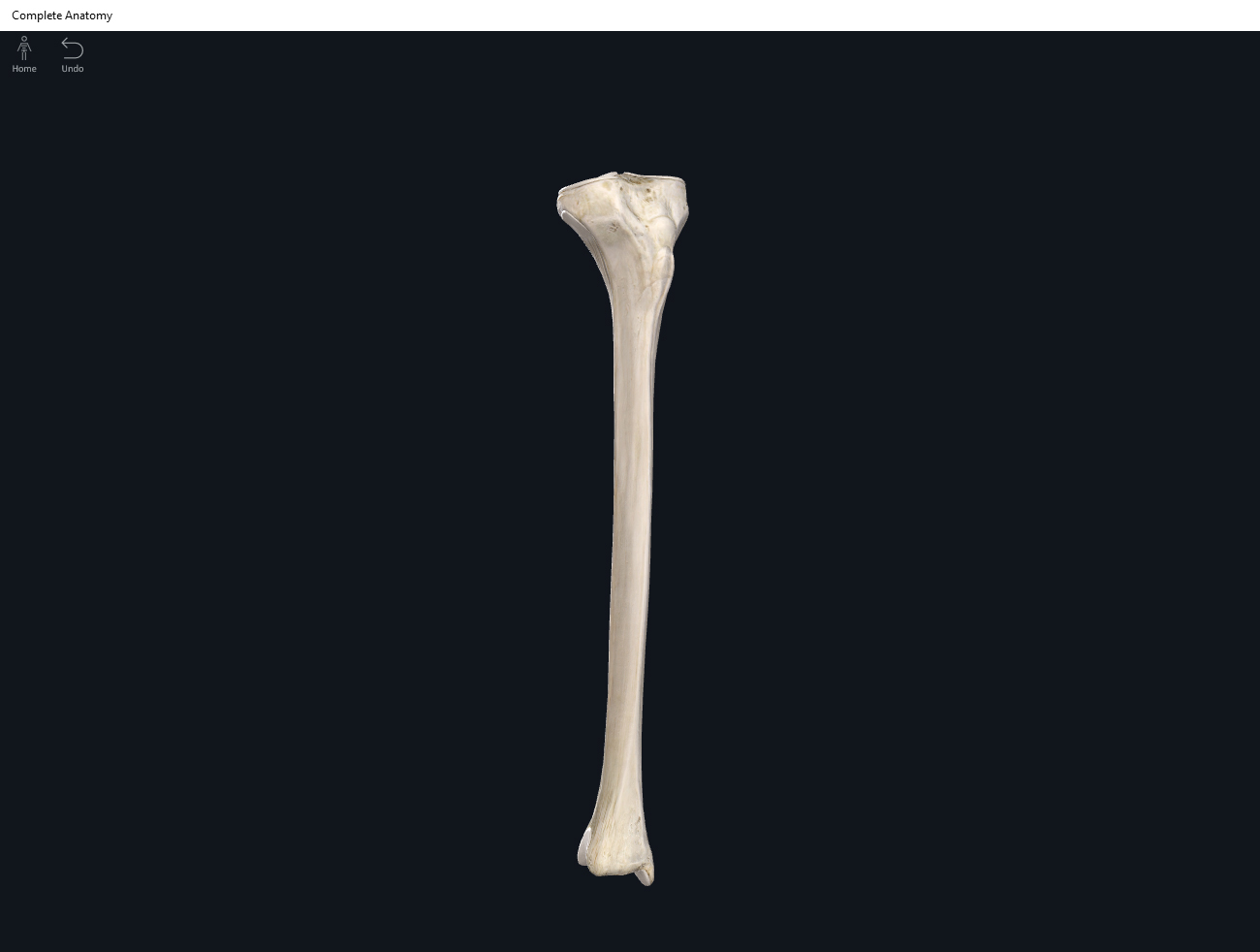
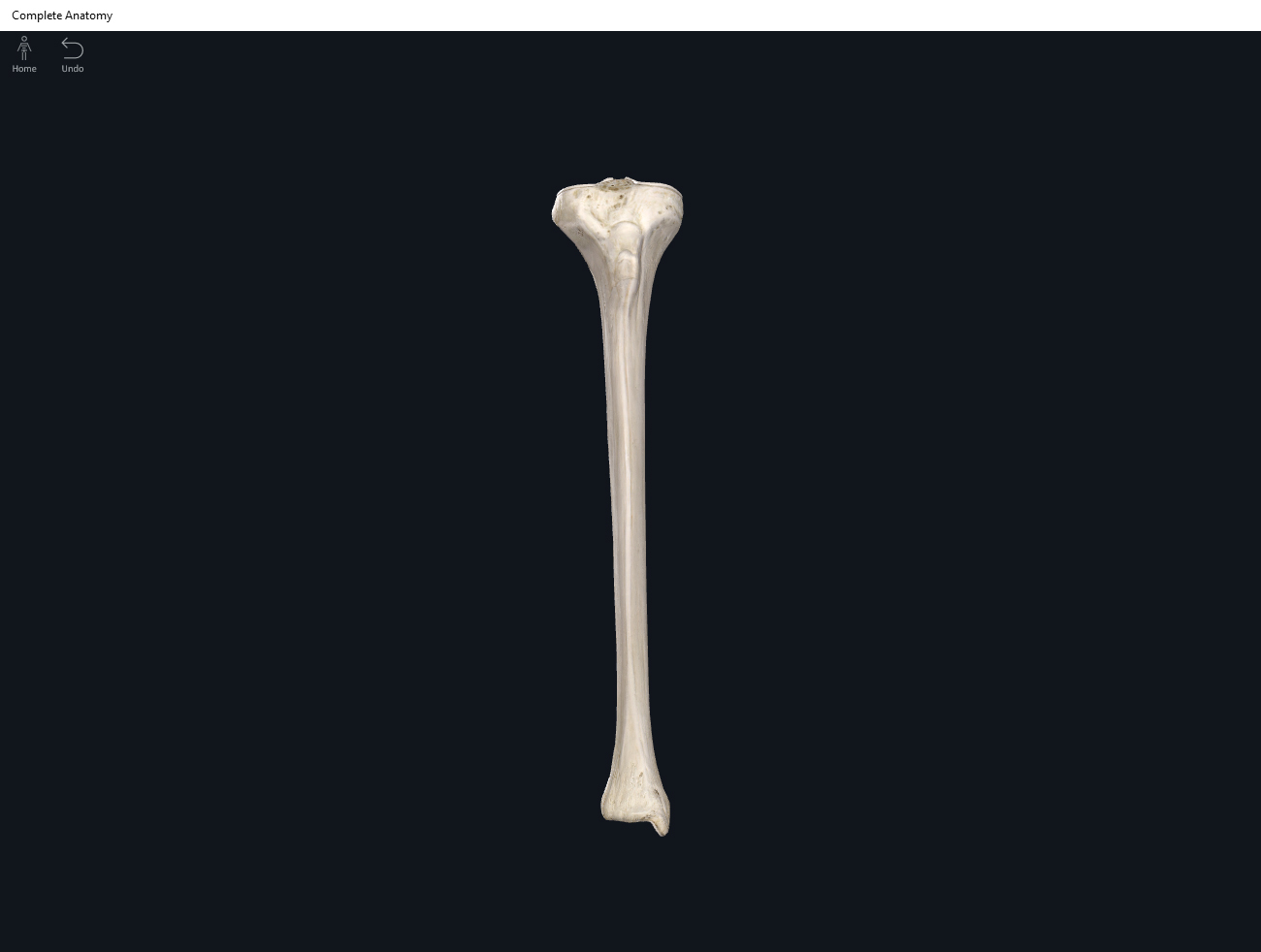
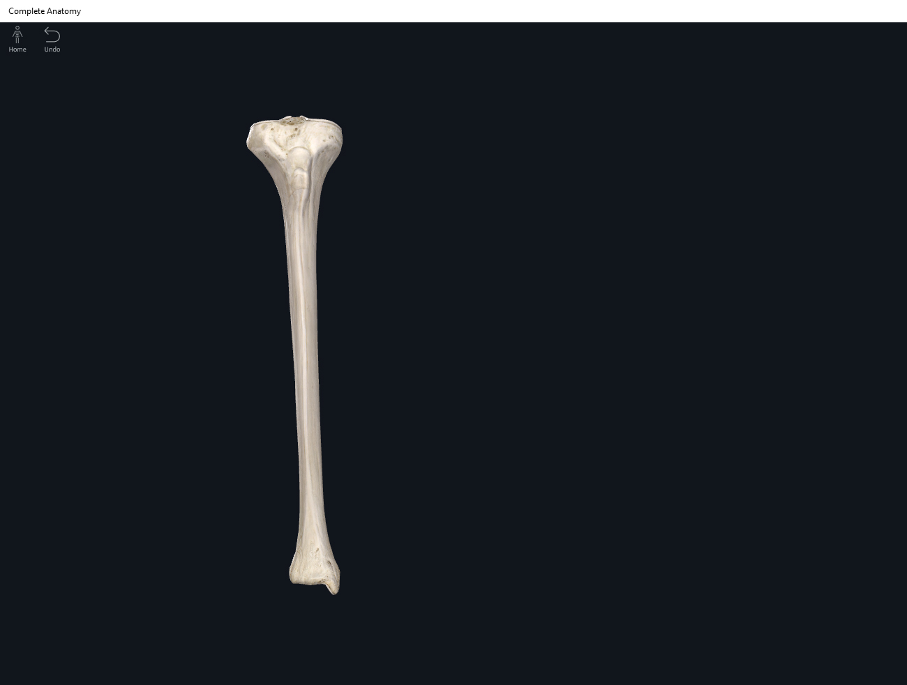
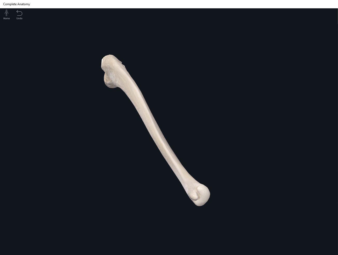
Anatomy & Physiology: Bones—Ulna.
Structure.
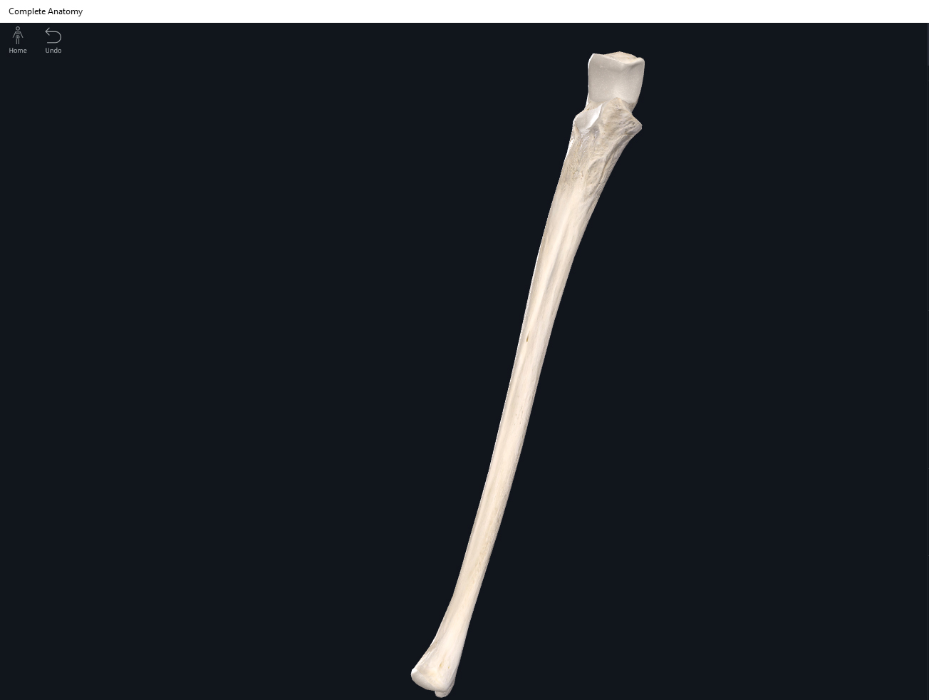
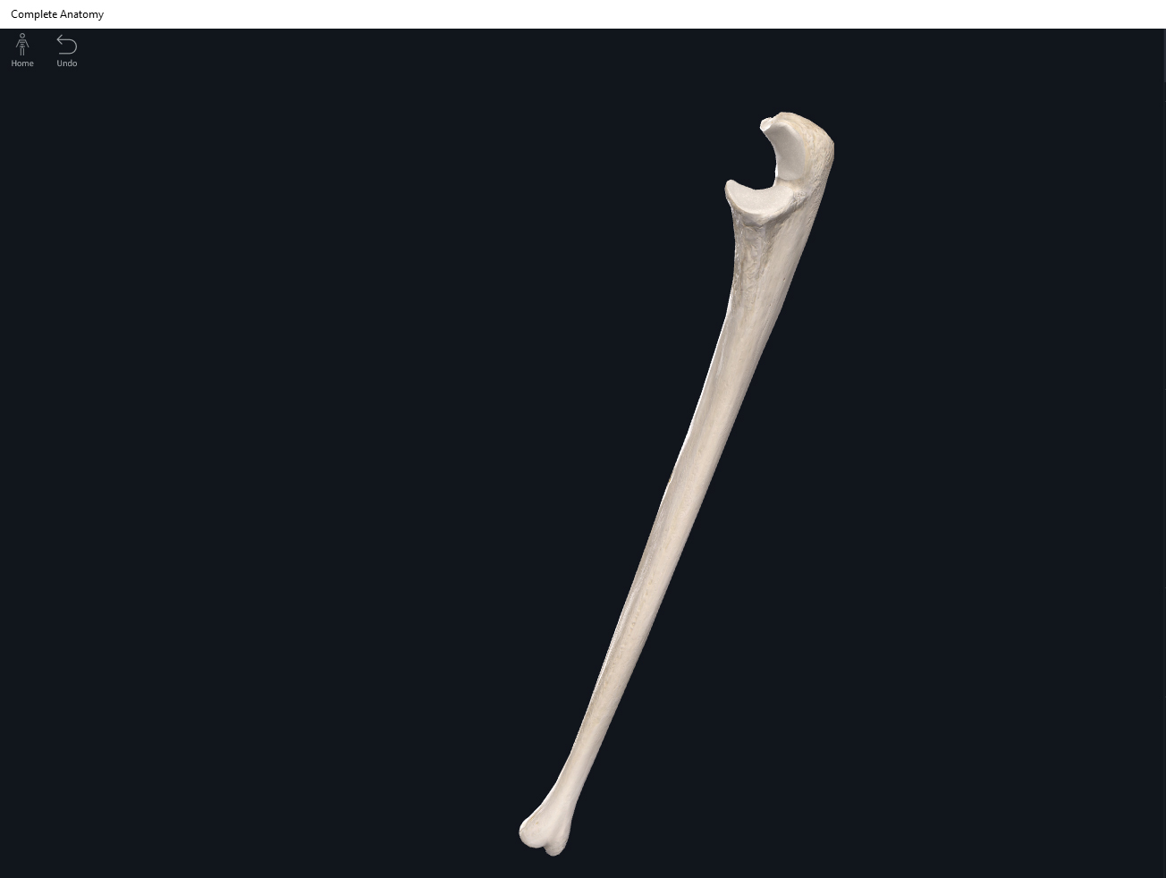
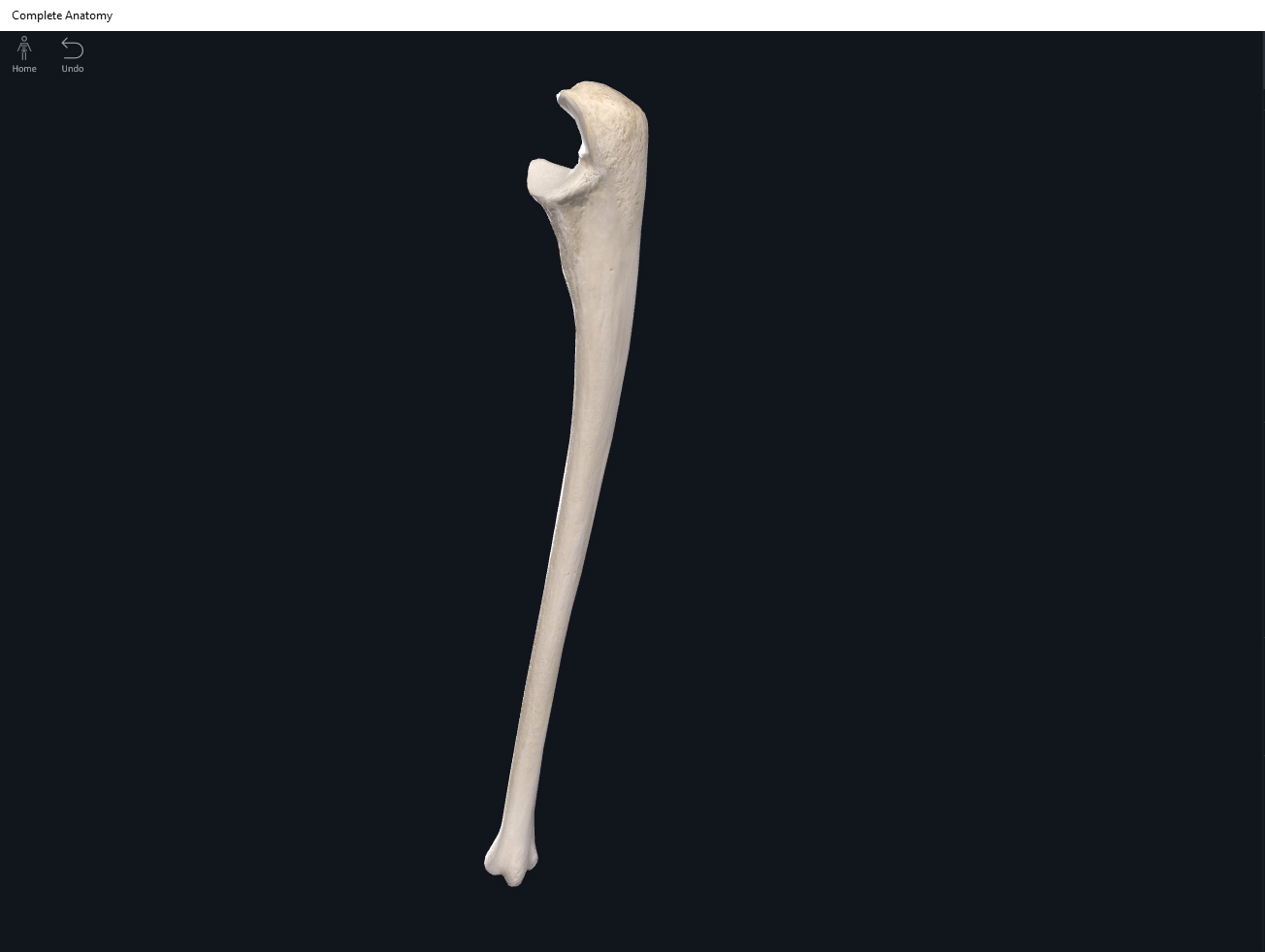
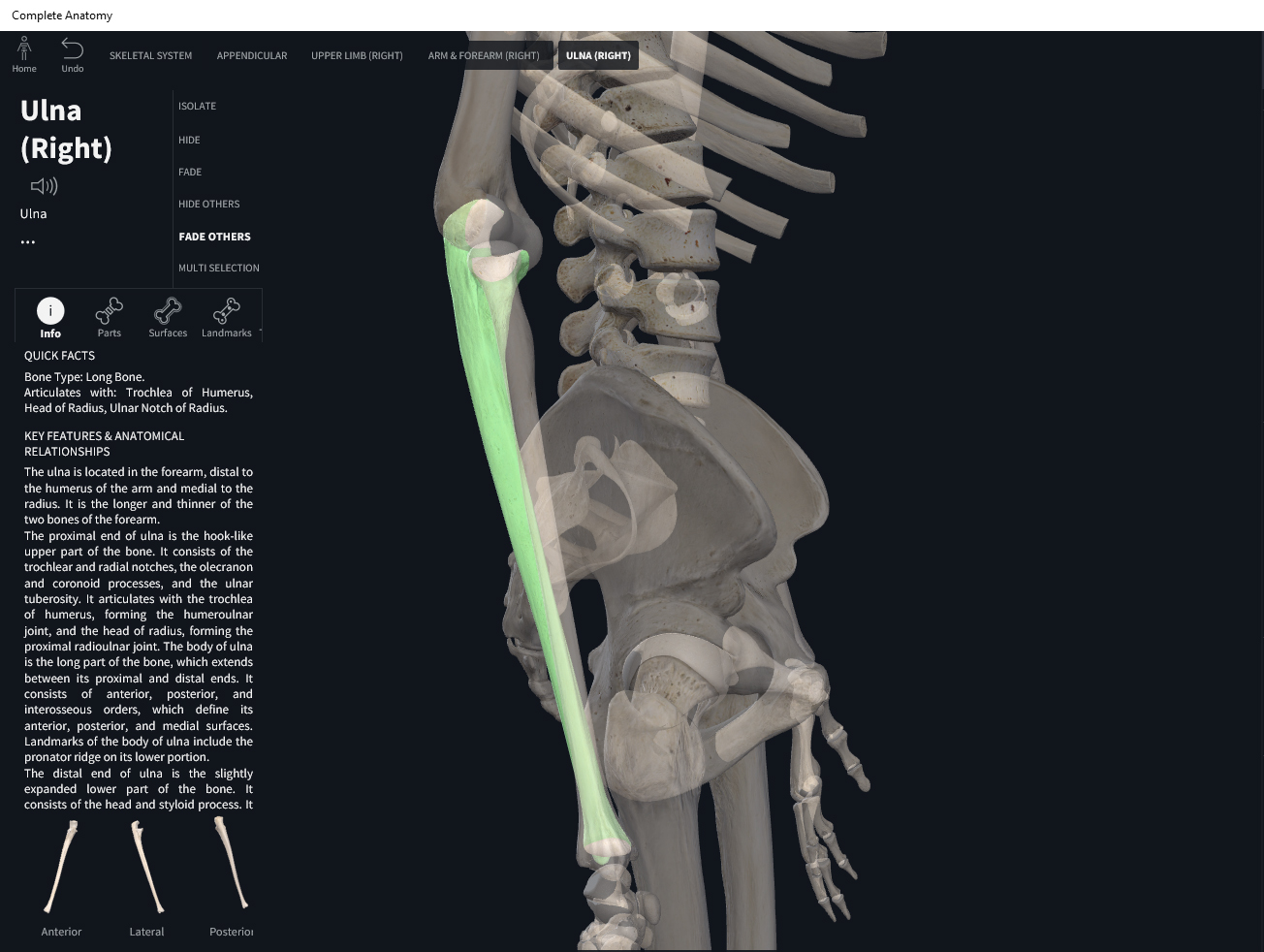
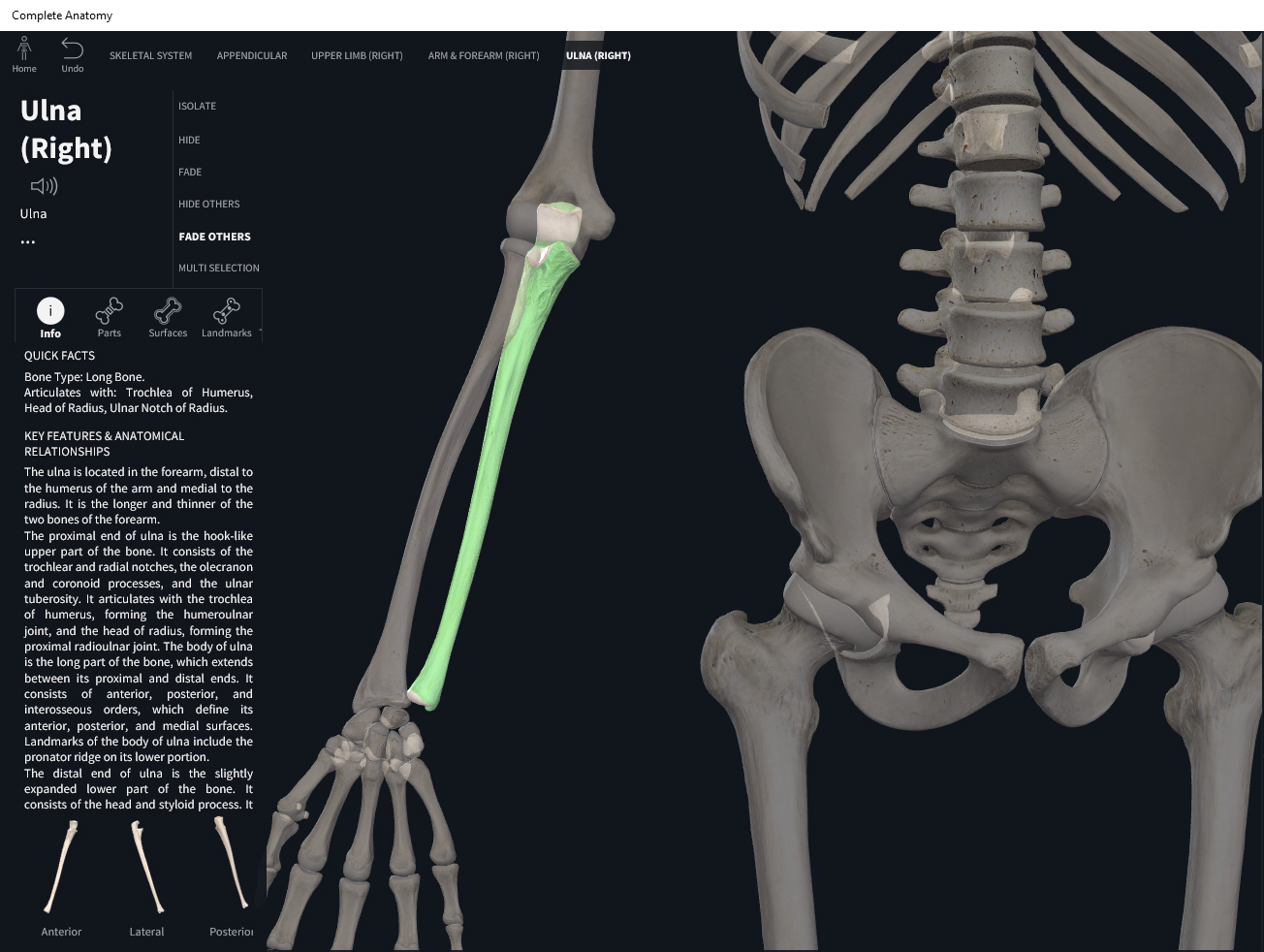
- The forearm bone on the “pinky” little finger side.
- Is larger, longer, and more massive than the radius.
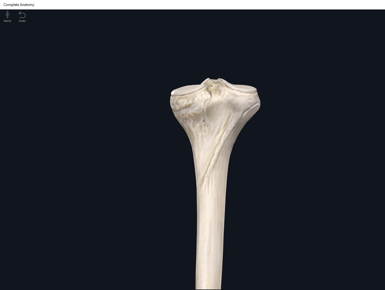
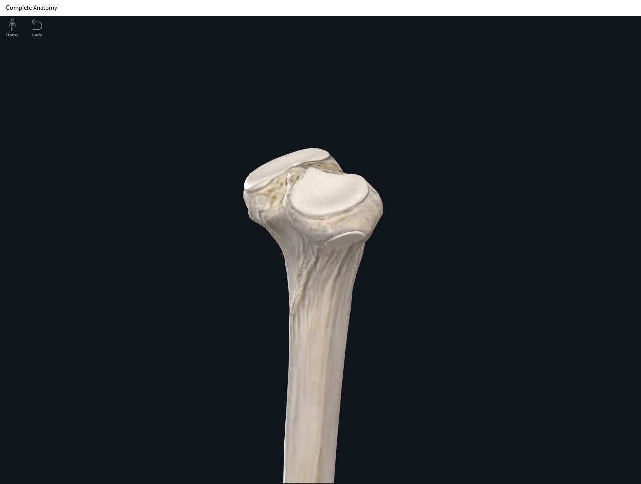
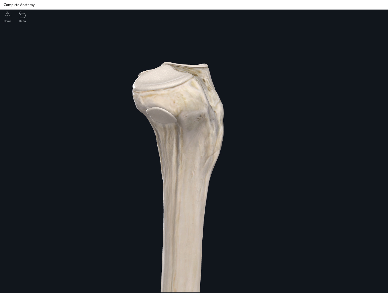
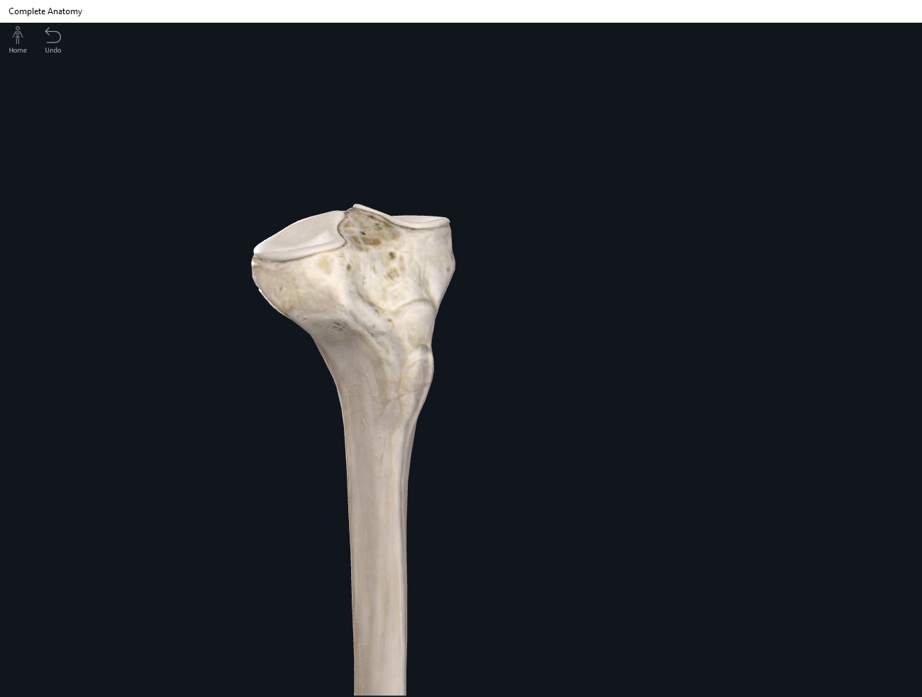
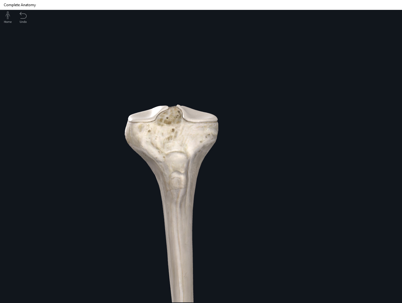
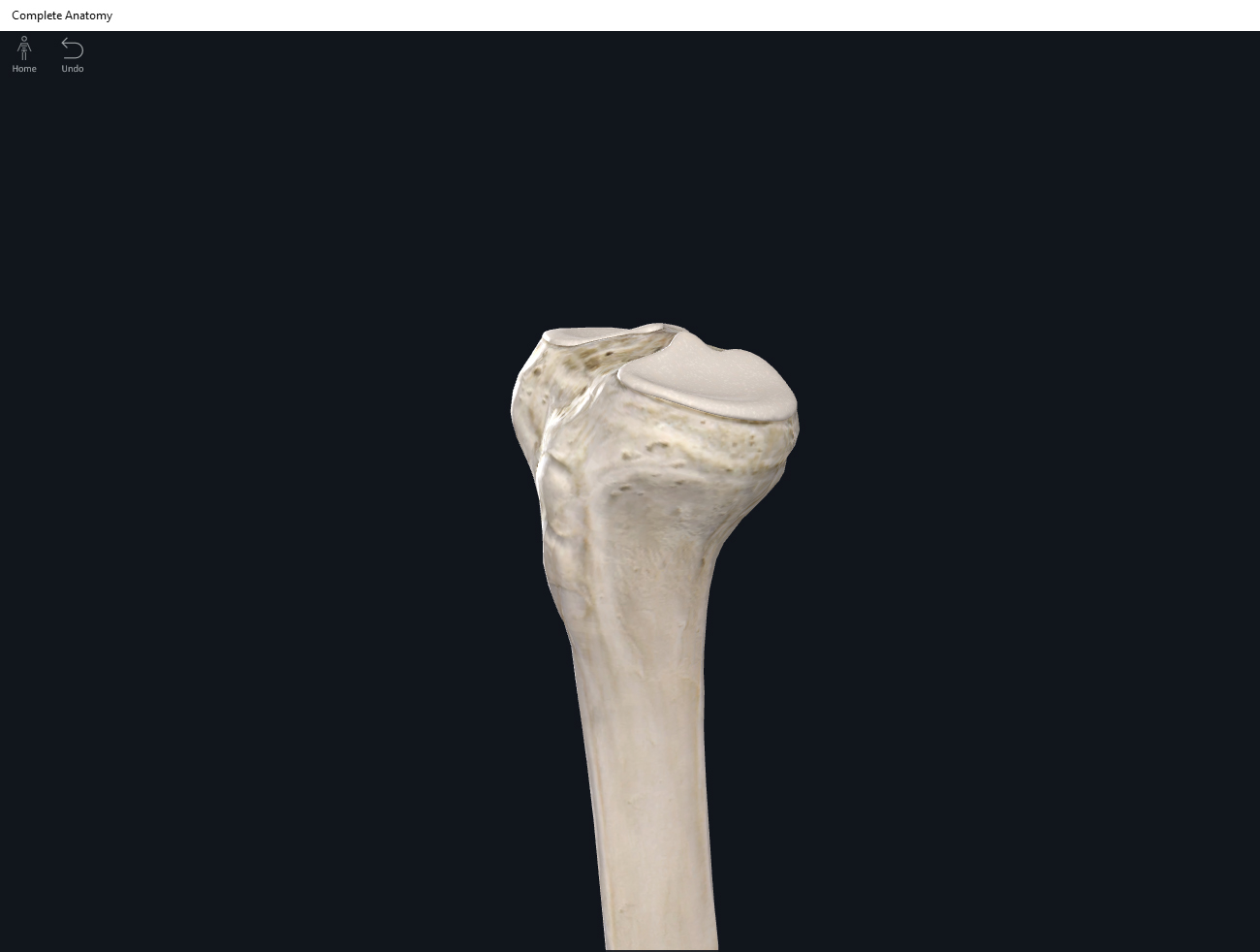
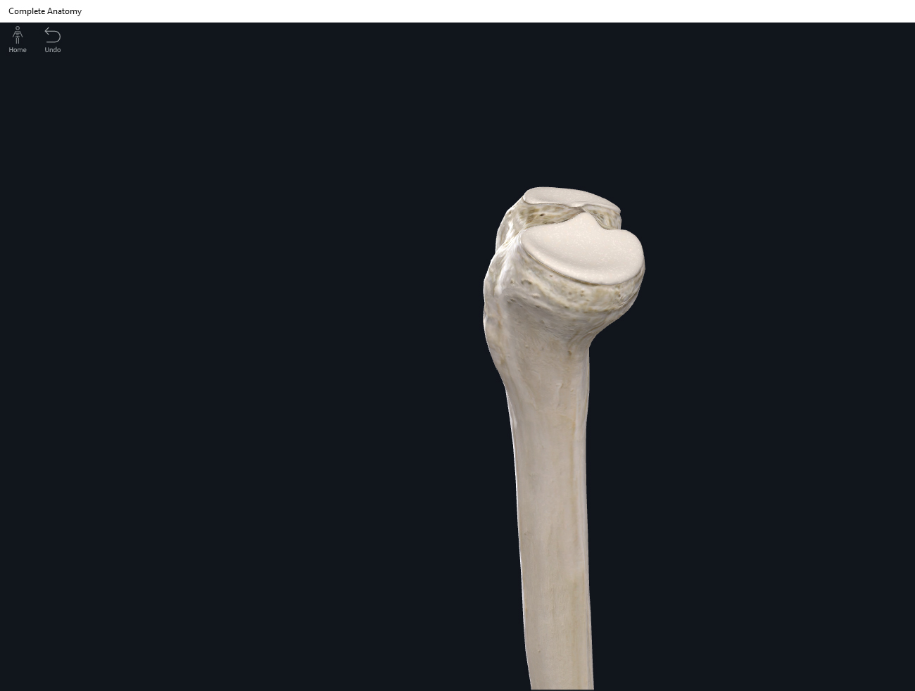
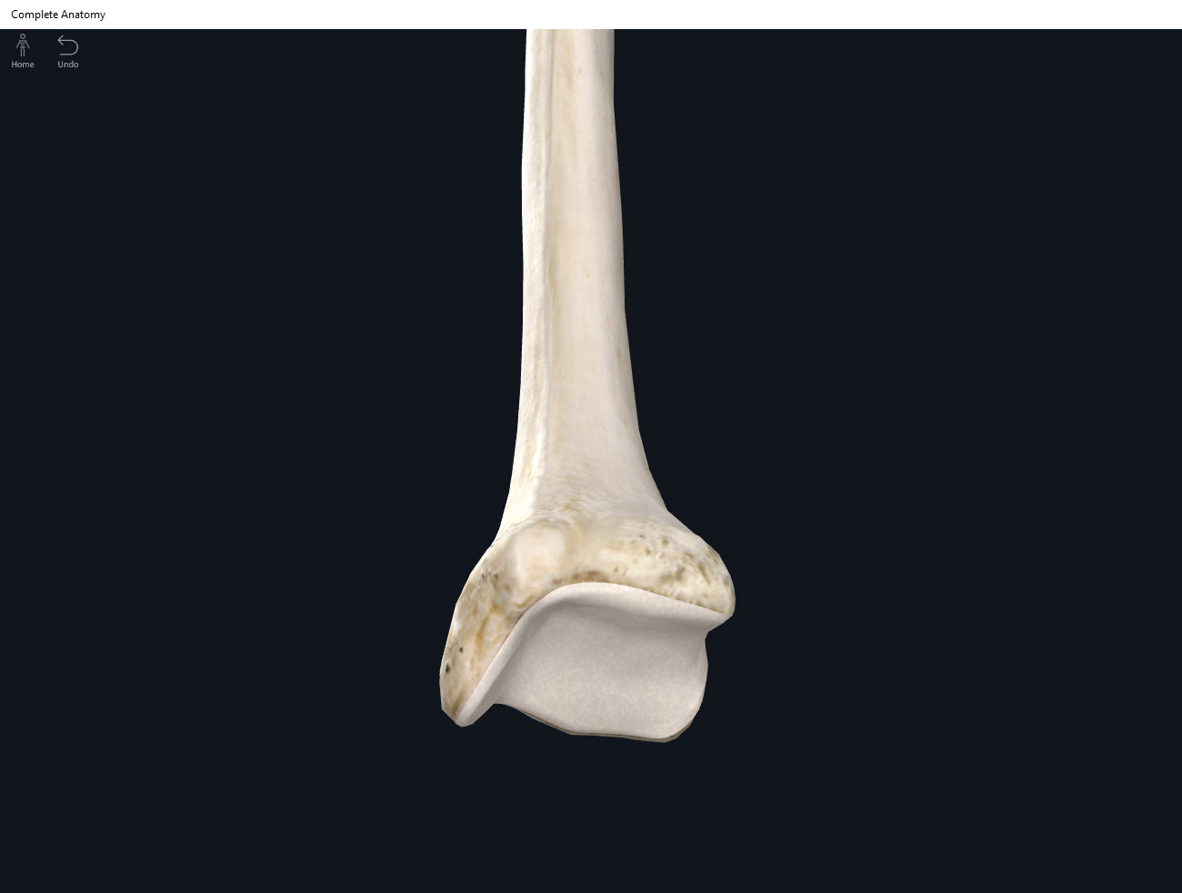
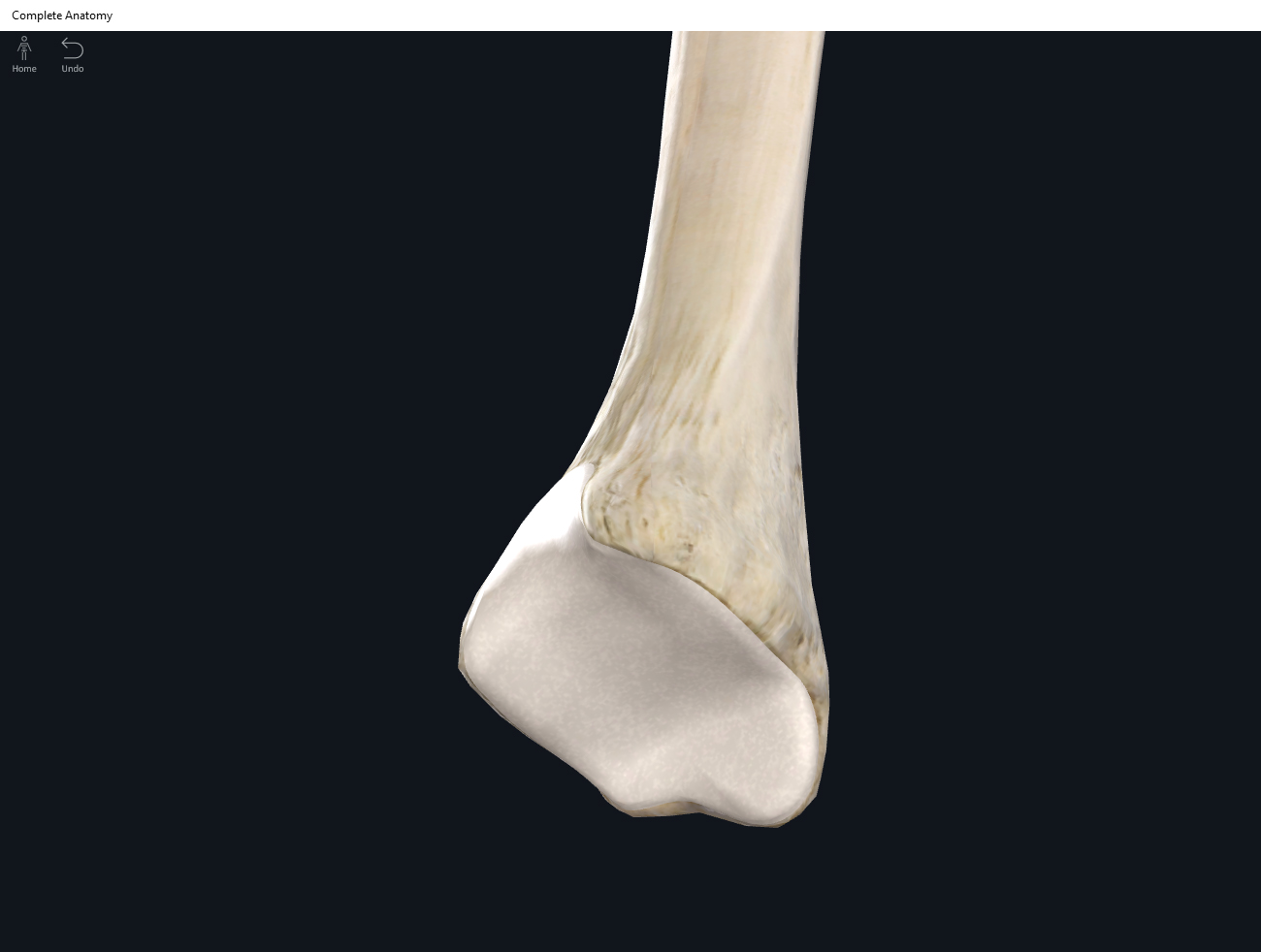
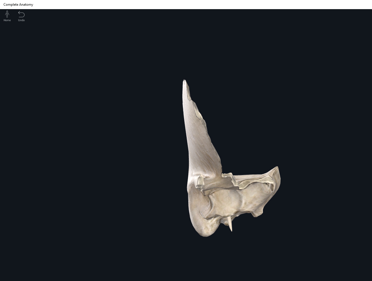
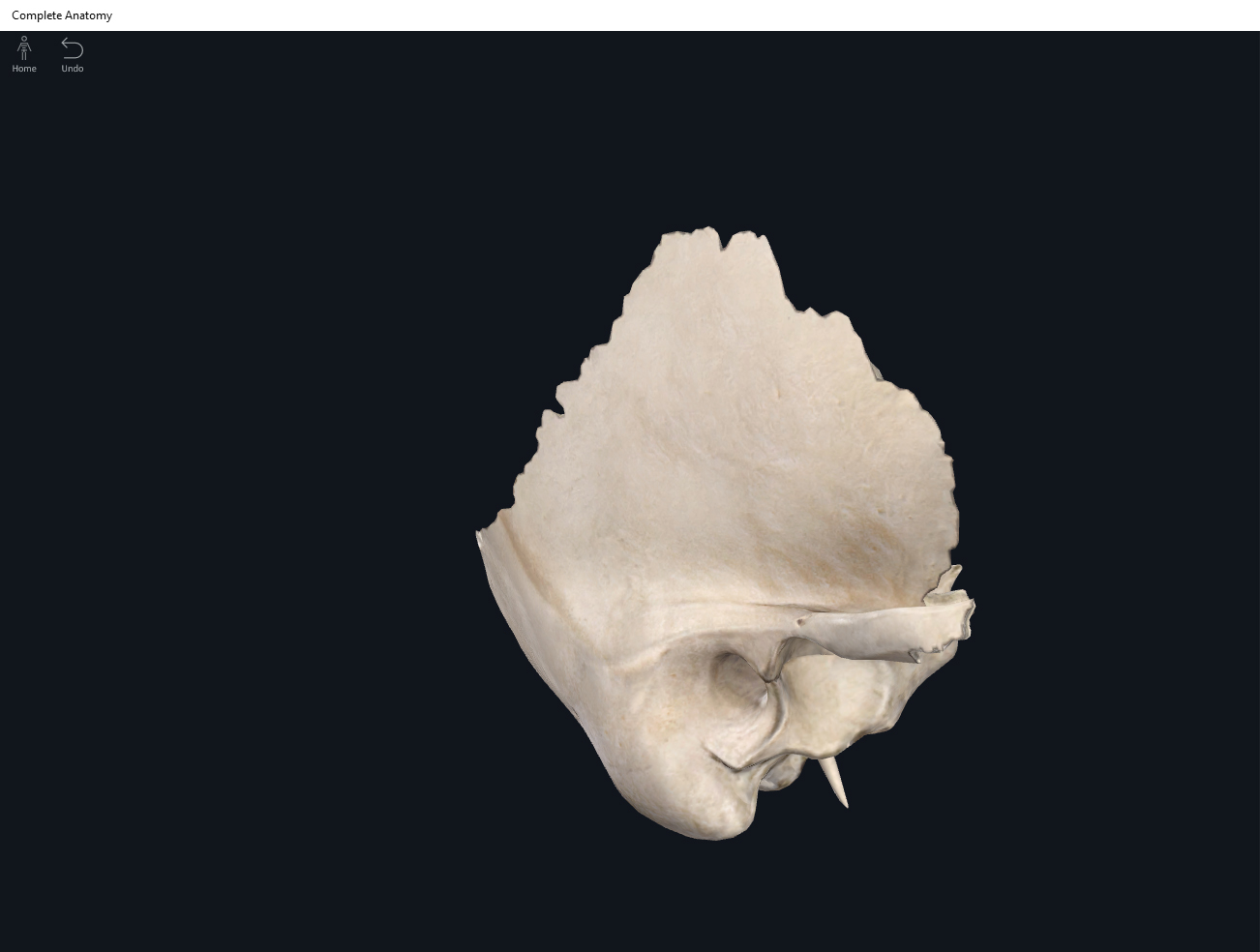
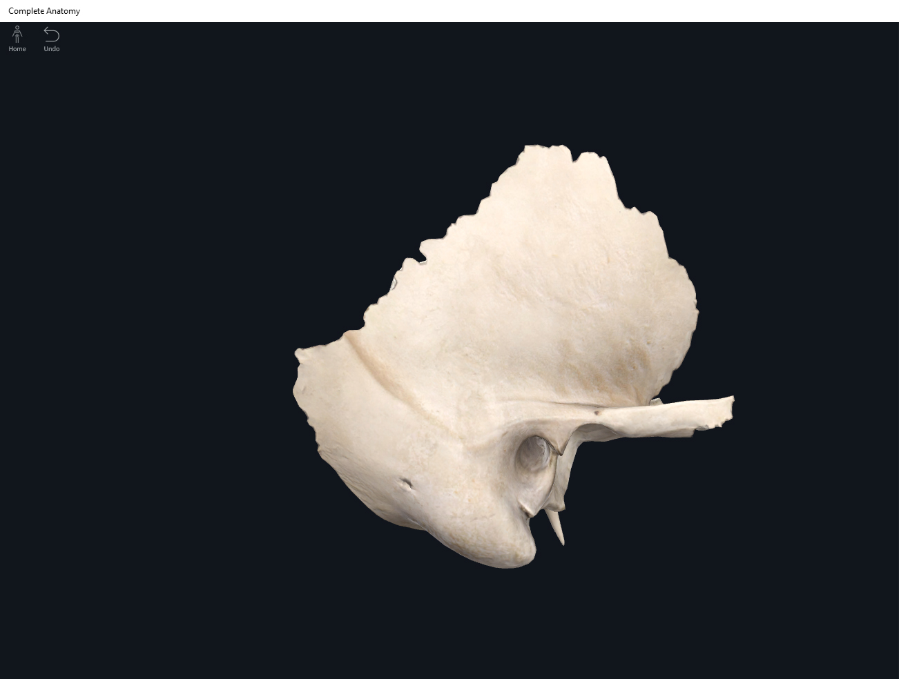
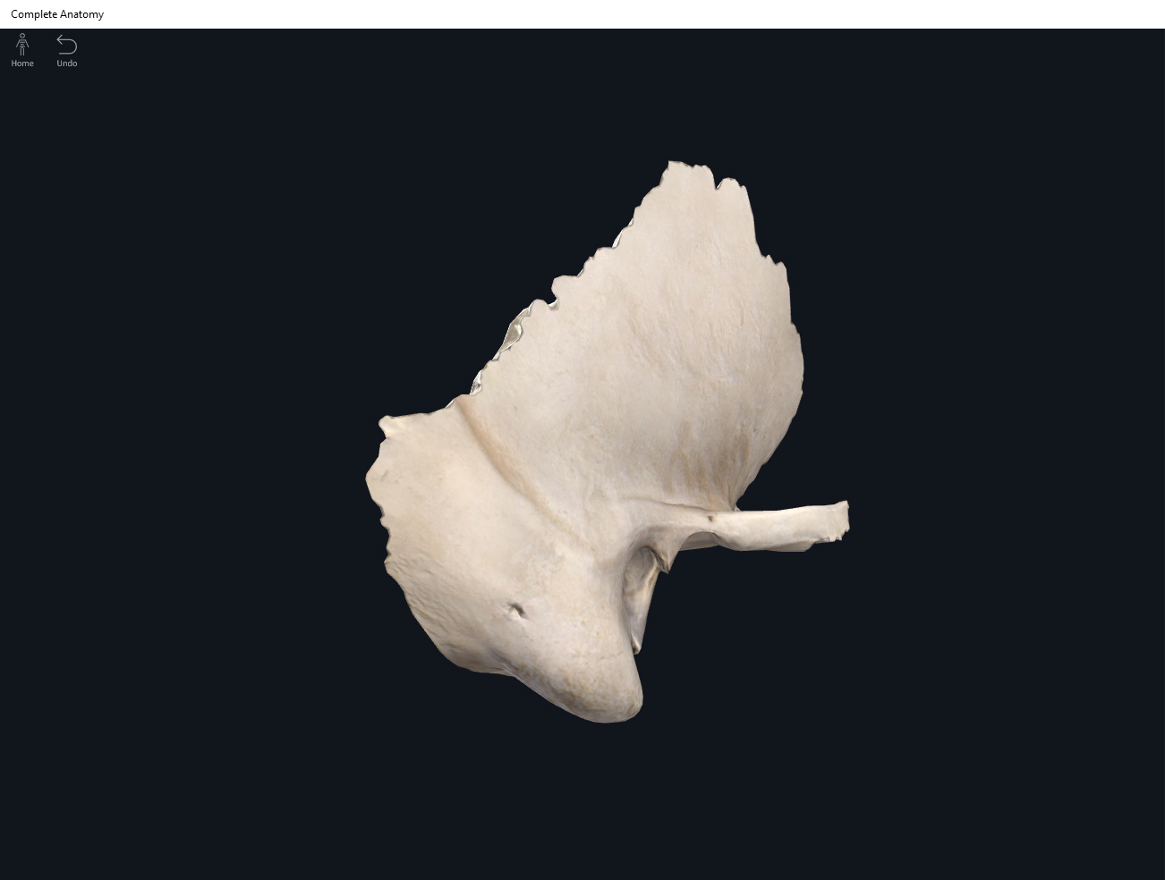
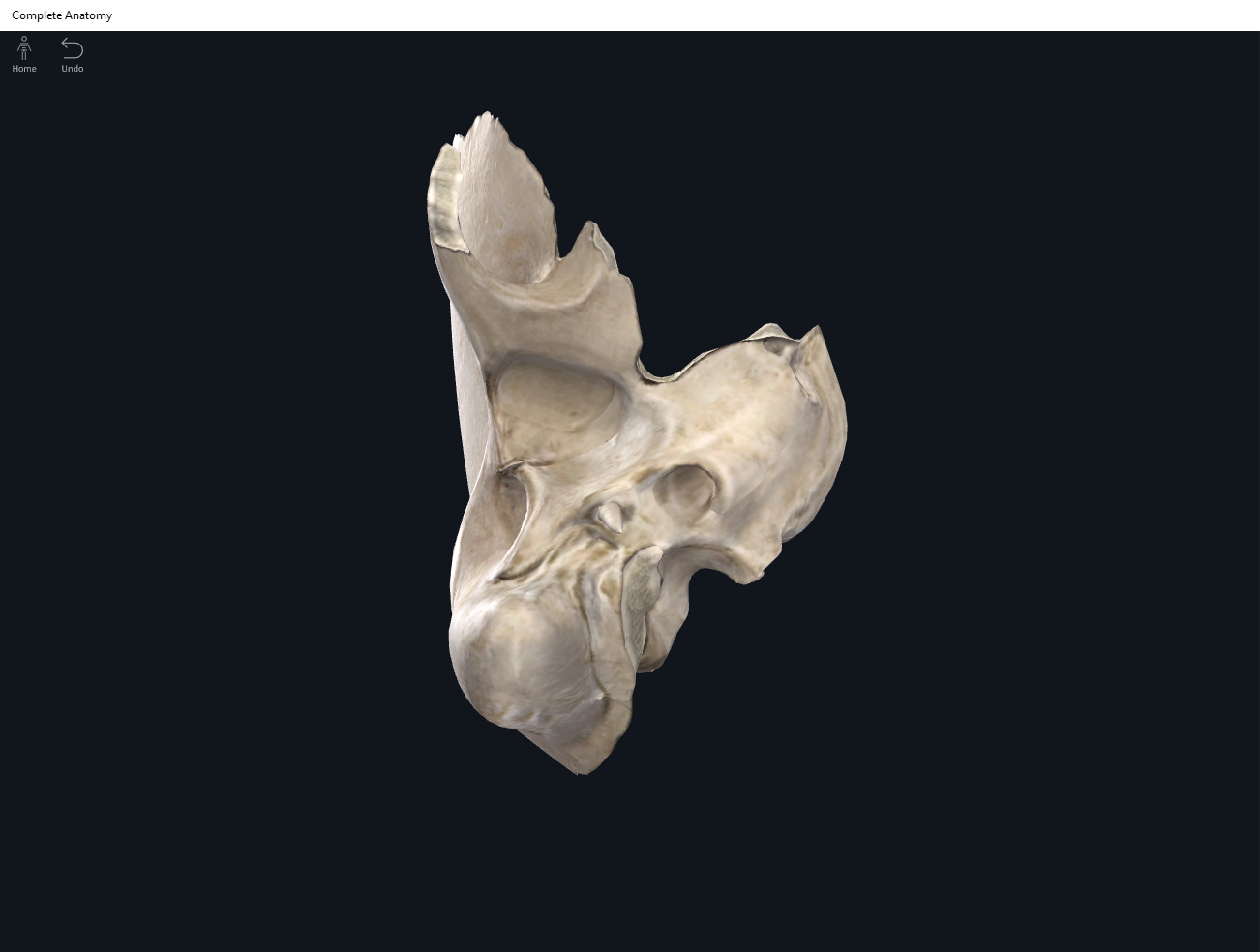
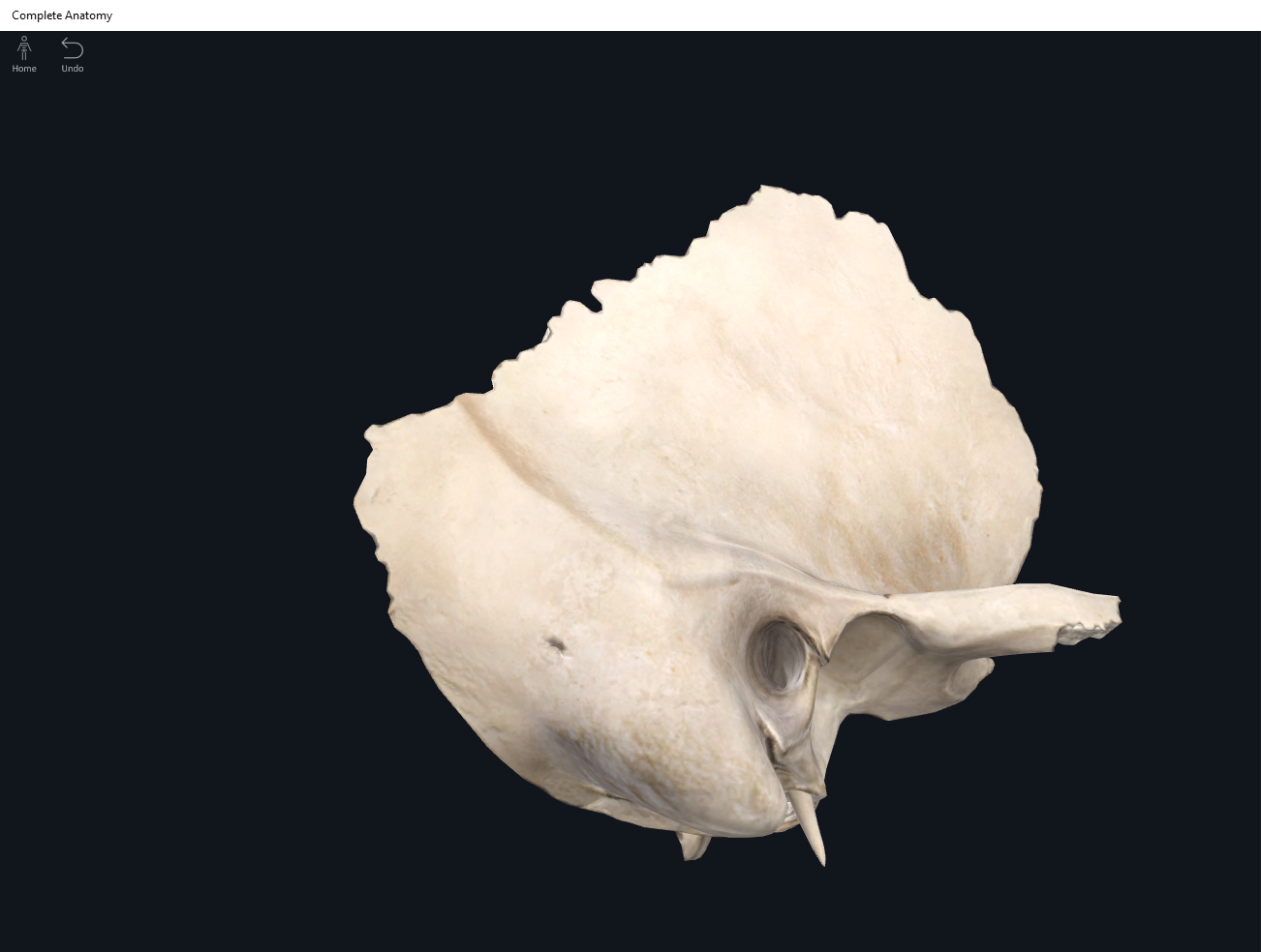
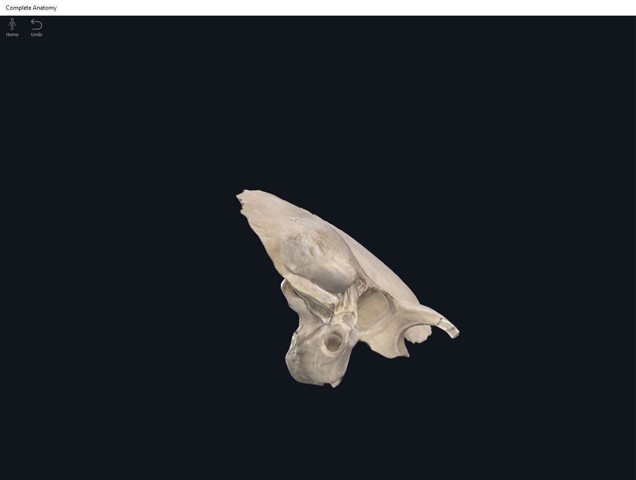
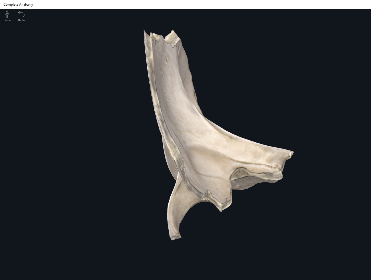
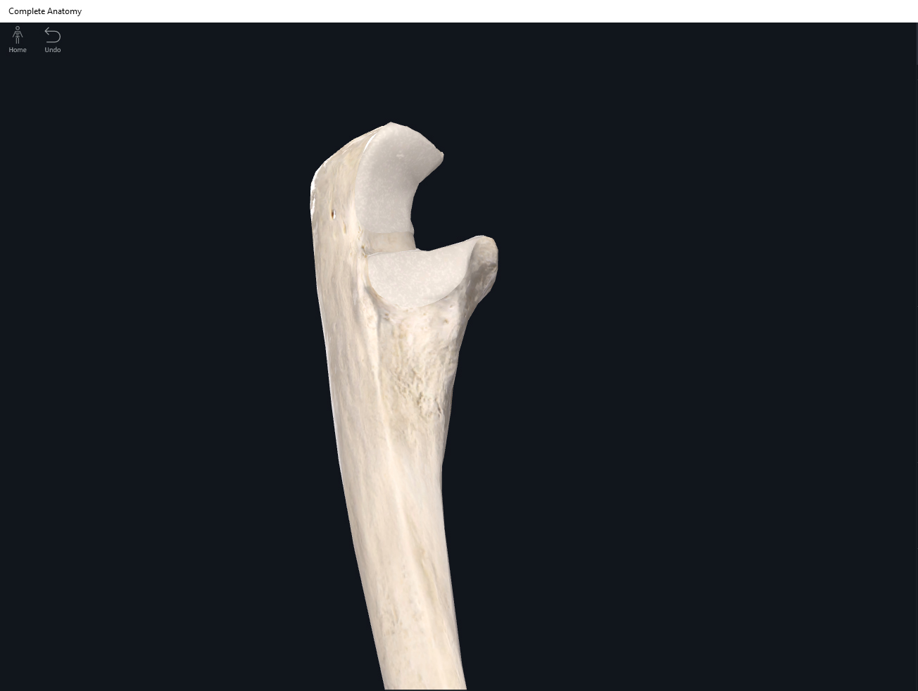
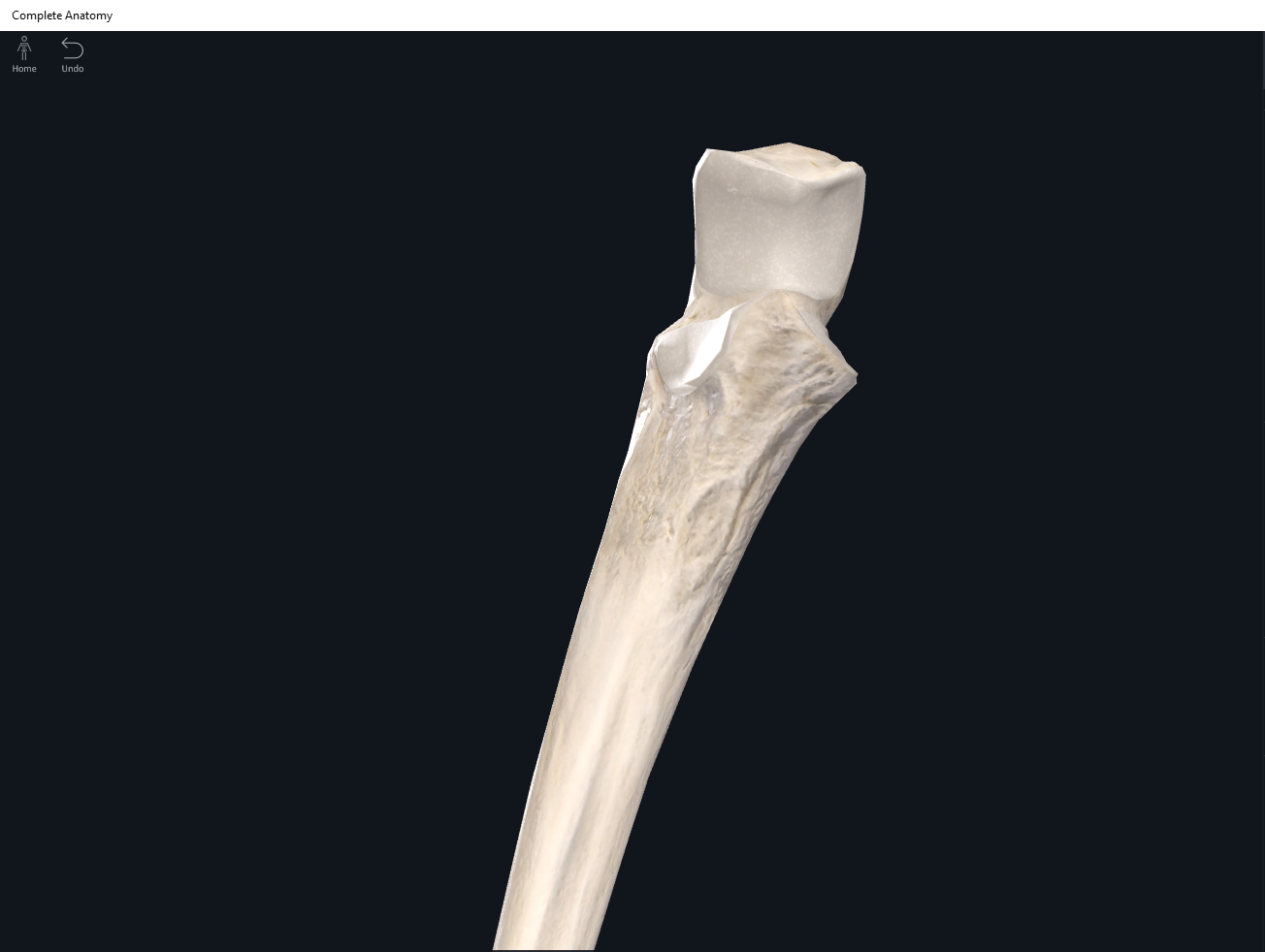
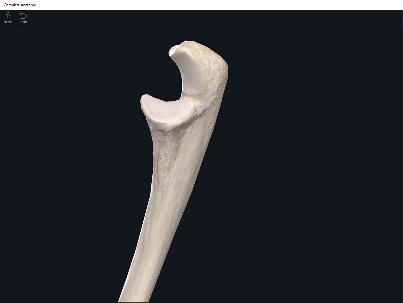
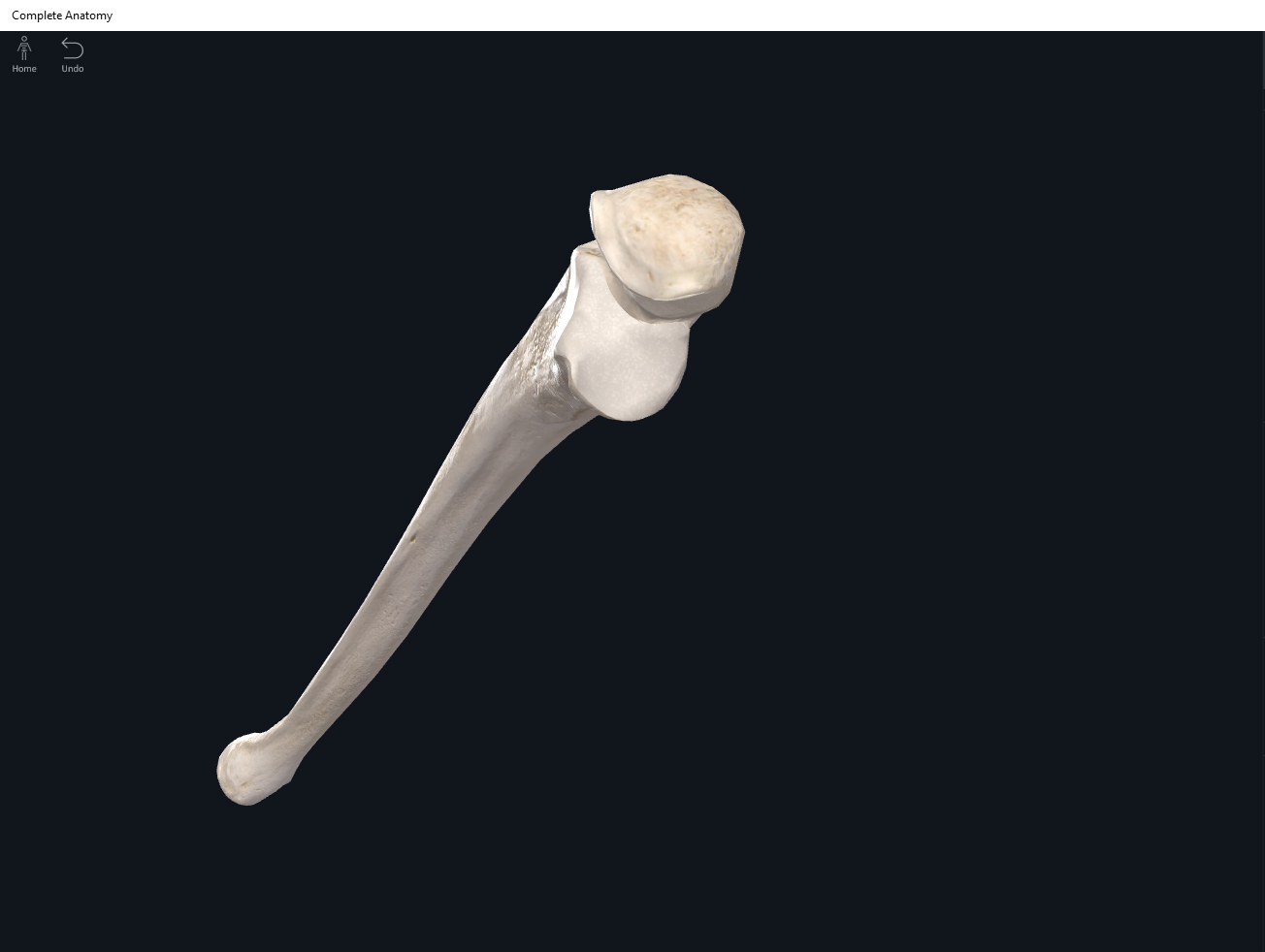
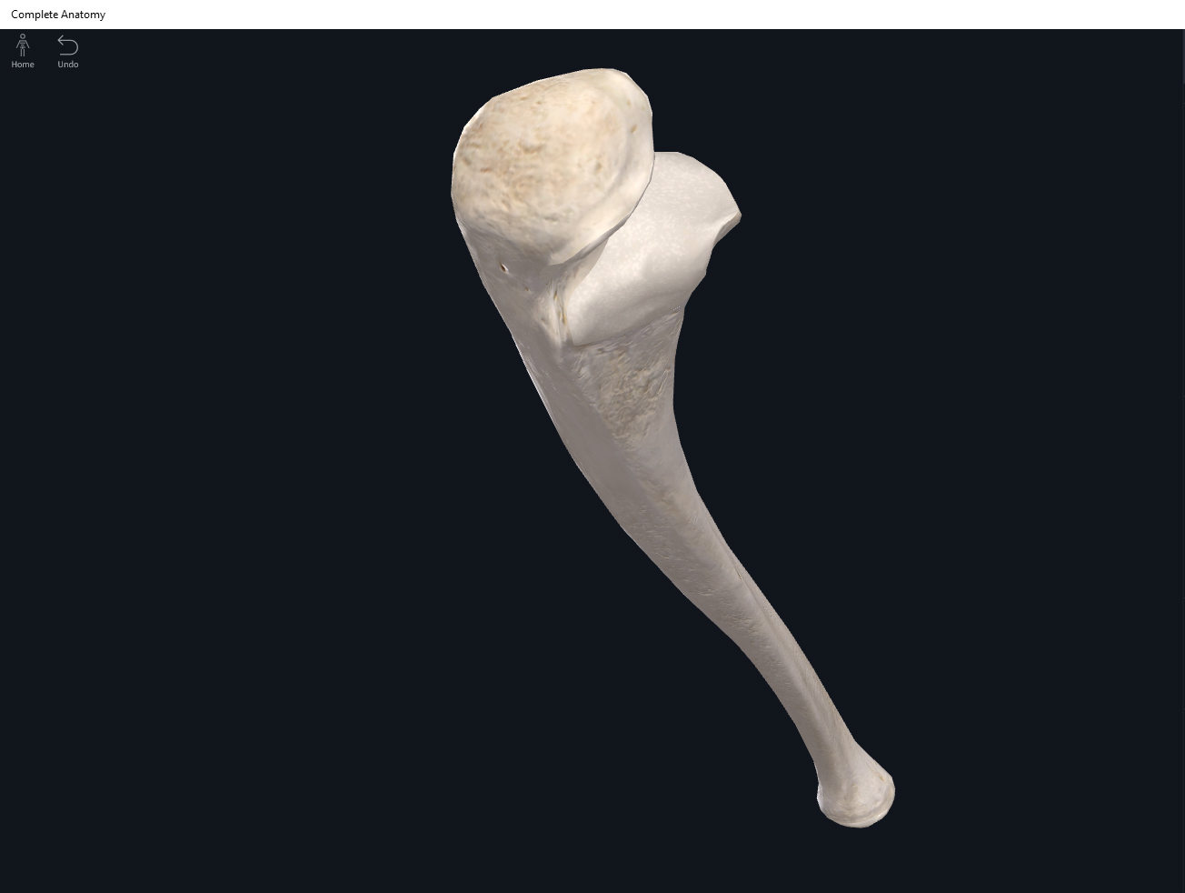
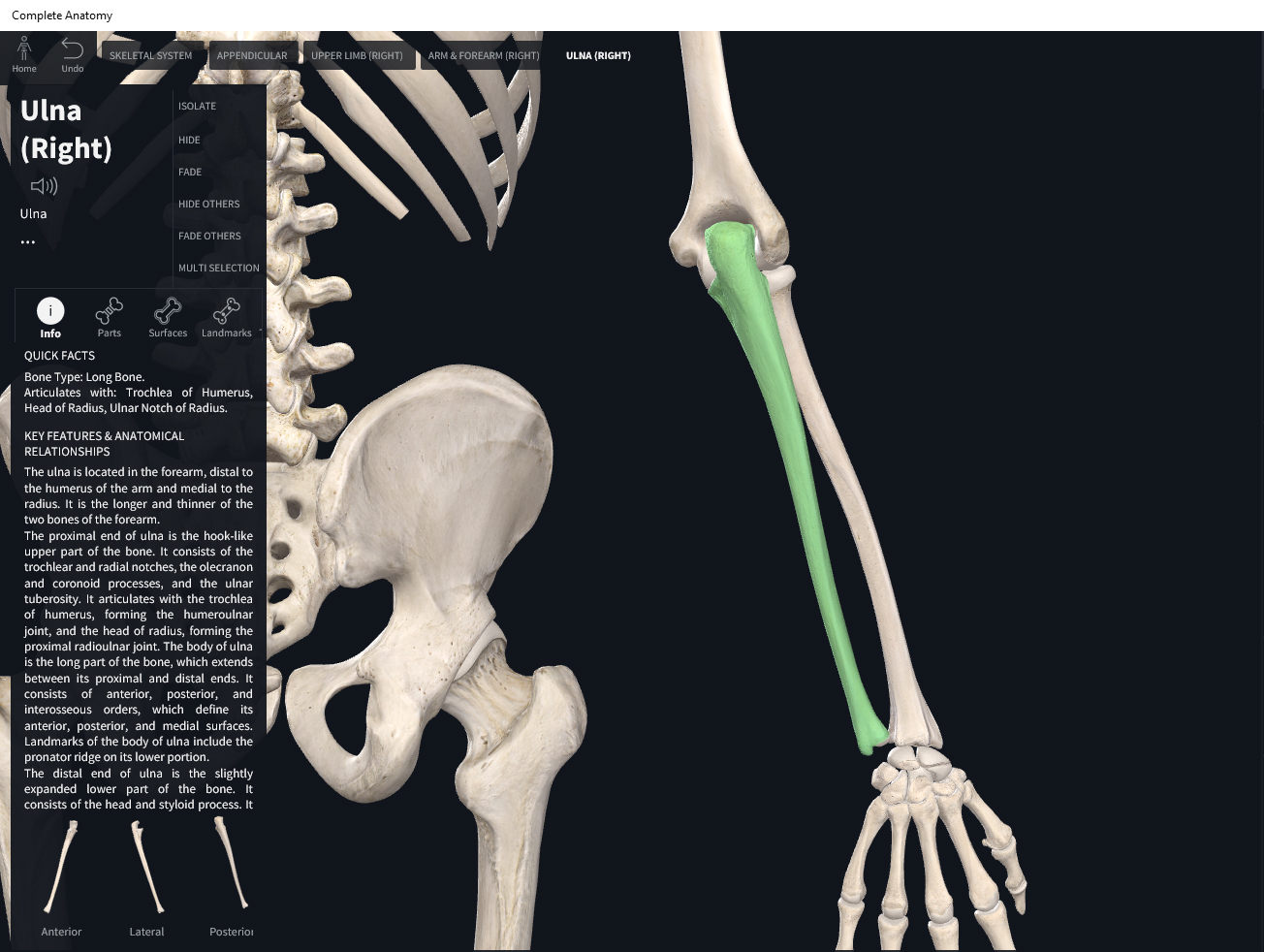
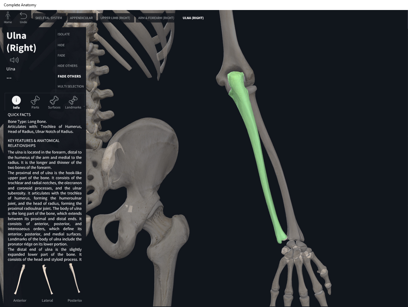
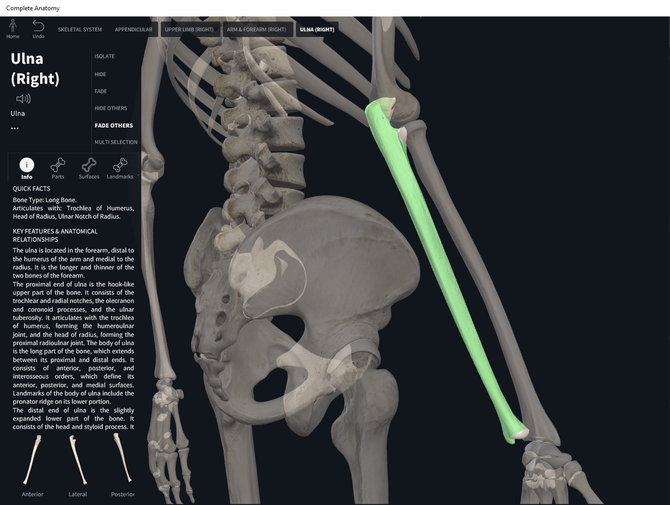
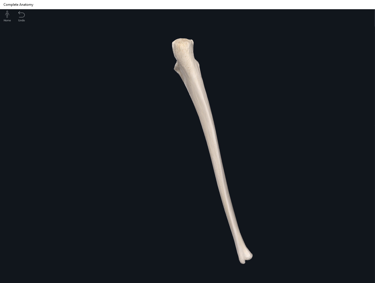
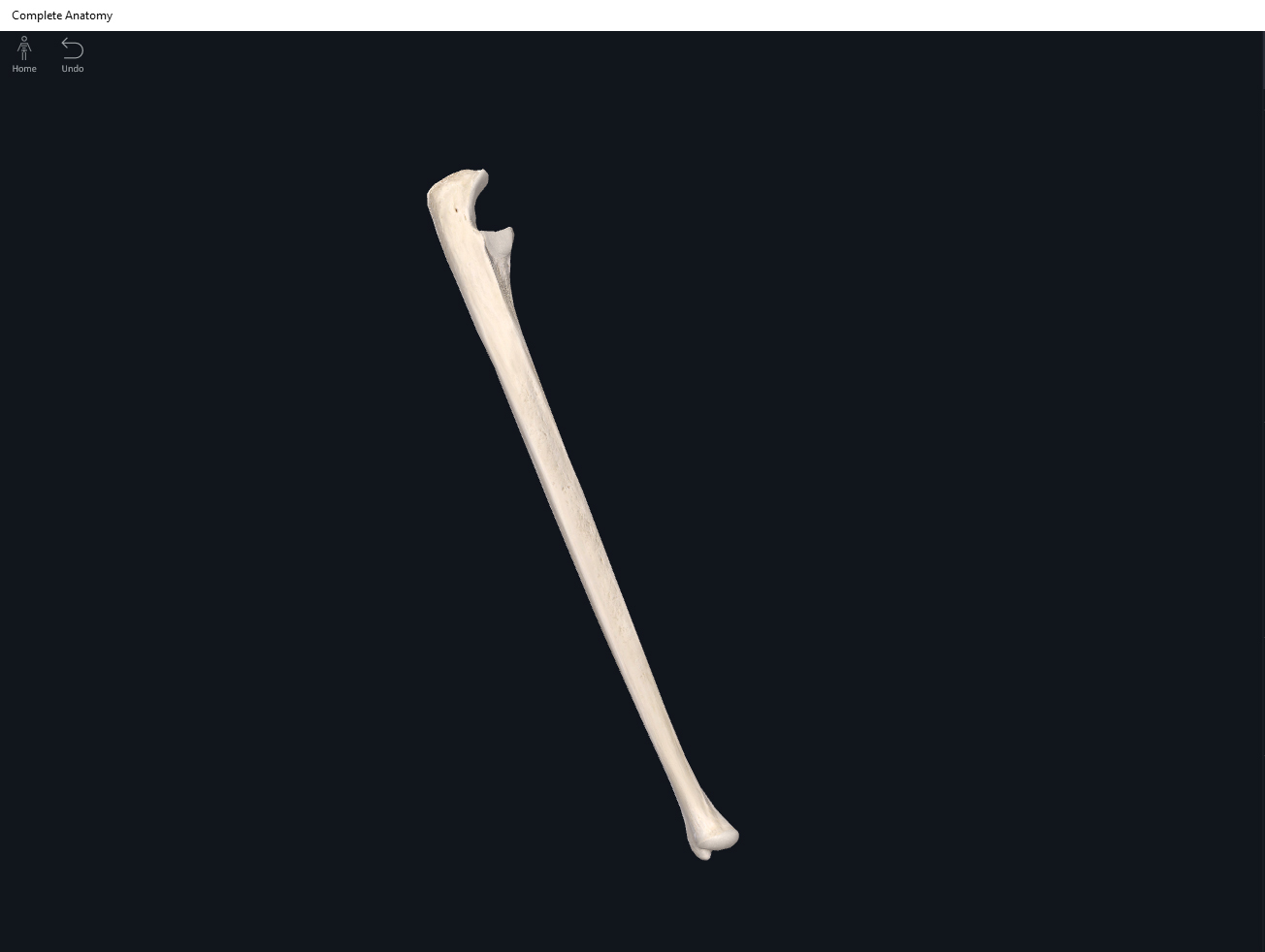
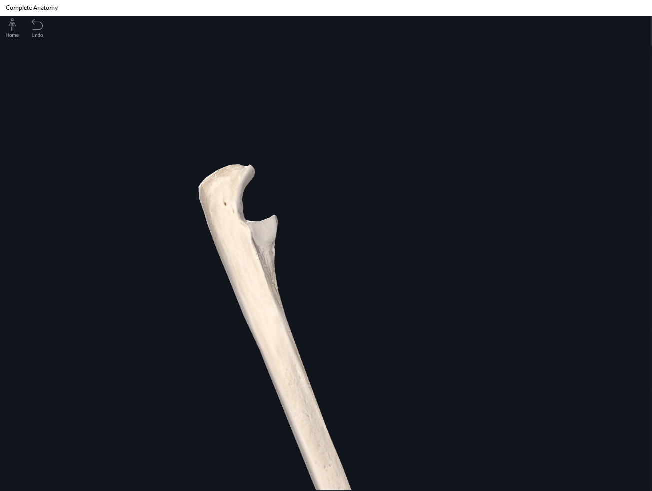
- Olecranon: located proximally forms the elbow, and is easily palpateable. When the elbow is extended the olecranon tucks into the olecranon fossa of the humerus.
- Coronoid process: a projection on the anterior proximal end of the ulna. This process tucks into the coronoid fossa of the humerus.
- Trochlear notch: articulates with the trochlea of the humerus. It lies between the olecranon and coronoid process.
- Radial notch: located lateral and inferior to the coronoid process, this notch articulates with the radial head.
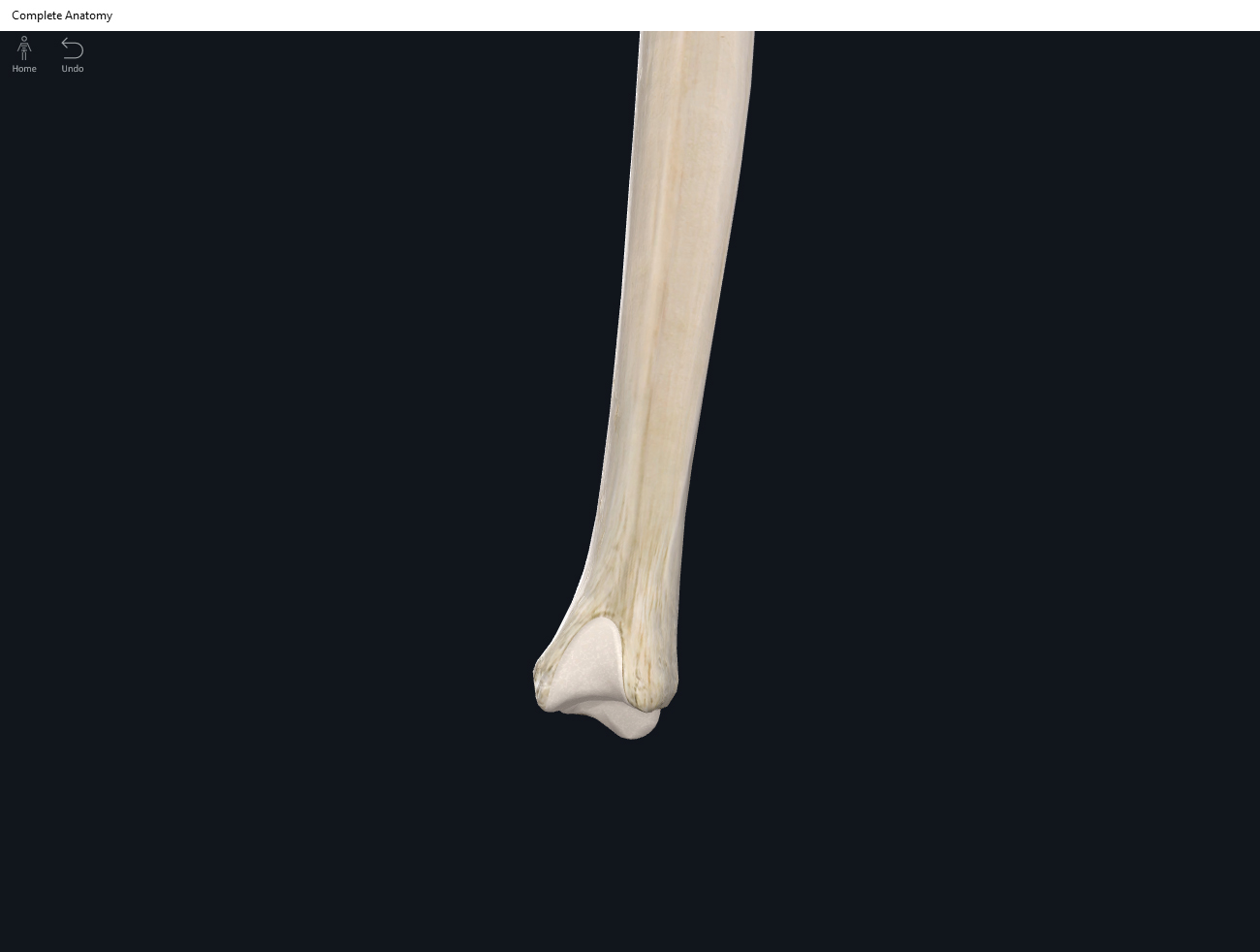
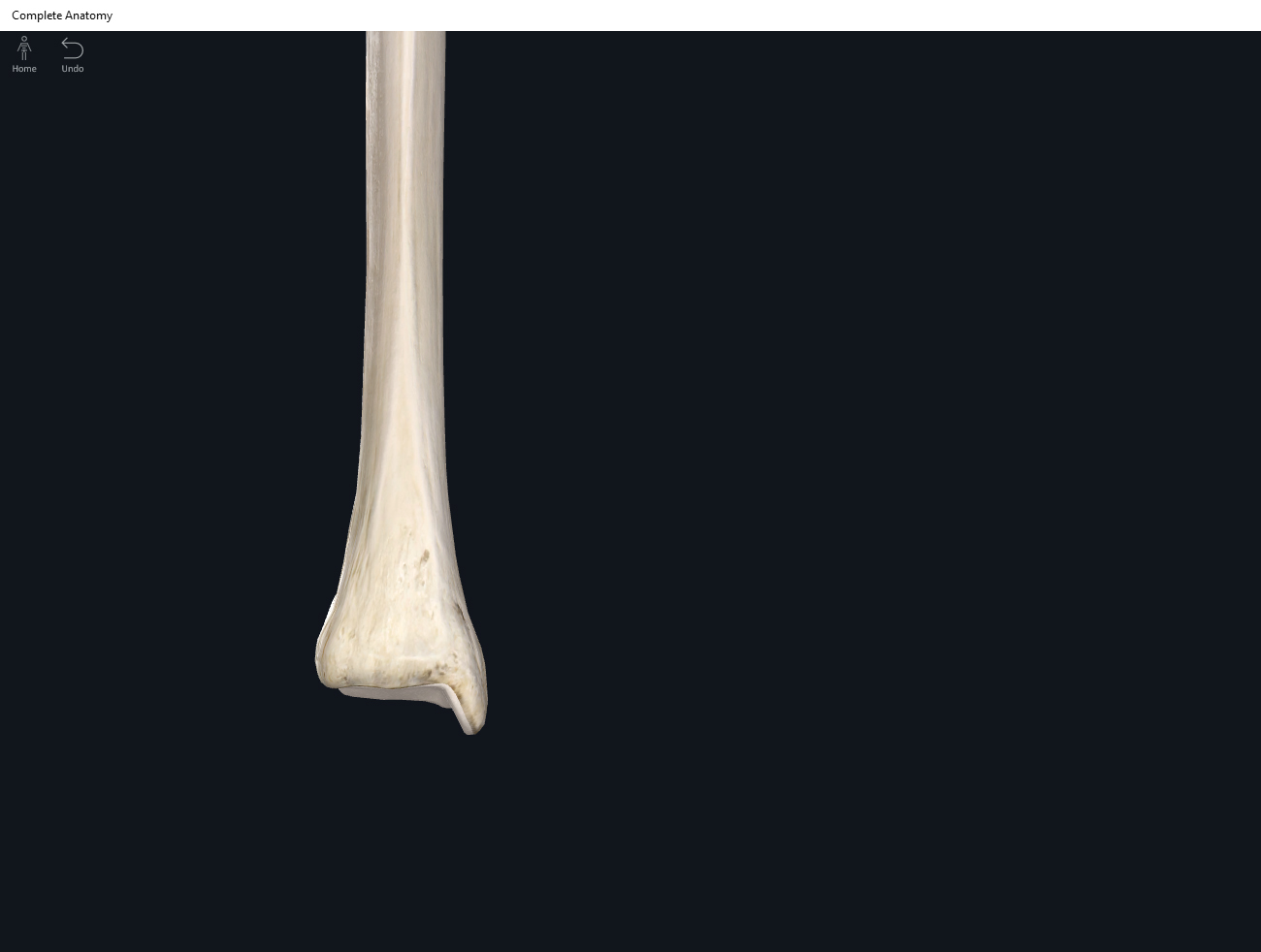
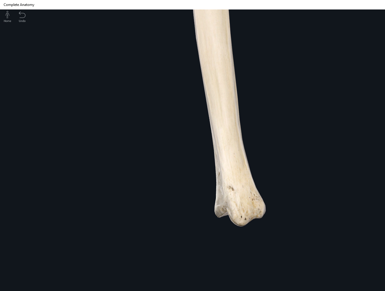
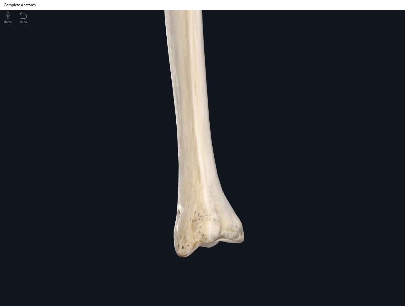
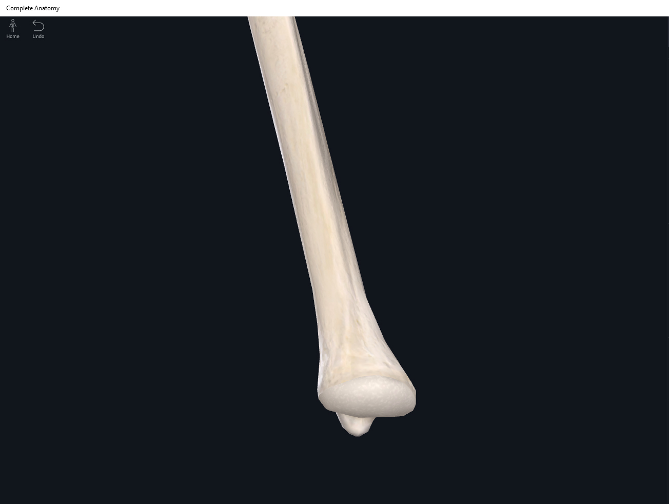
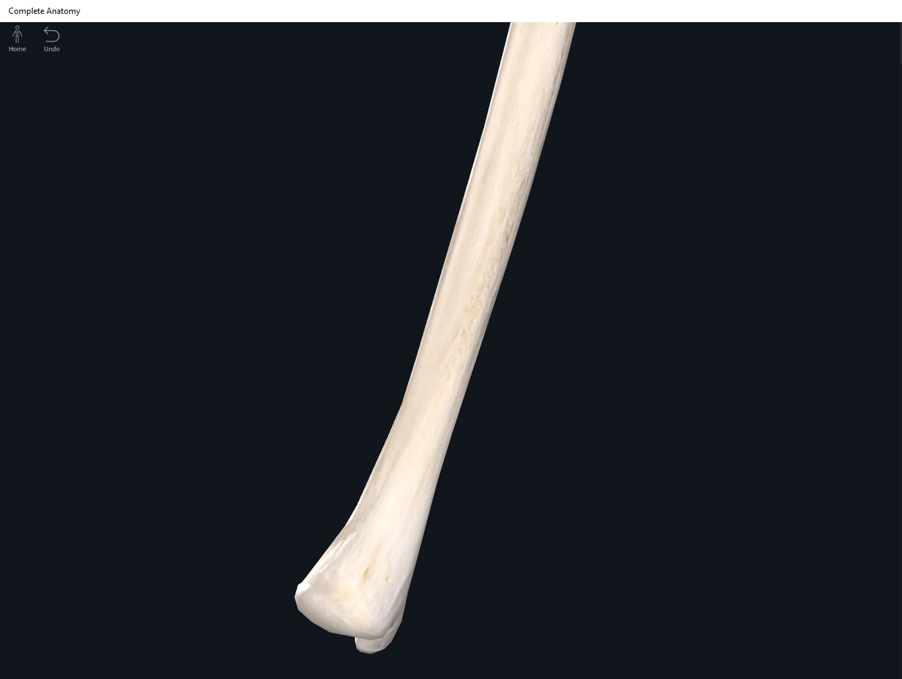
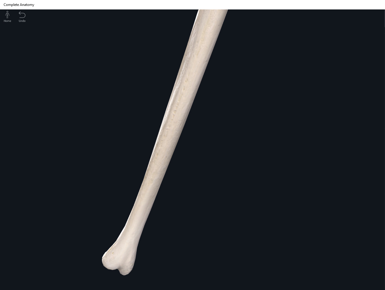
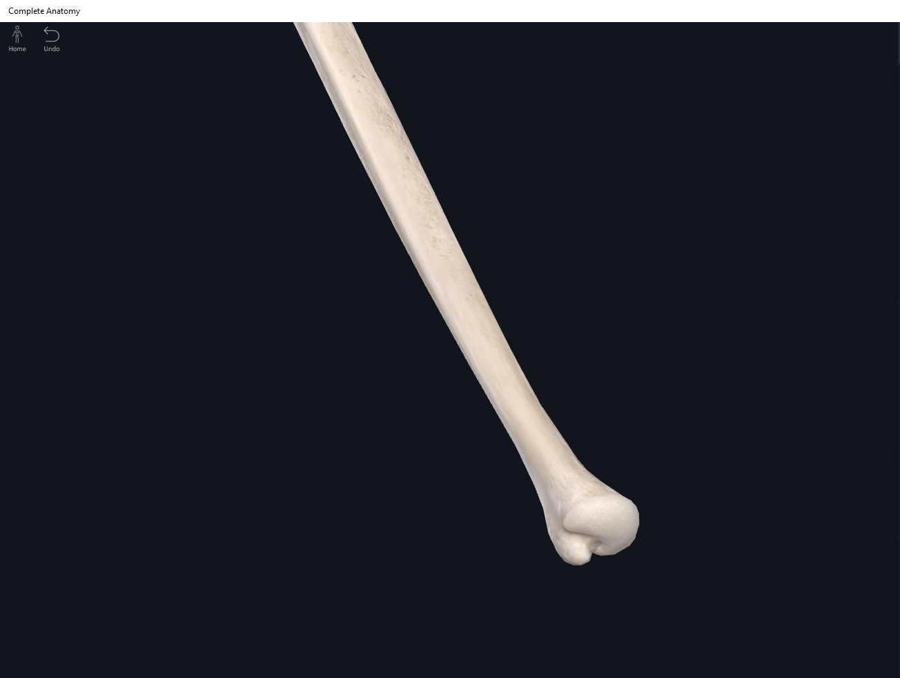
- Head: located on the distal end of the ulna.
- Styloid process: distal and posterior end of the ulna.
- Ulnar tuberosity.
Function.
Clinical Significance.
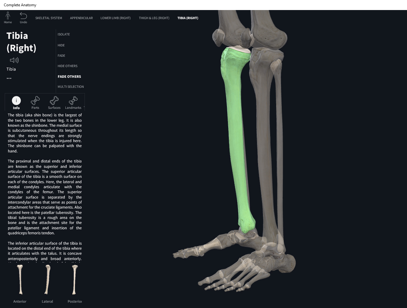
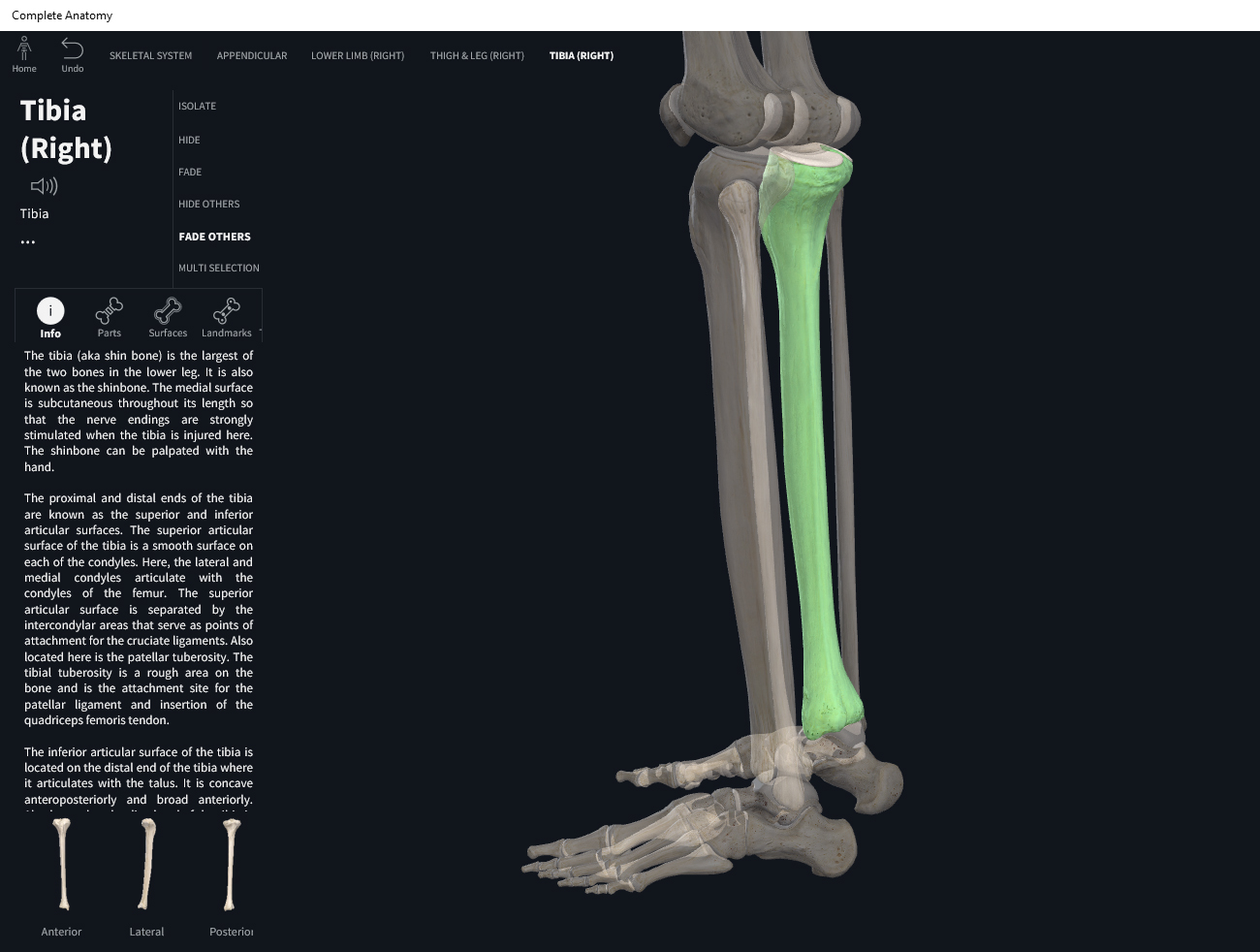
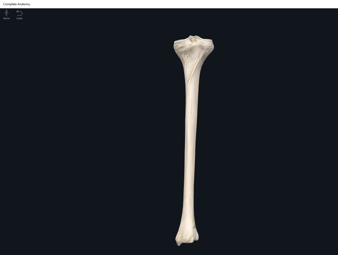
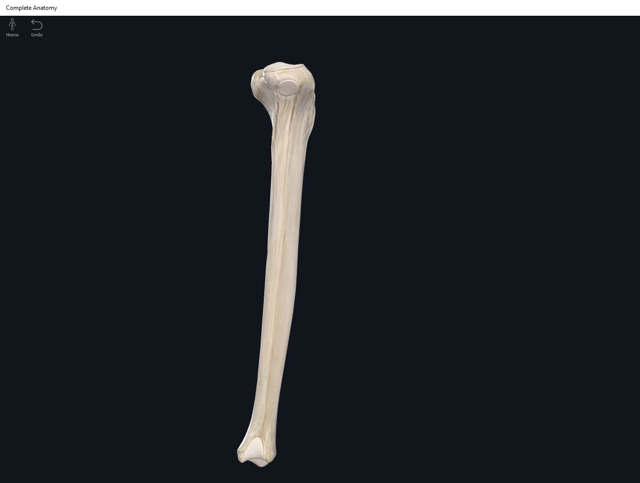
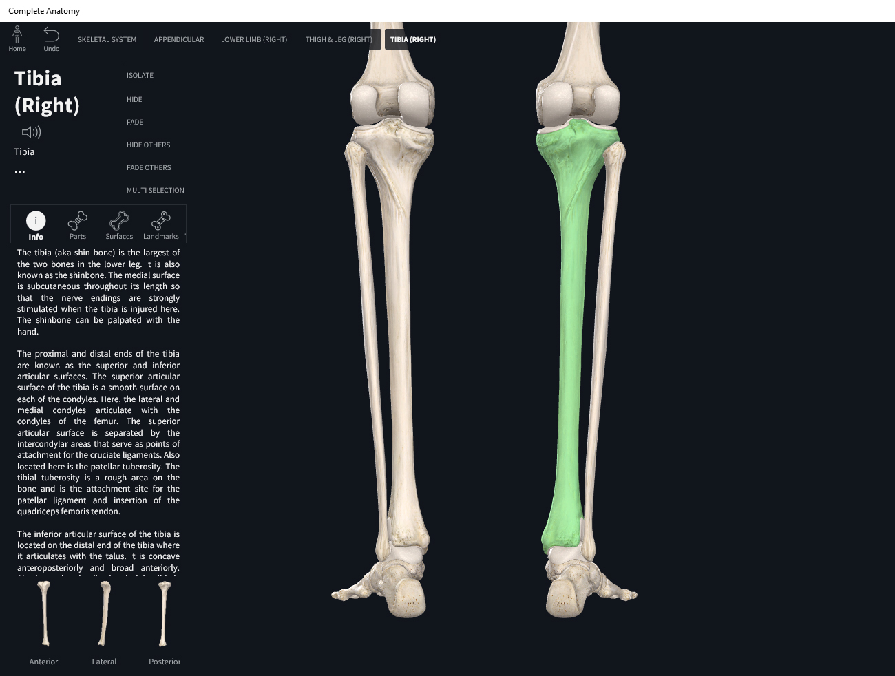
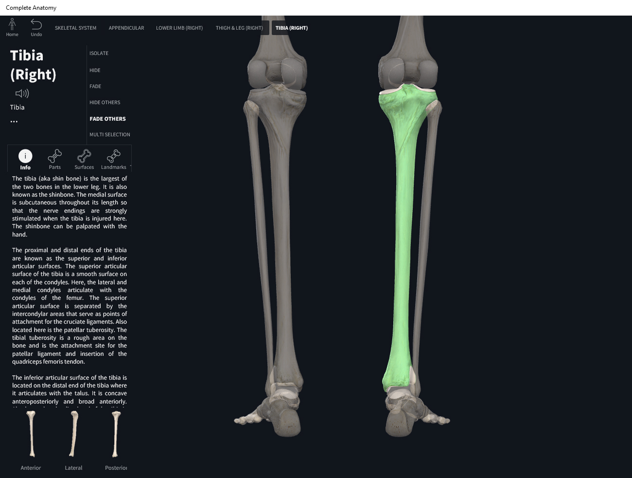
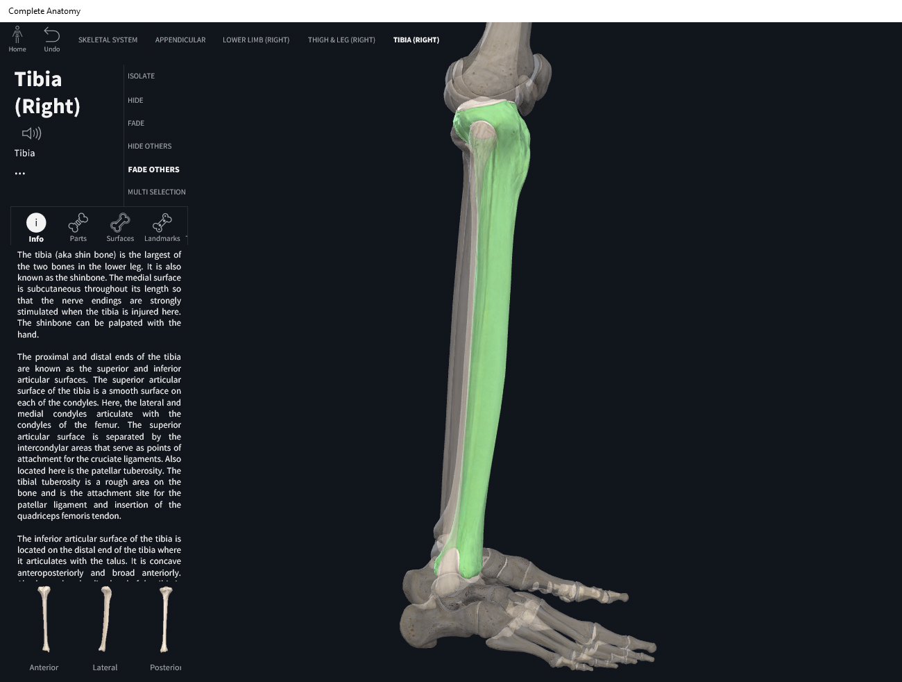
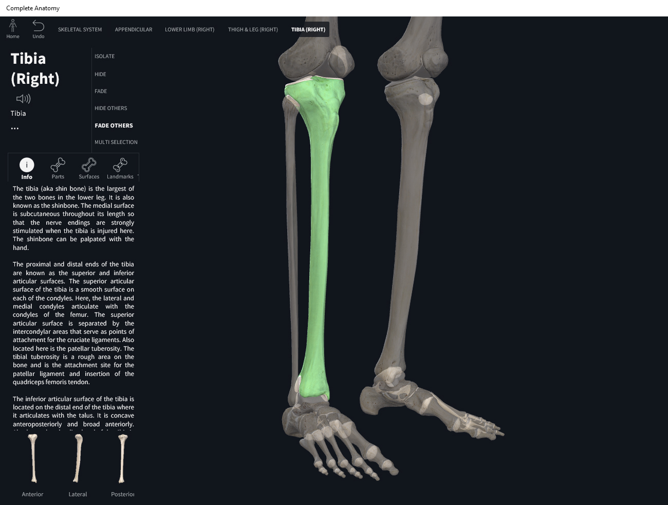
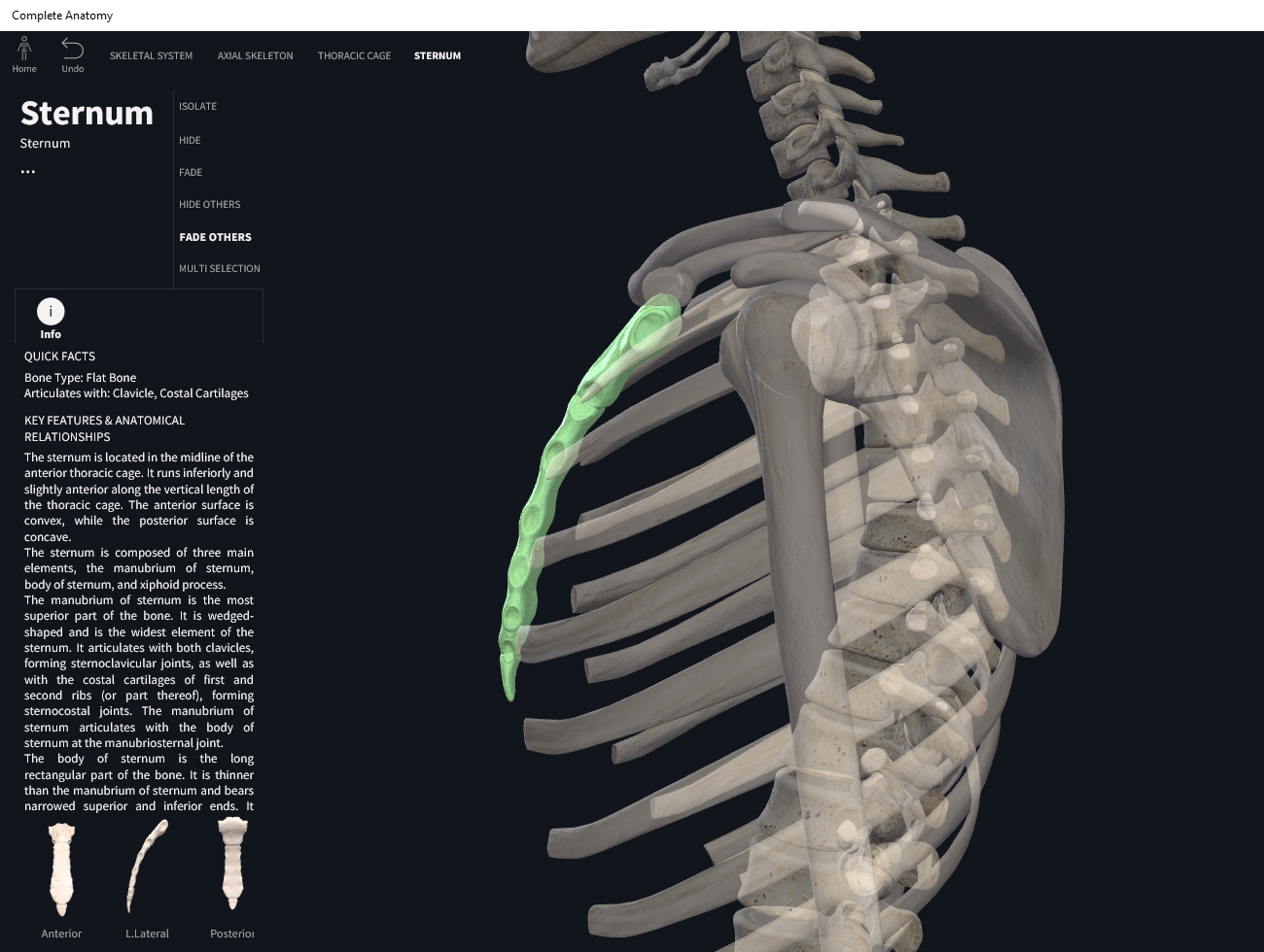
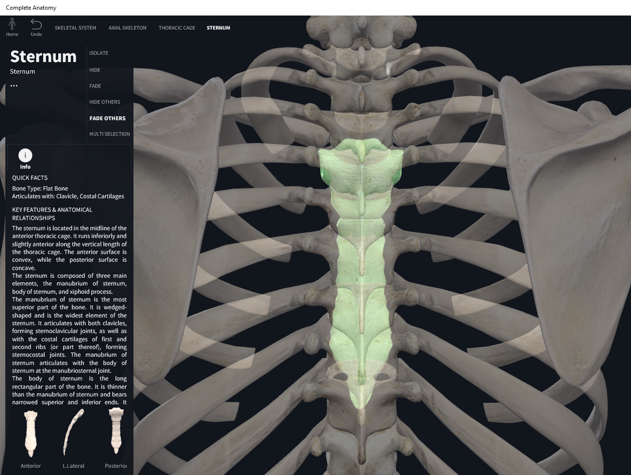
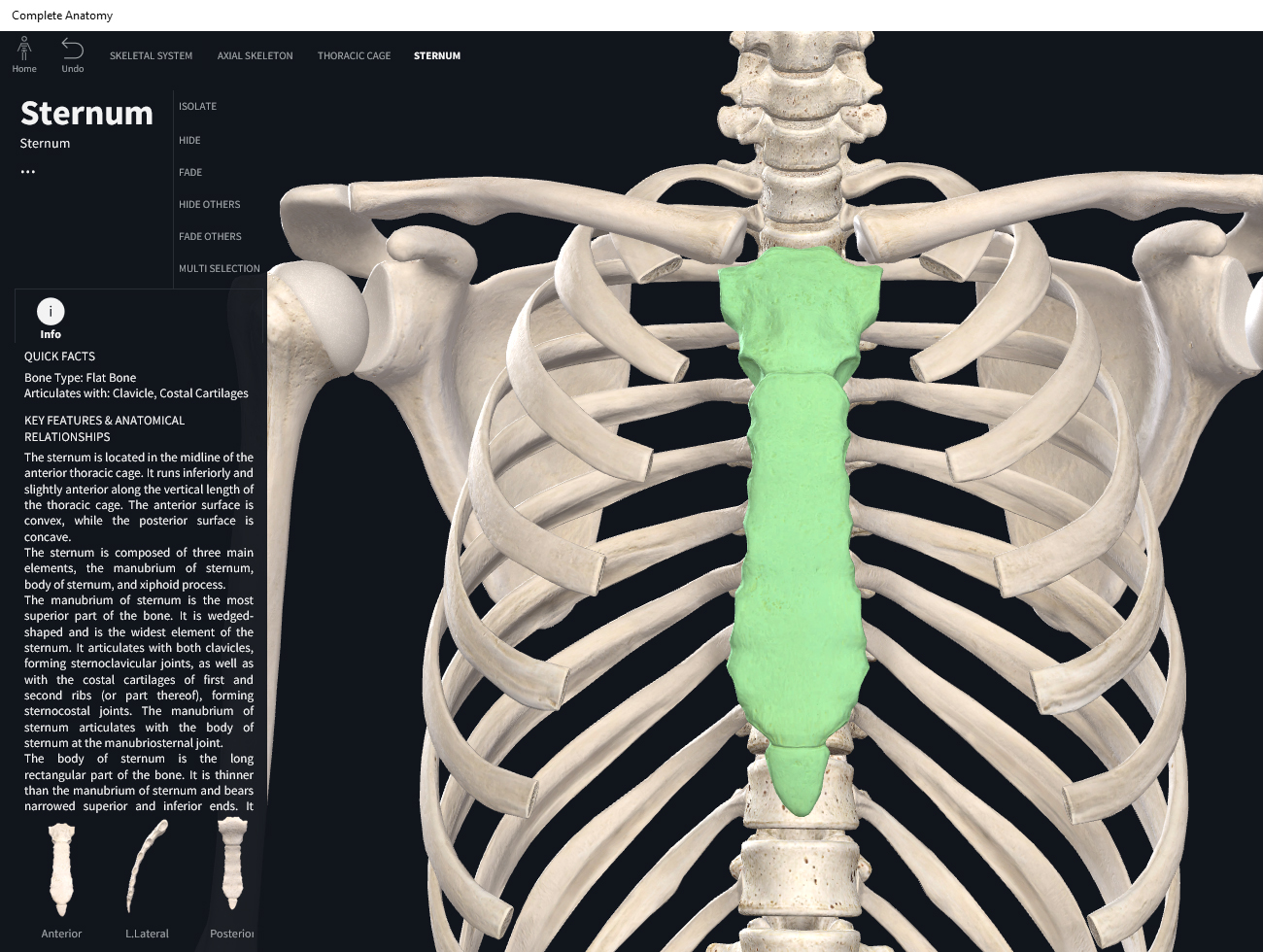
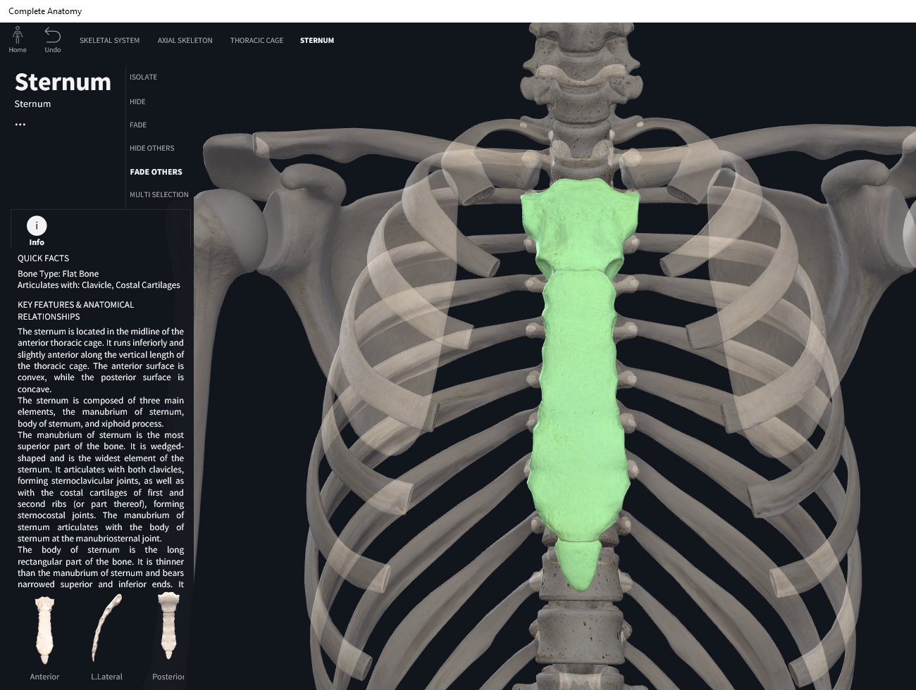
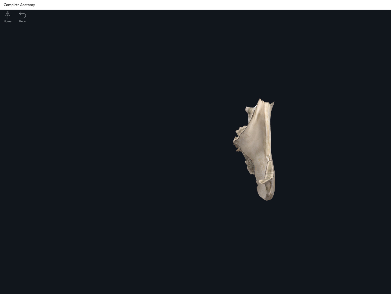
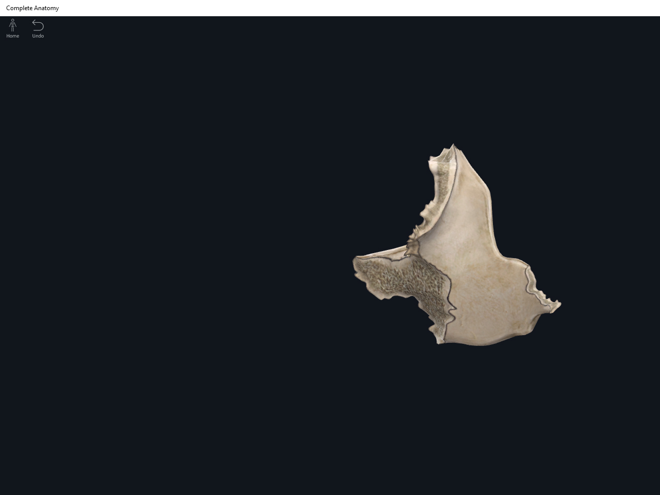
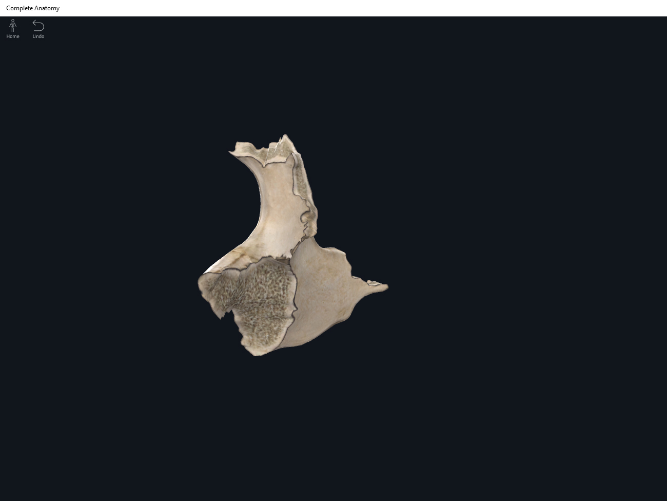
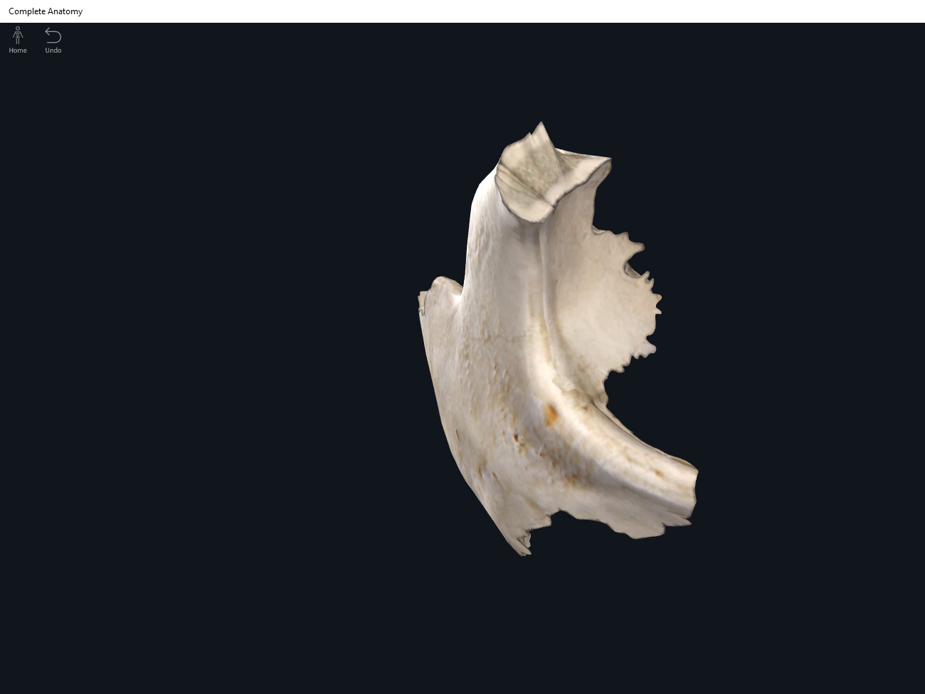
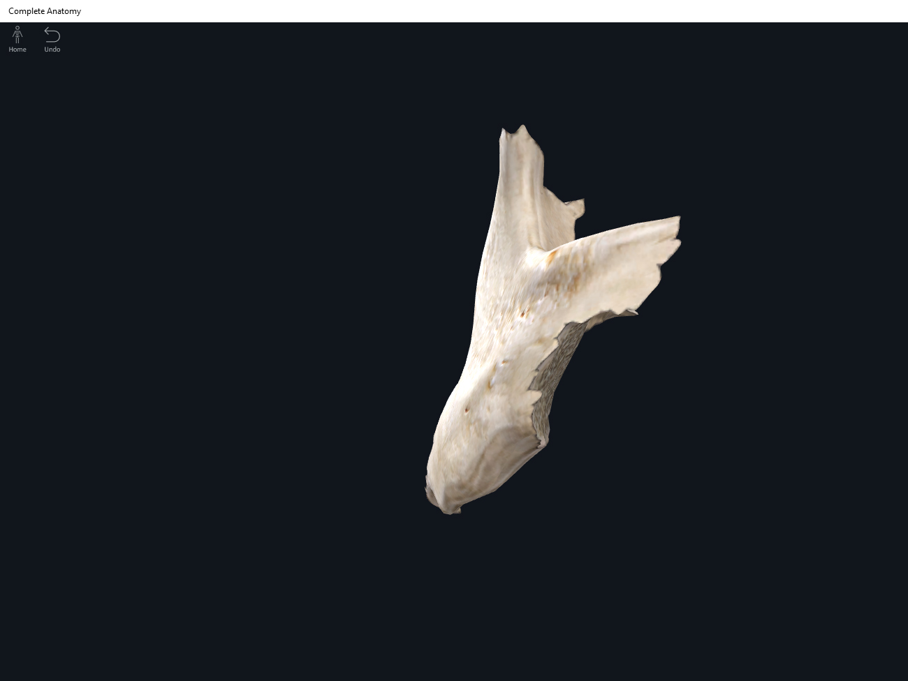
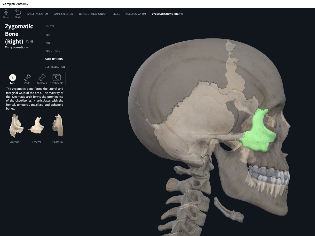
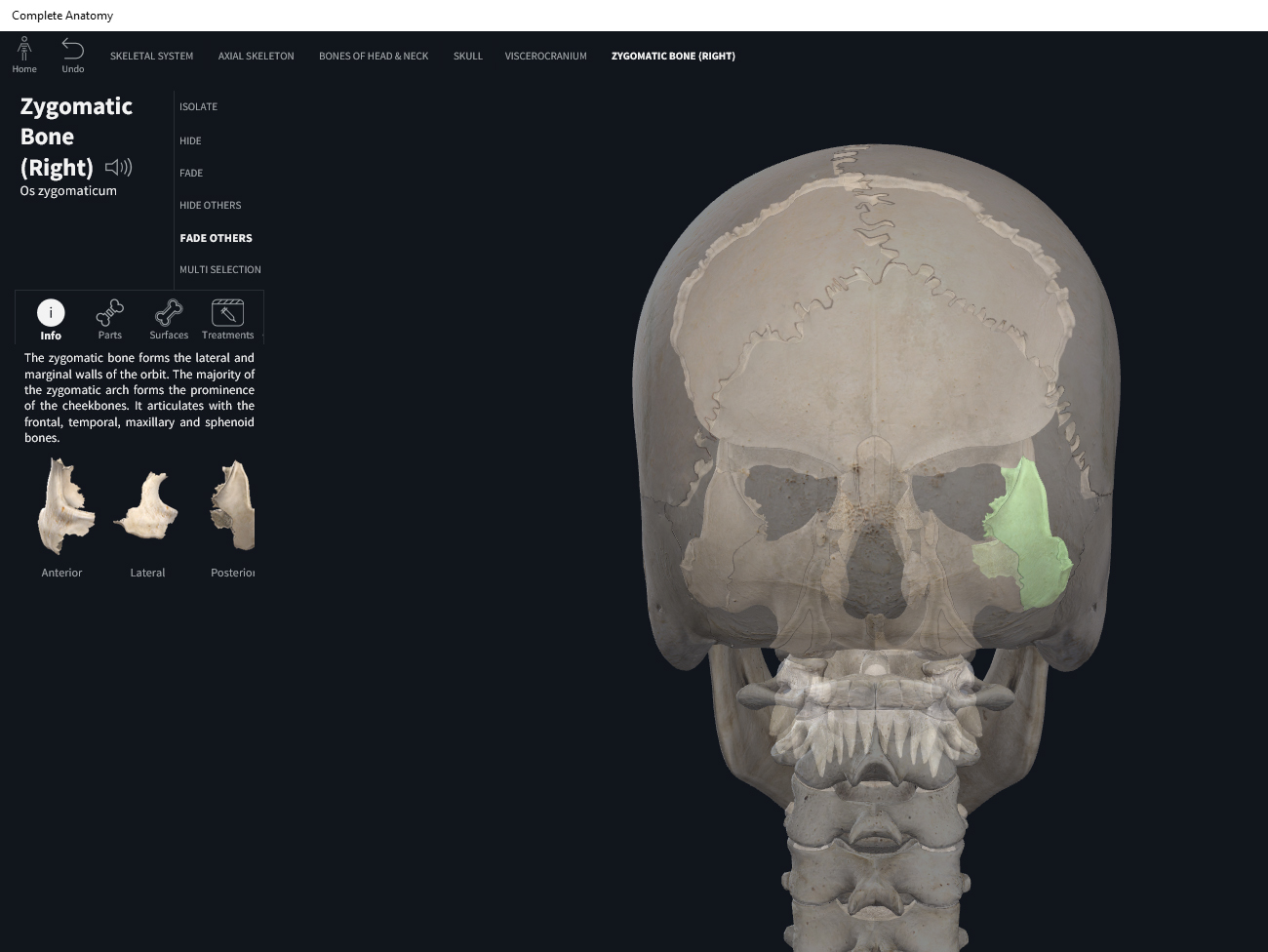
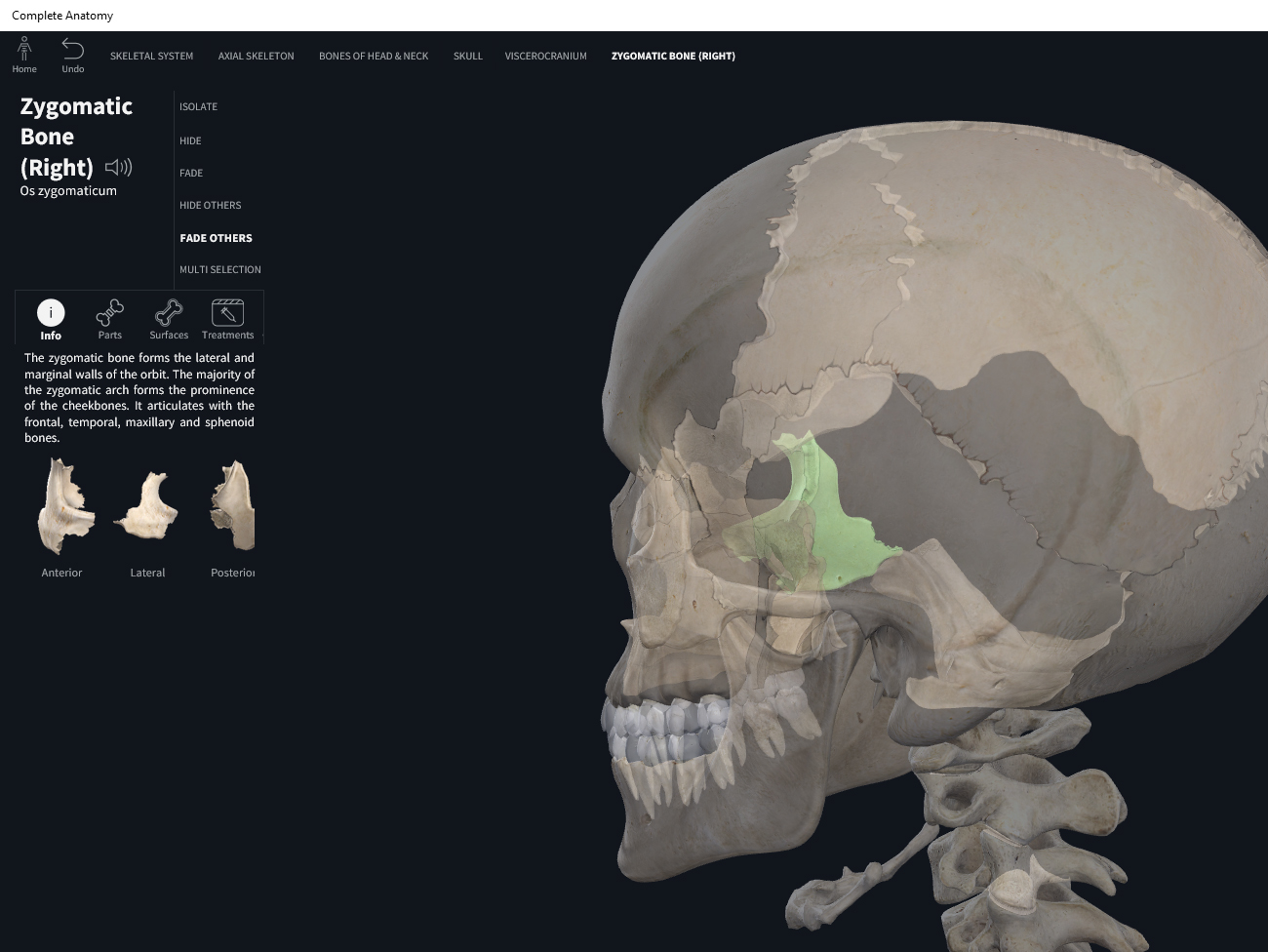
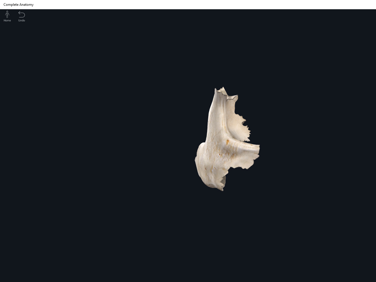
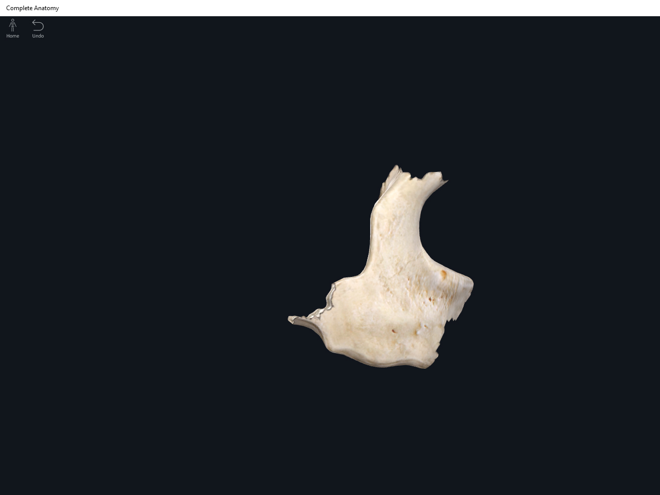
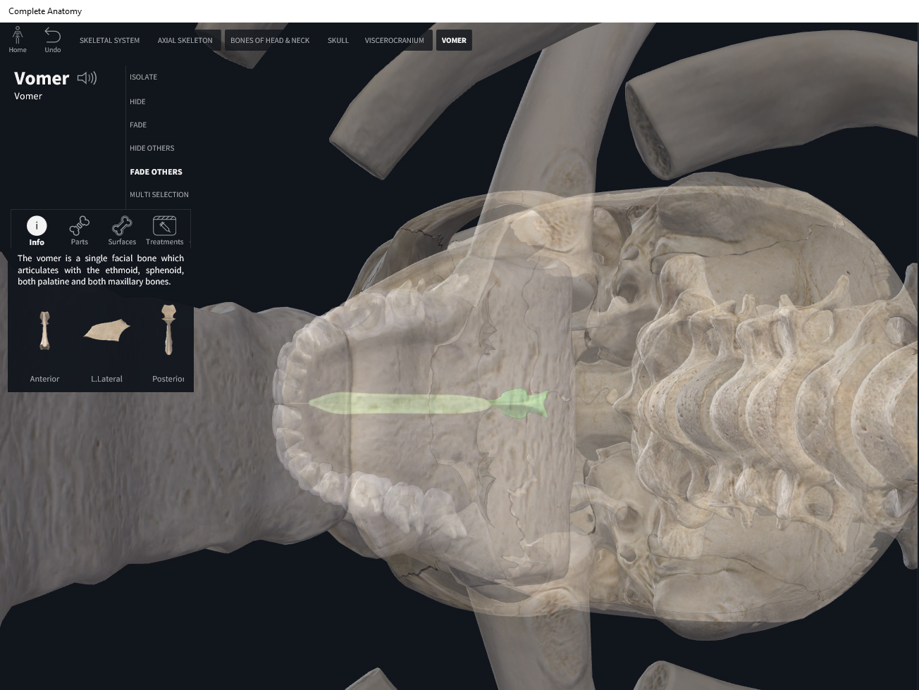
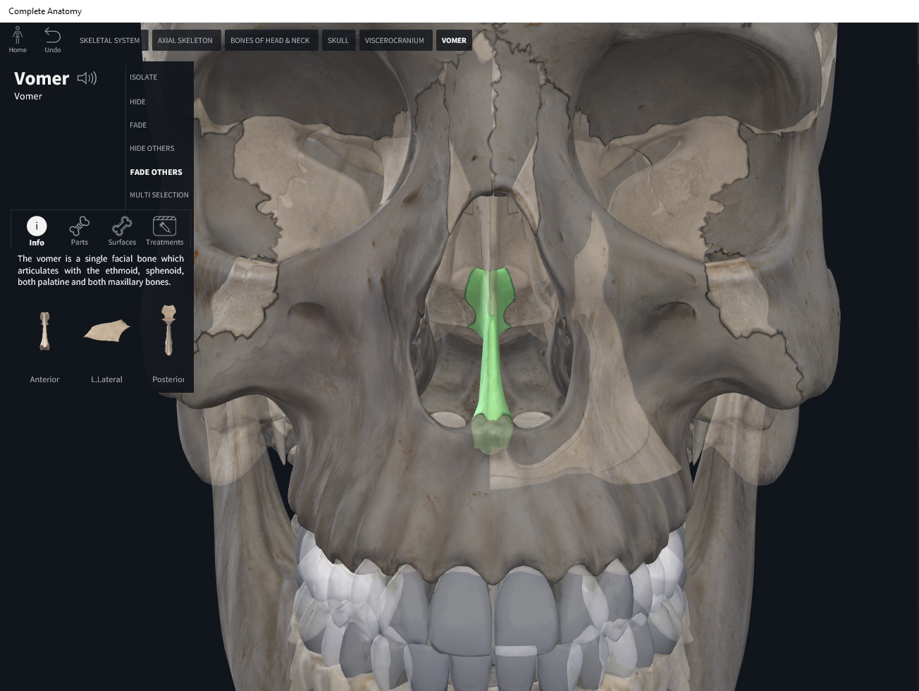
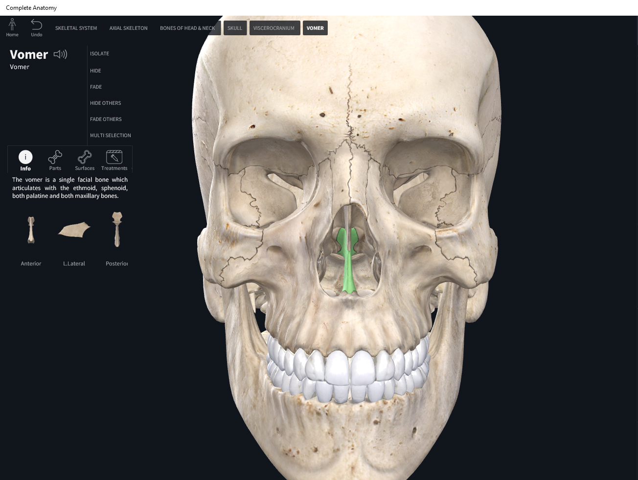
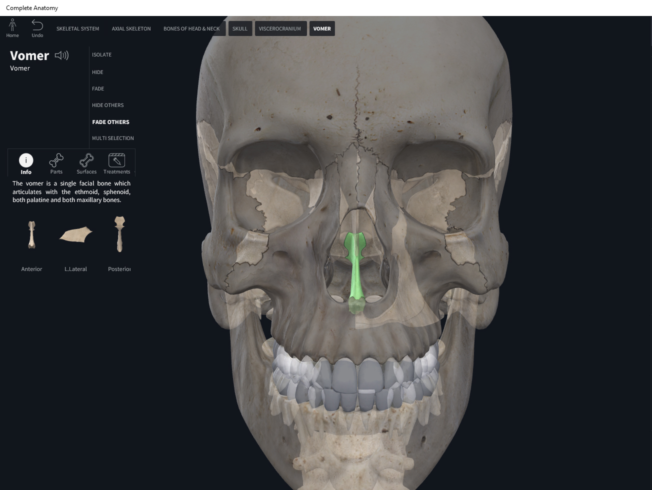
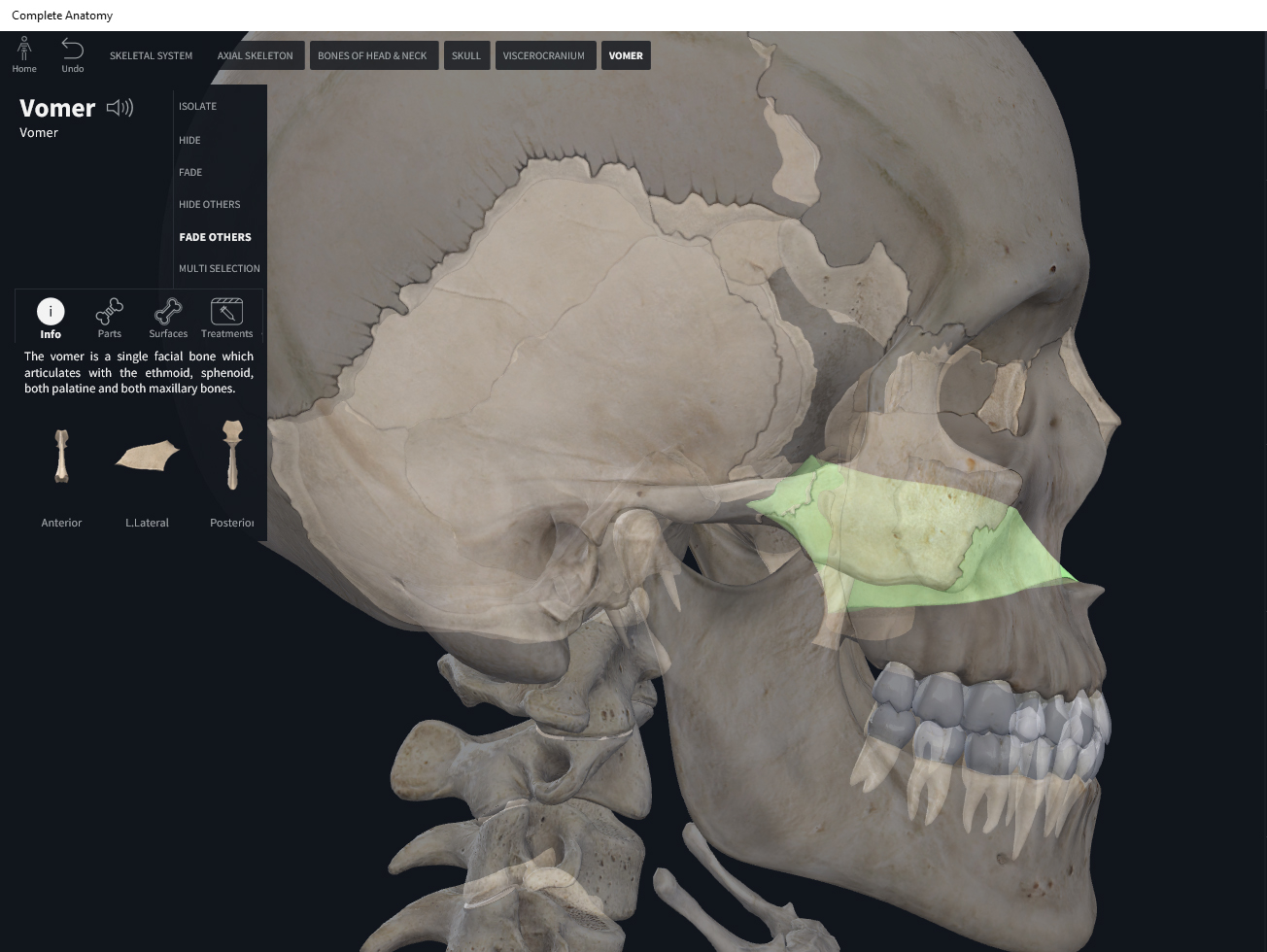
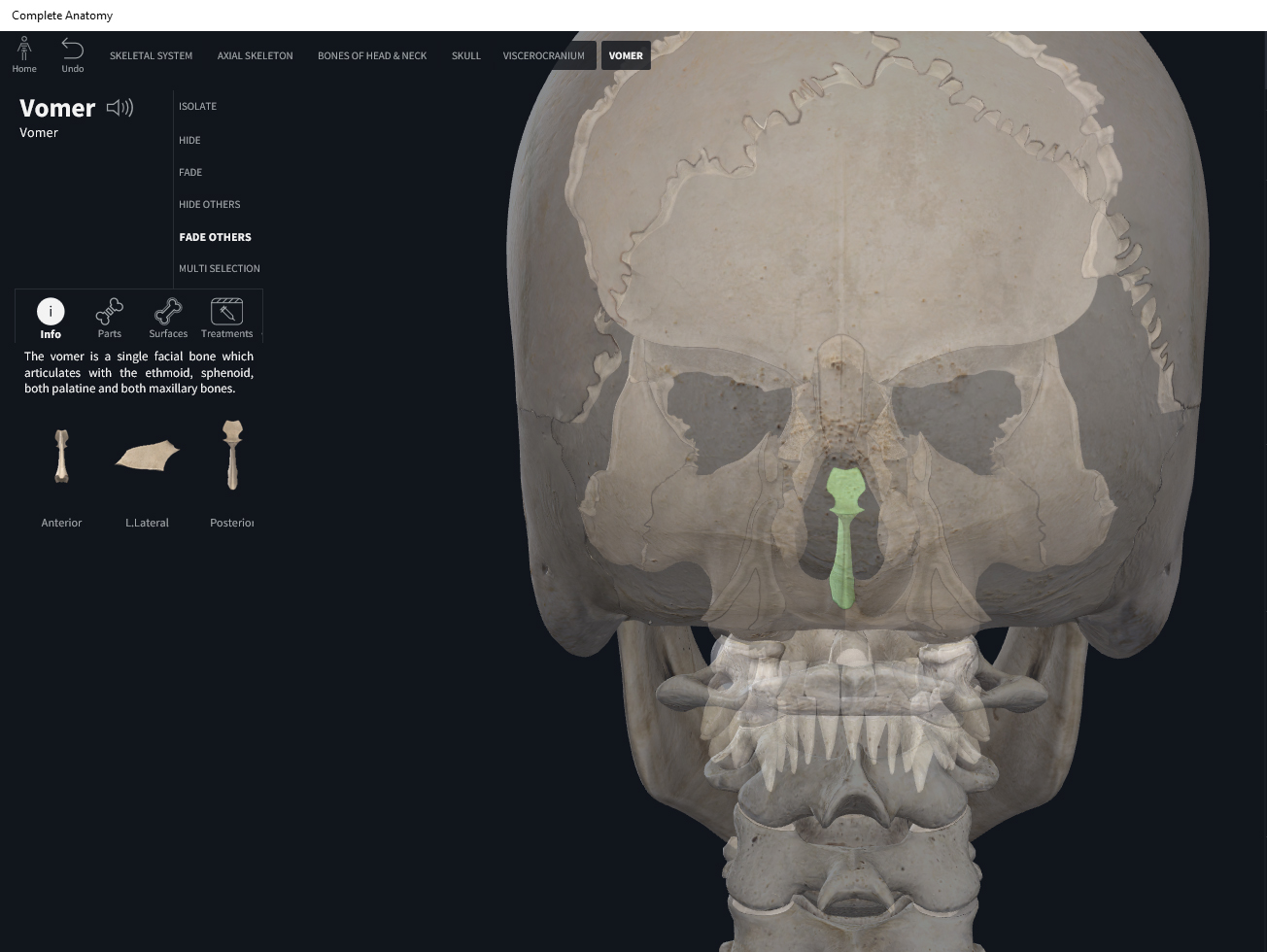
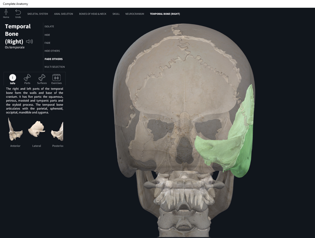
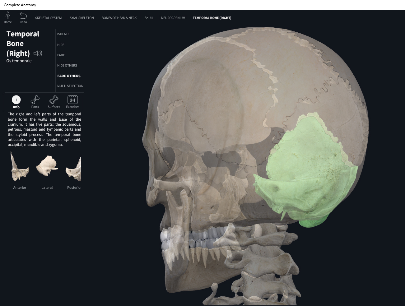
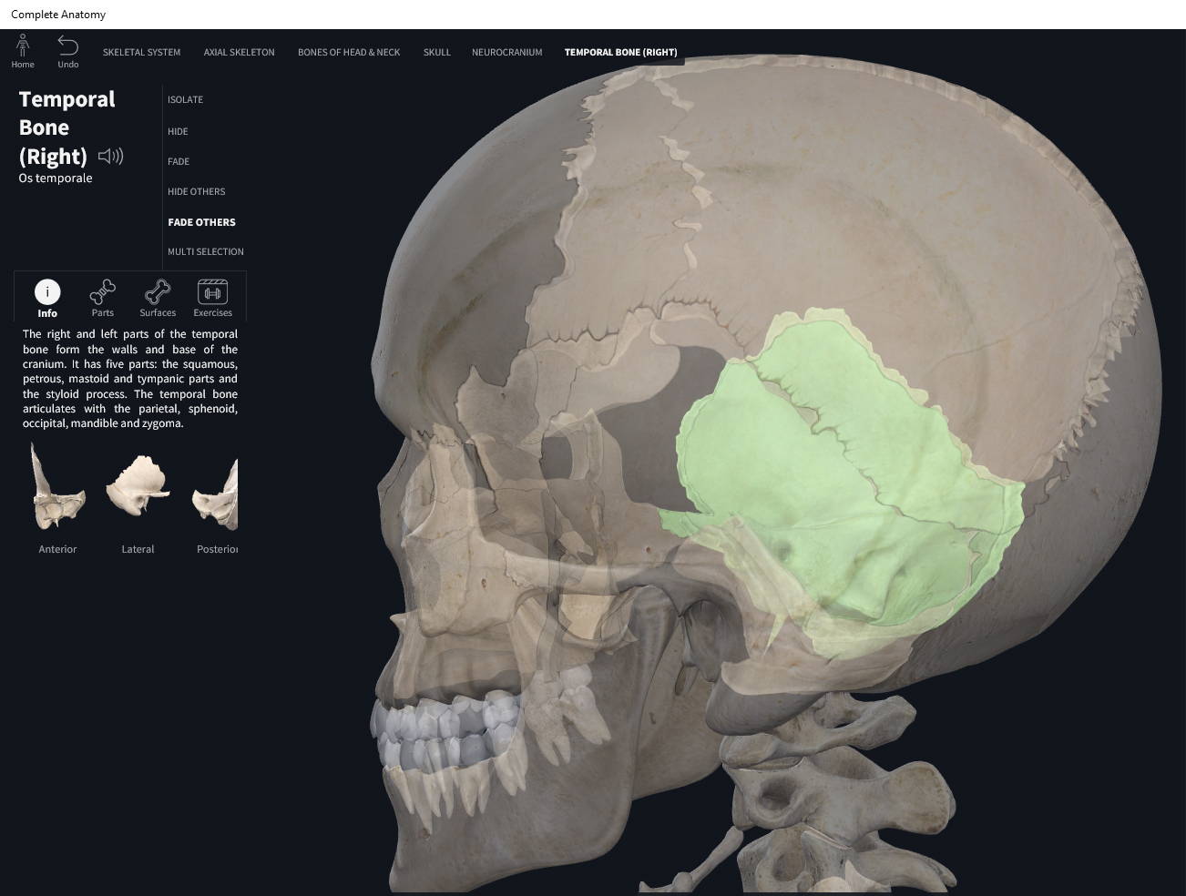
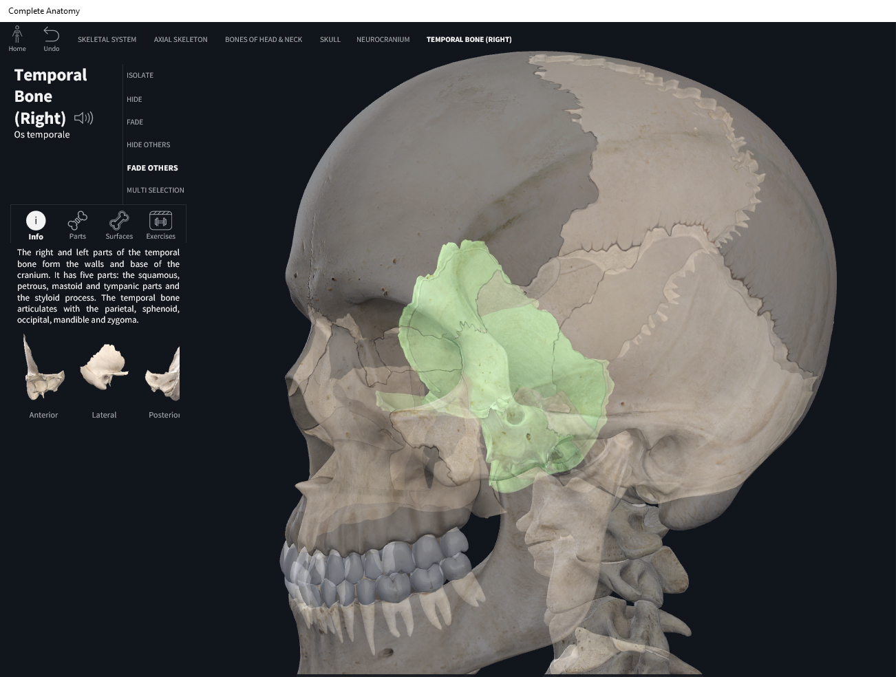
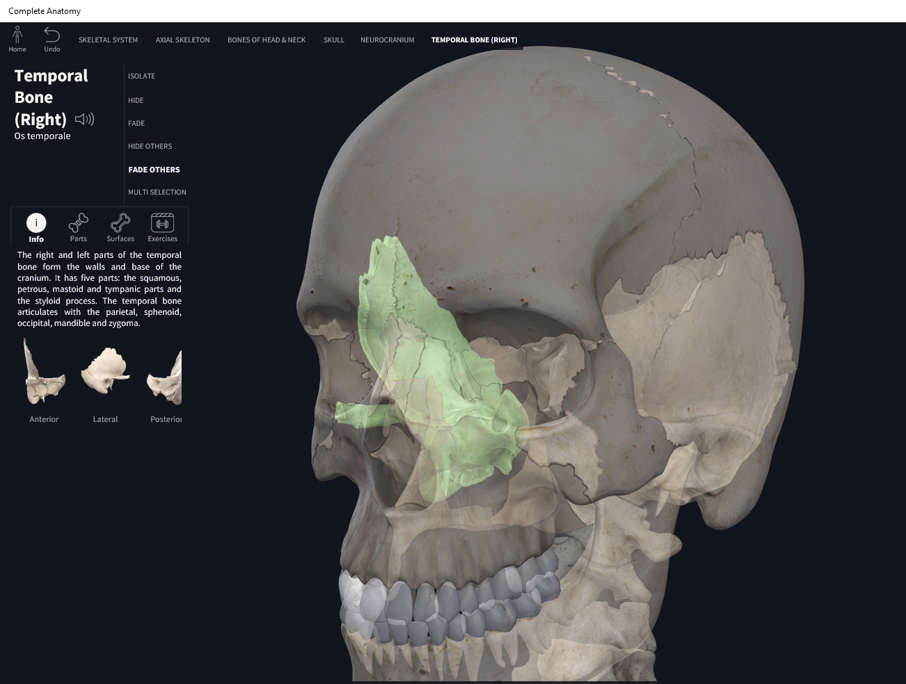
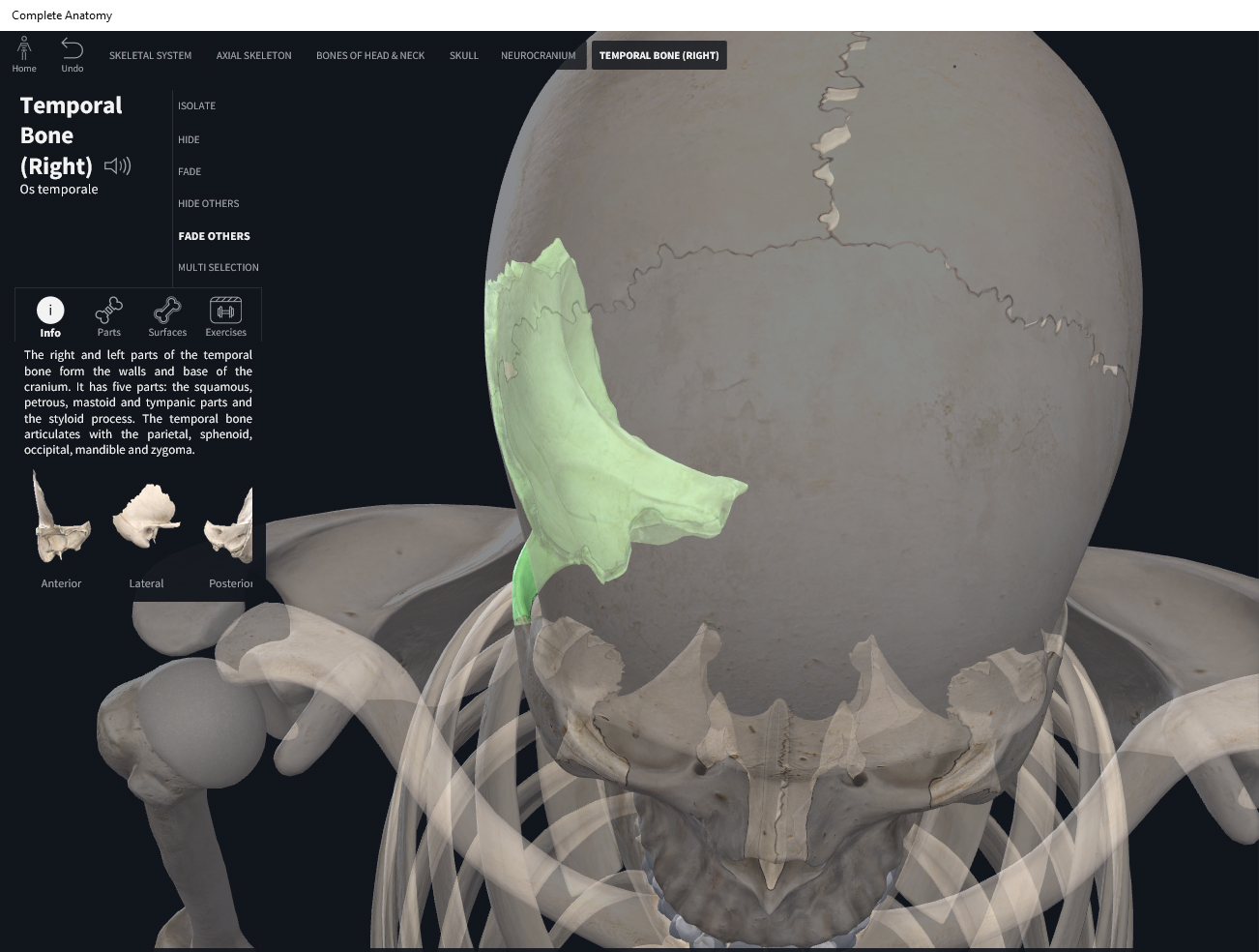
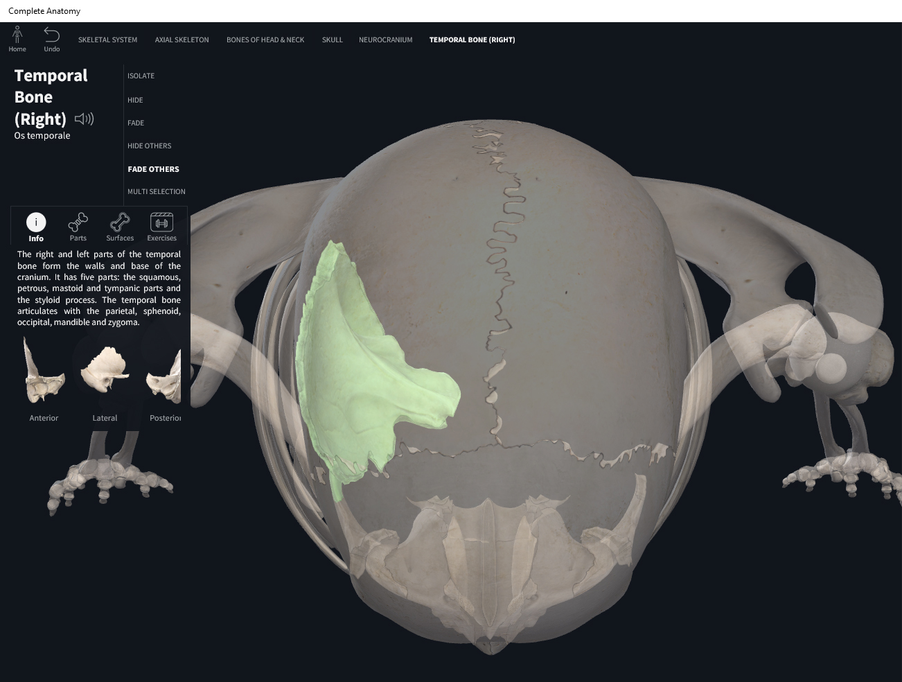
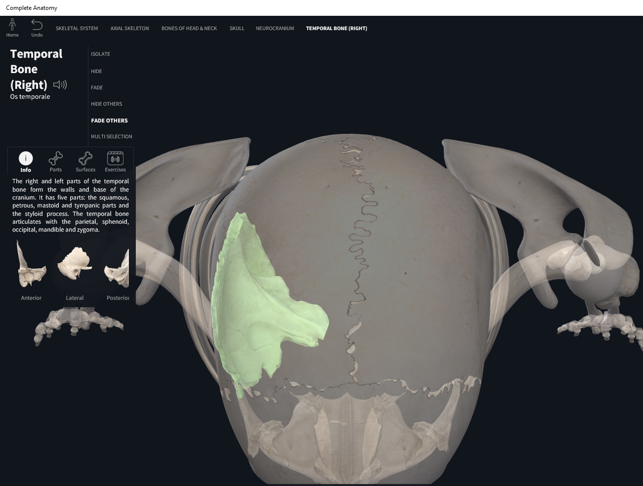
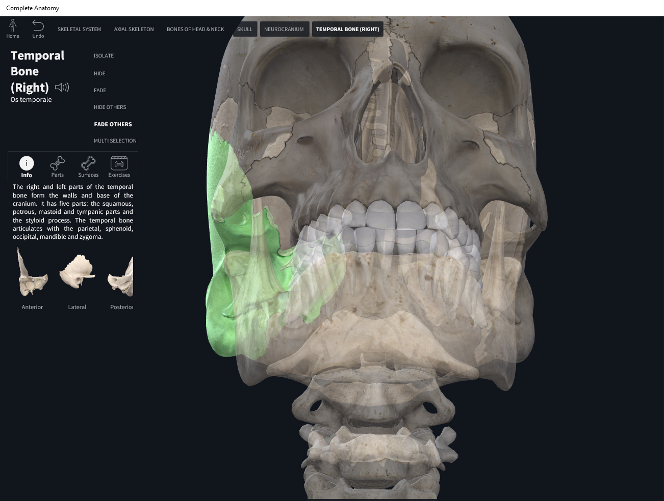
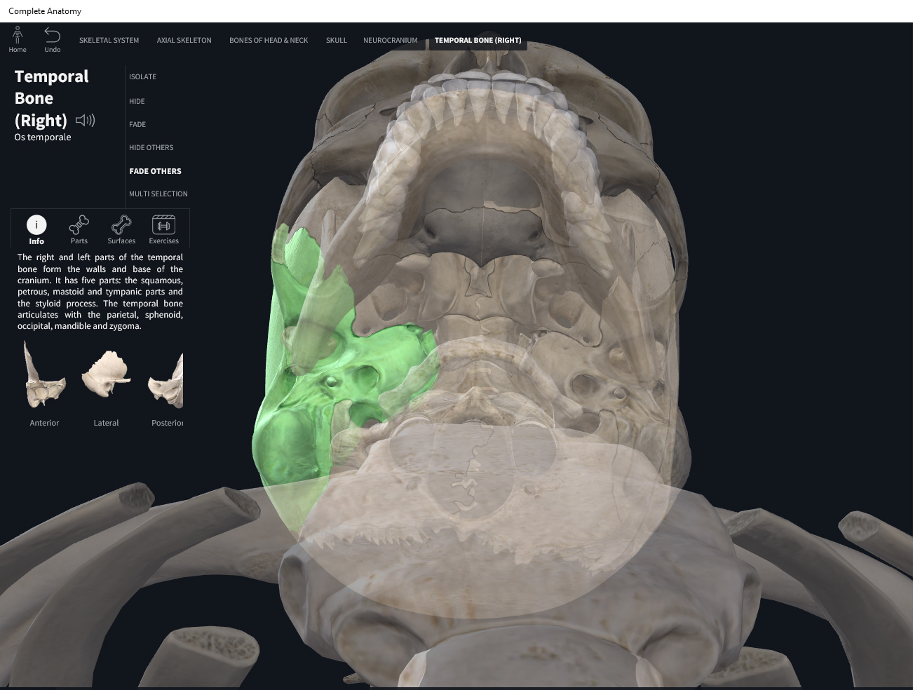
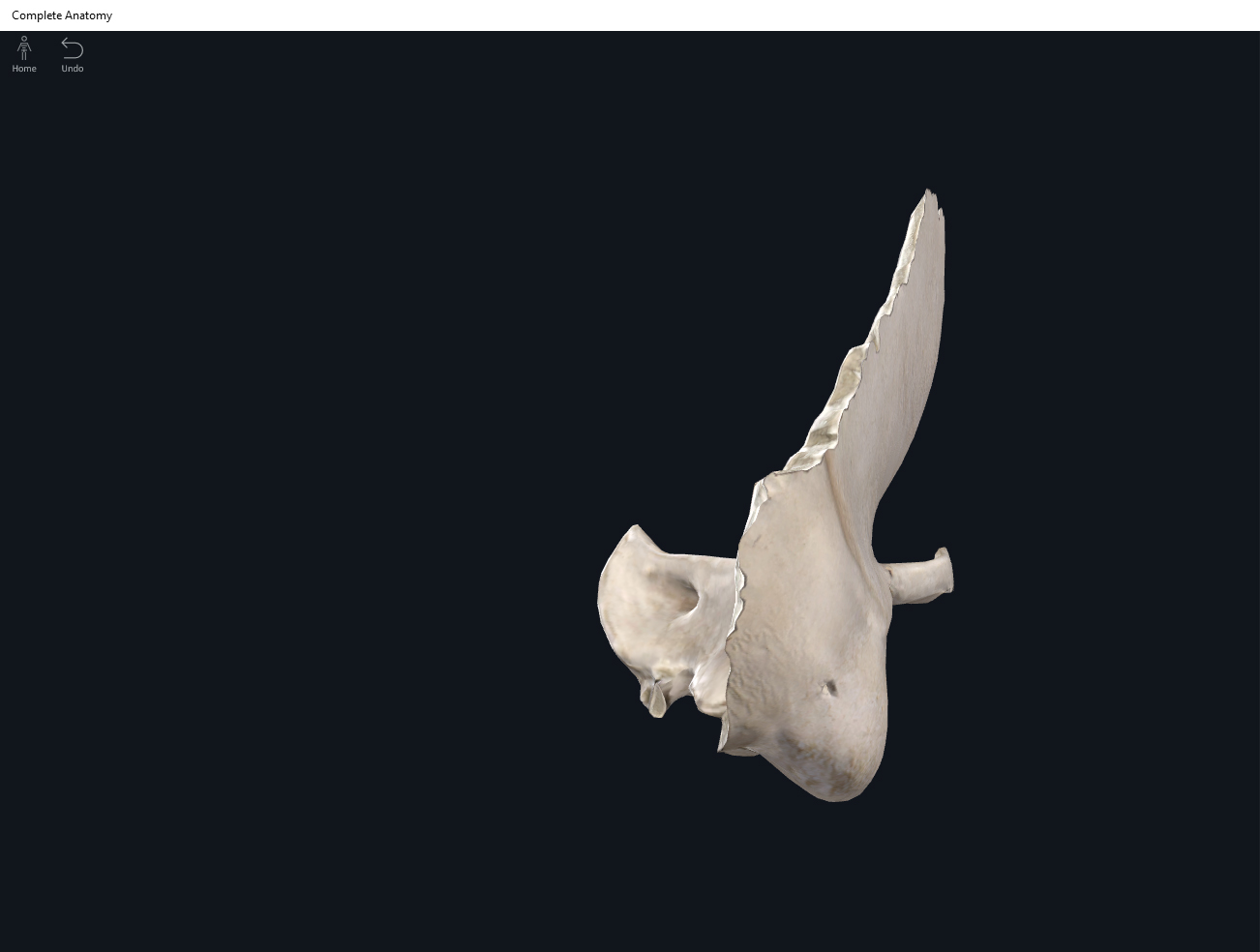
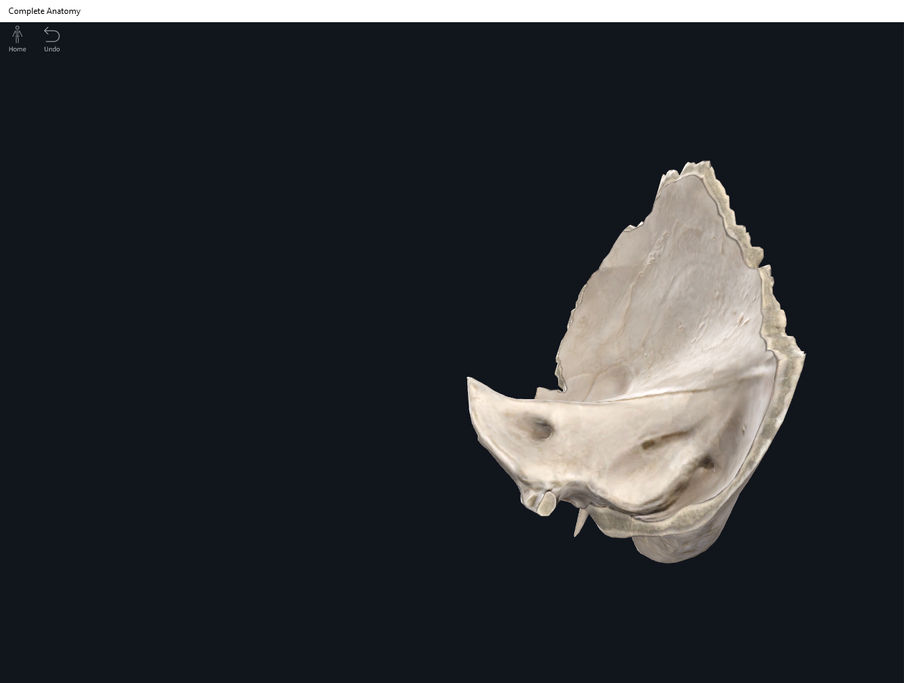
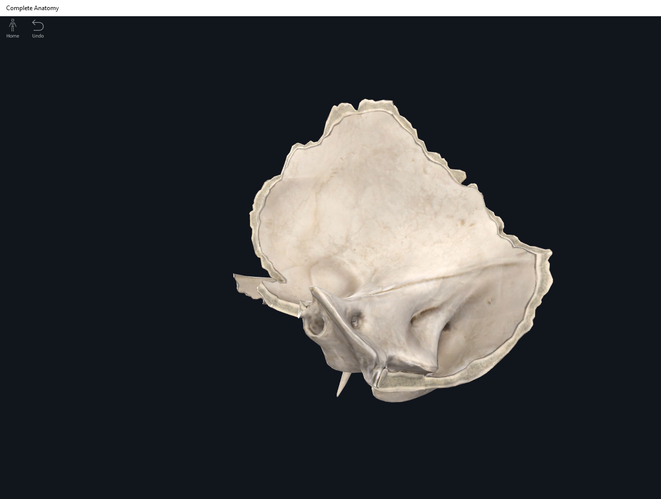
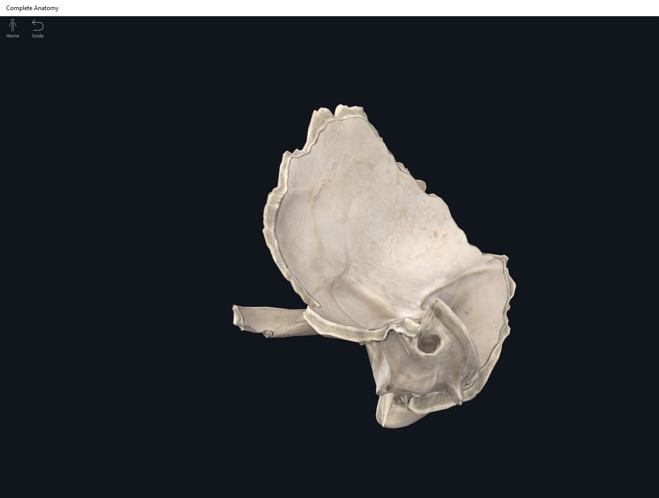
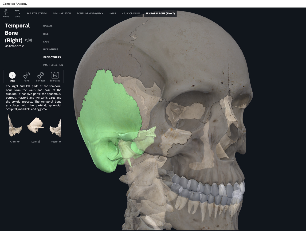
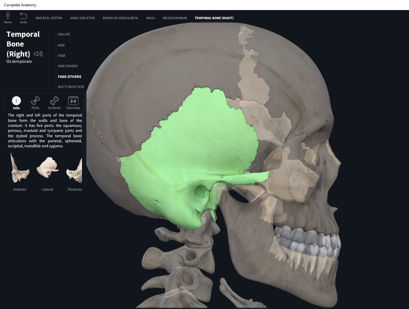
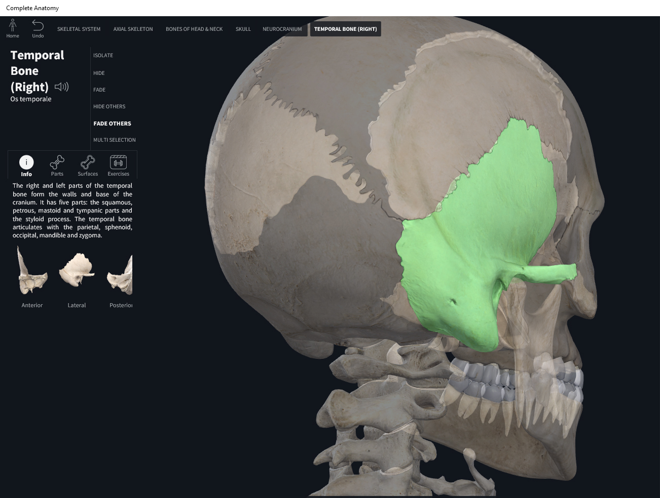
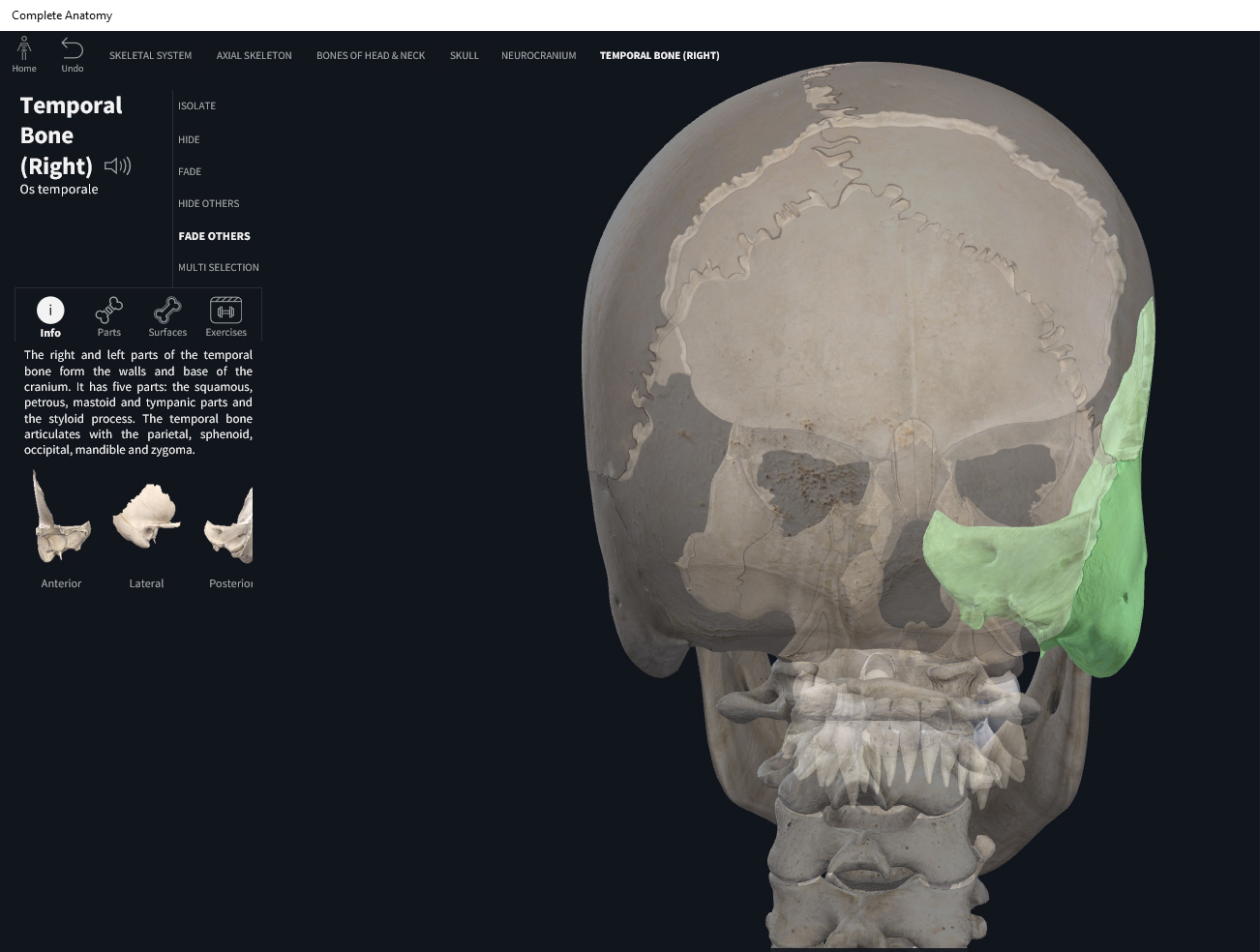
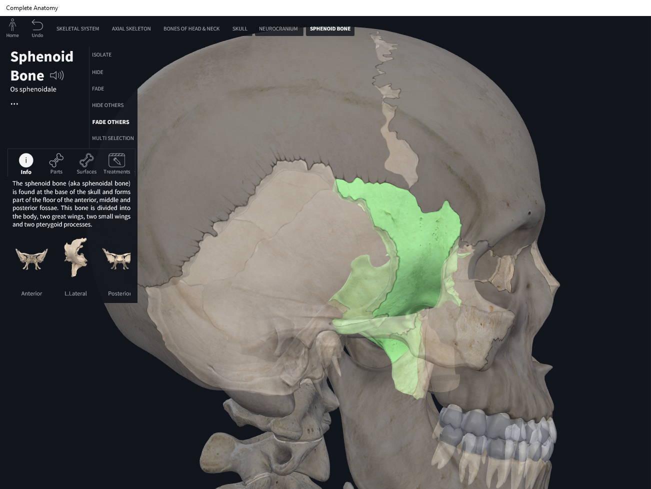
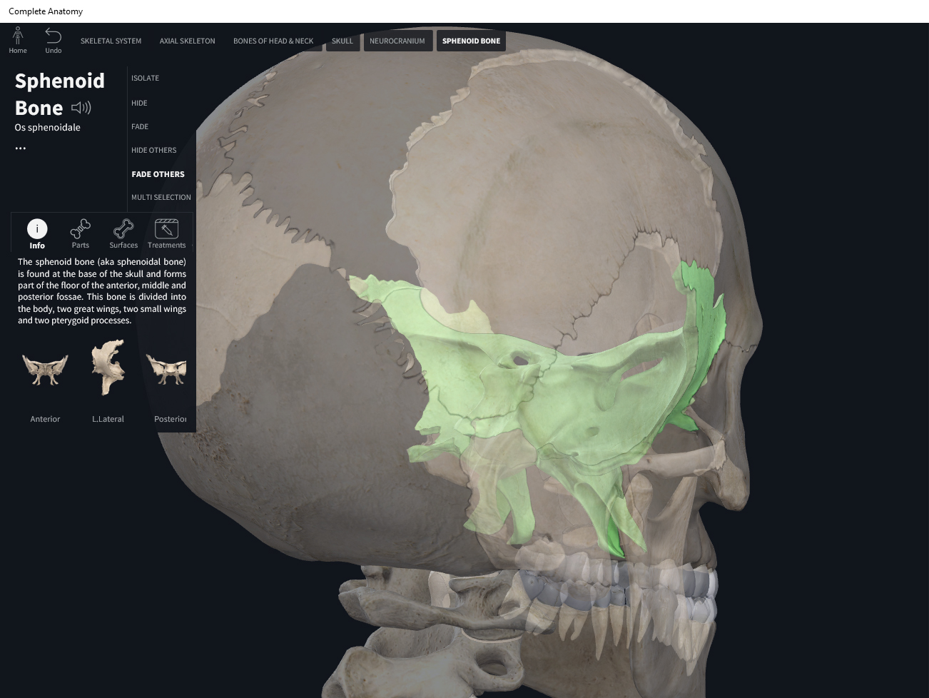
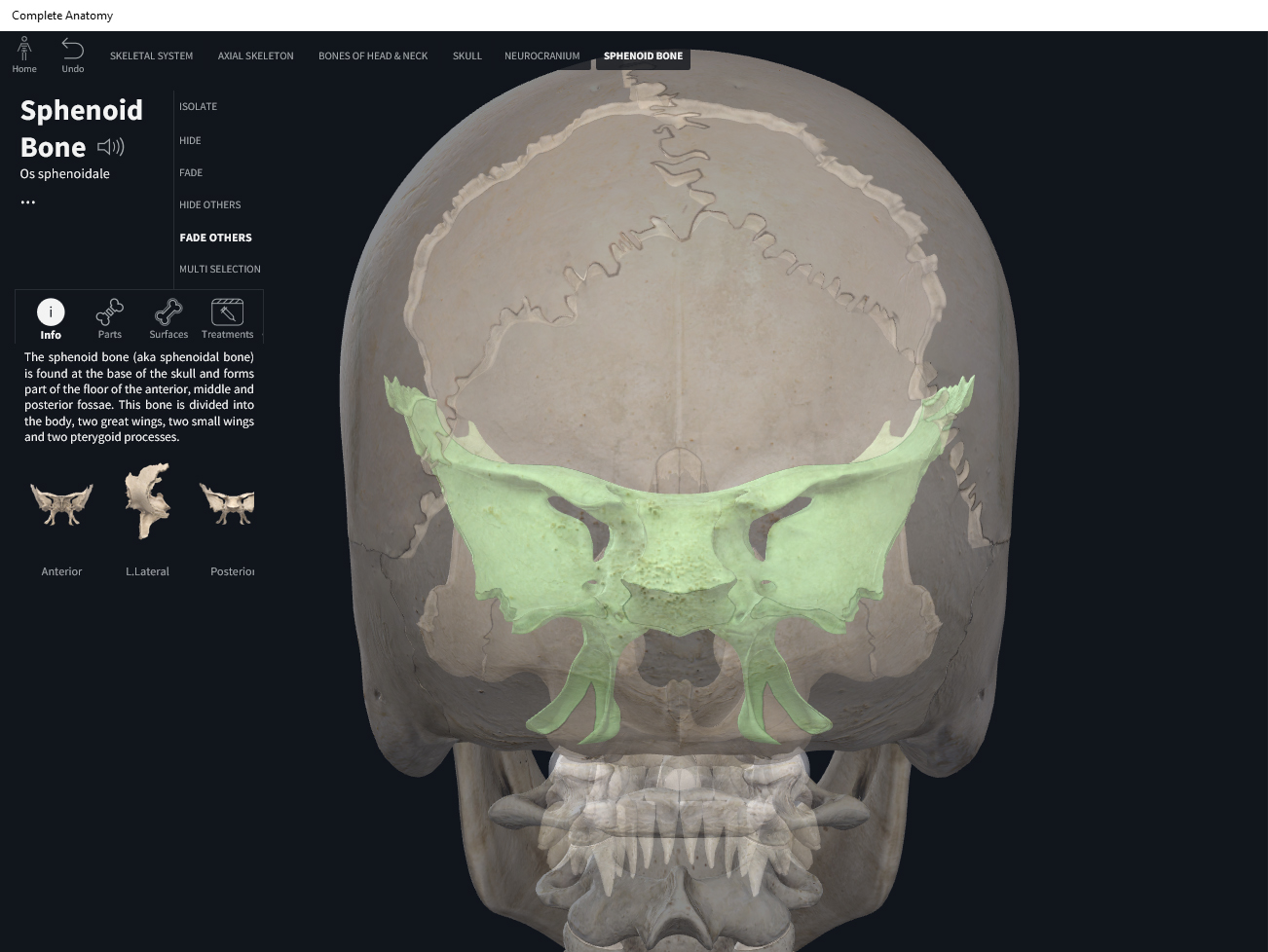
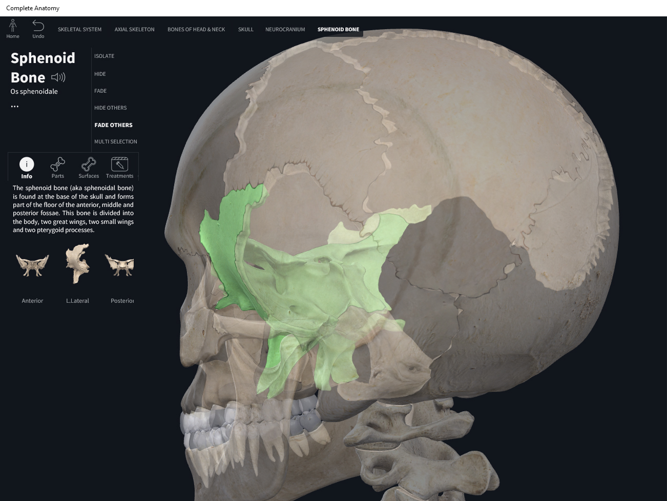
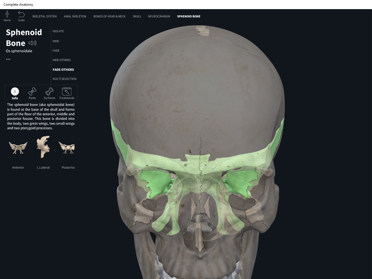
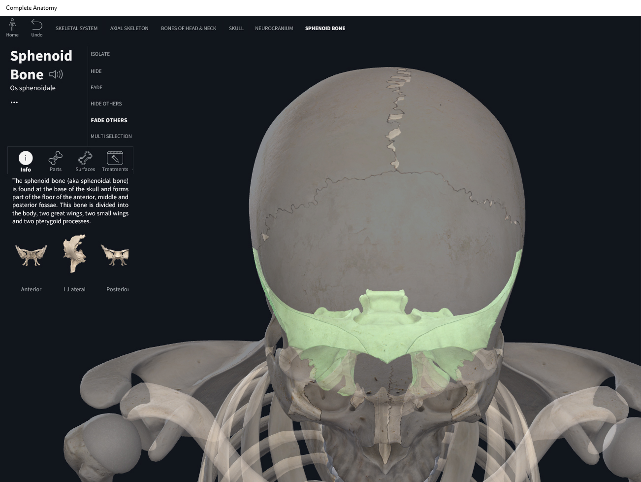
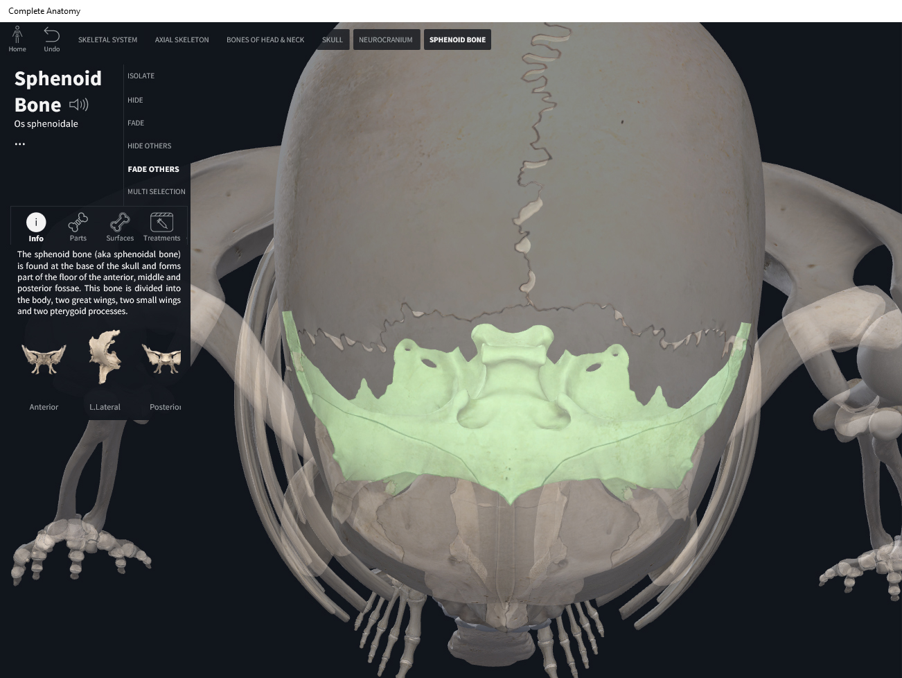
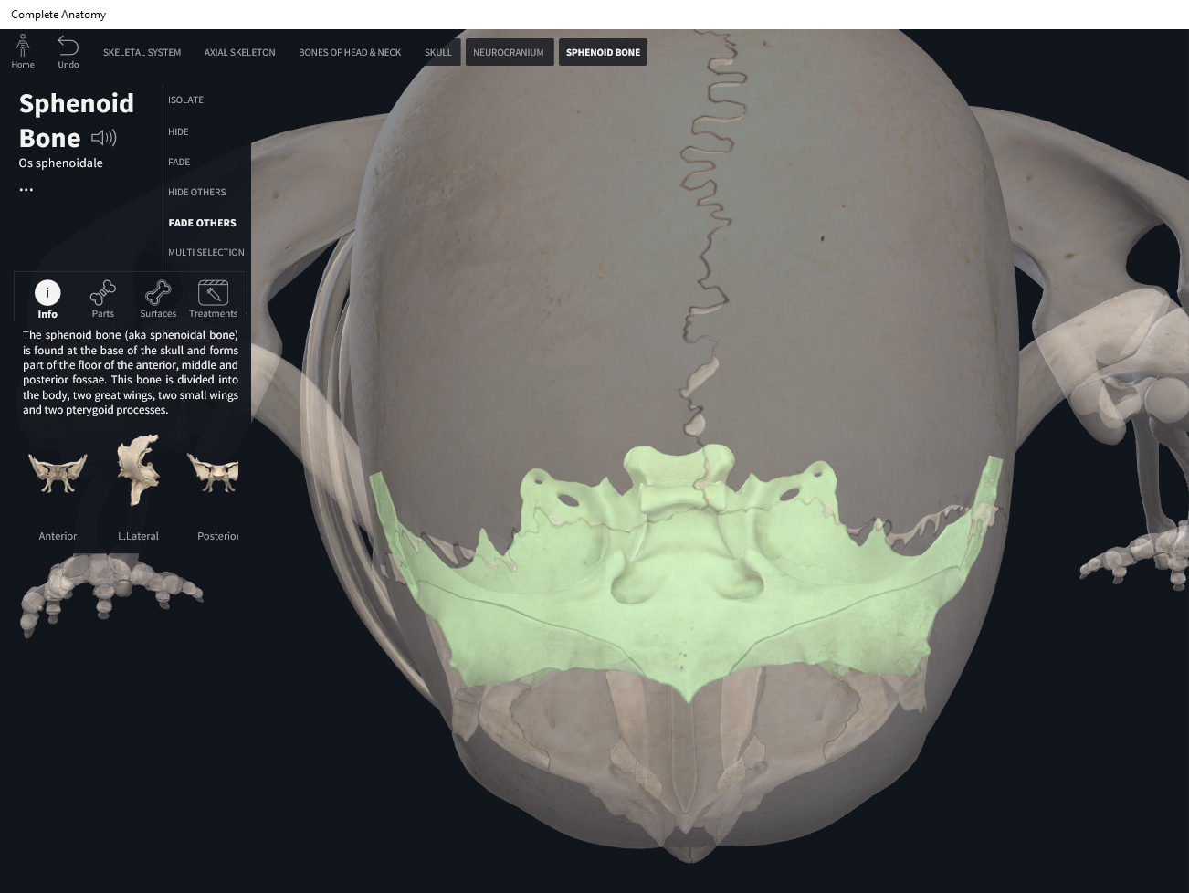
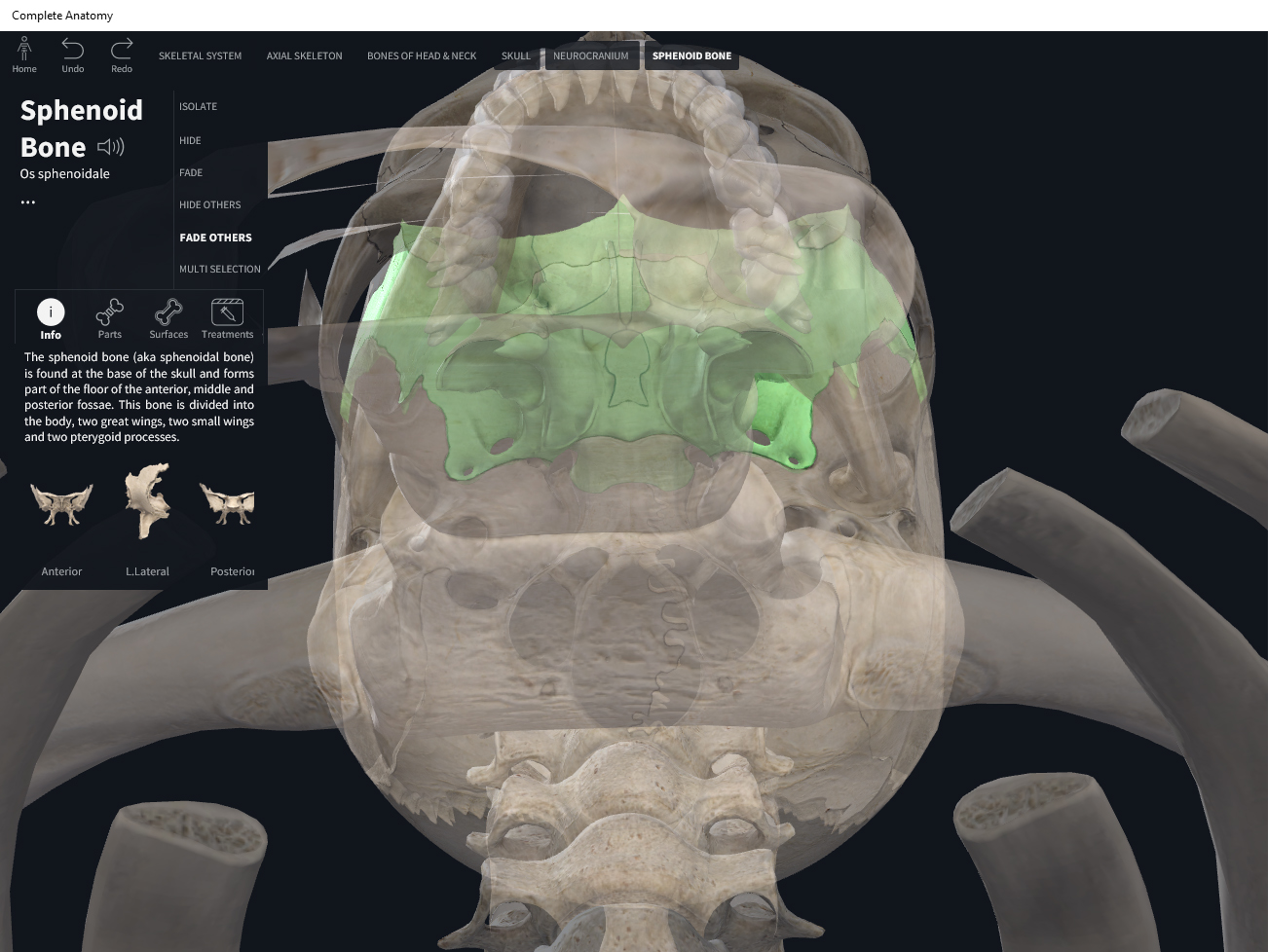
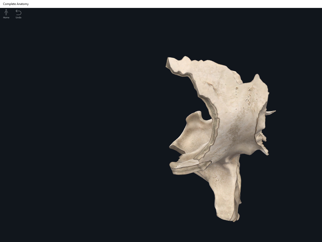
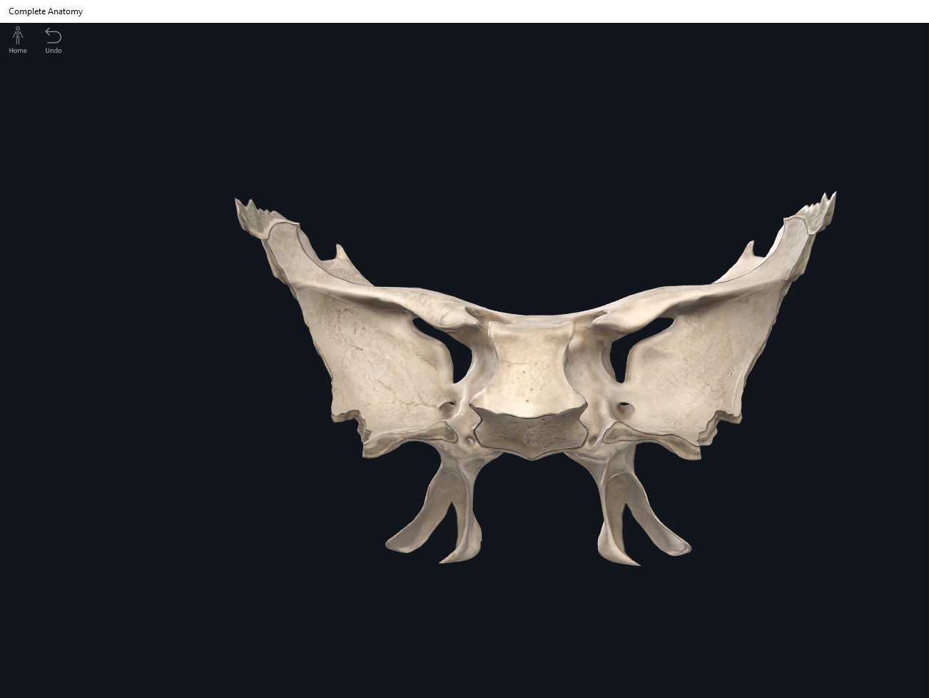
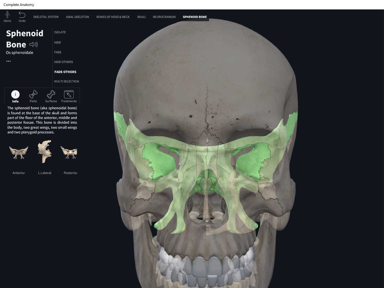
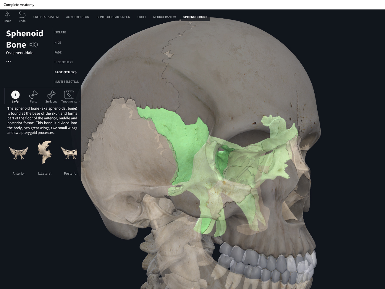
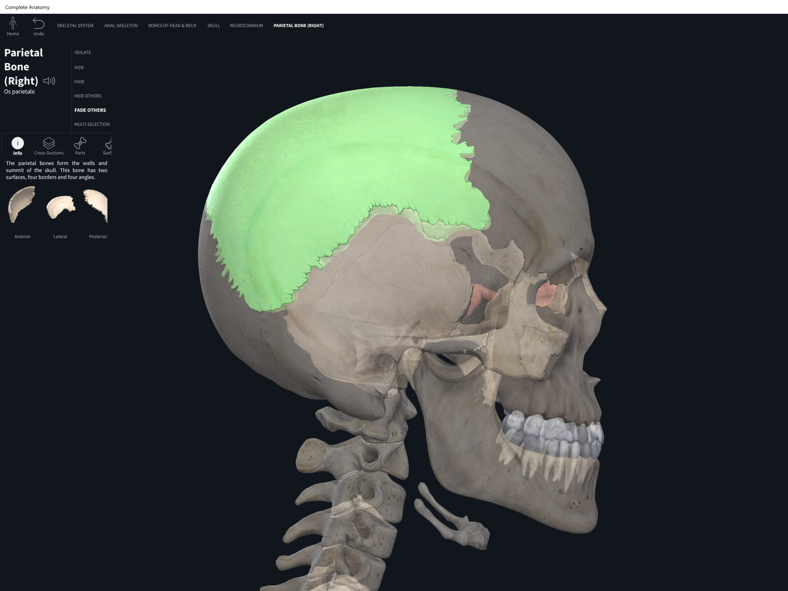
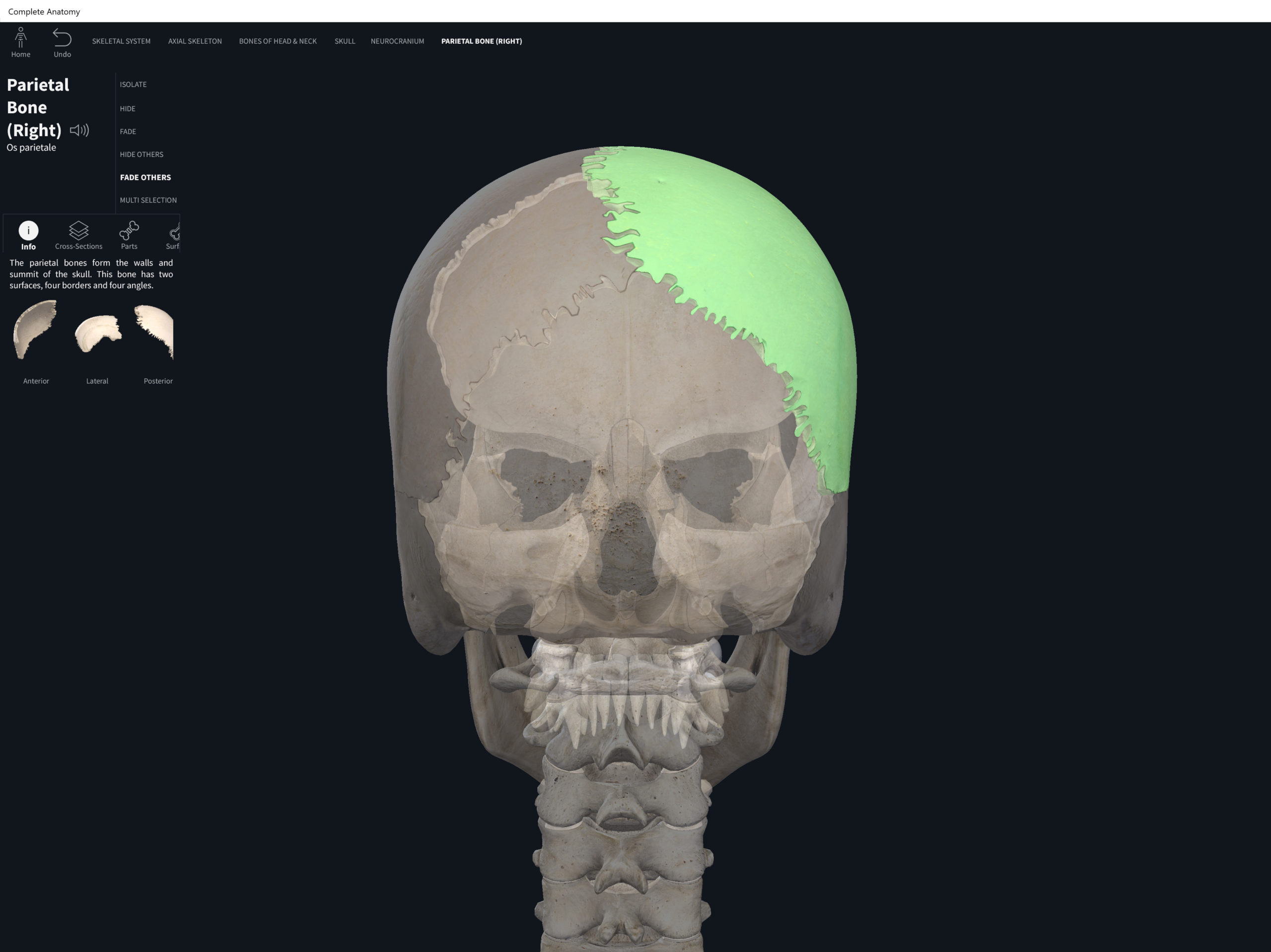
Ulna. Used with permission by 3D4Medical.


References
Biel, A. (2015). Trail guide to the body: A hands-on guide to locating muscles, bones and more.
Cedars-Sinai. (2018). Vertebrae of the spine. Retrieved from https://www.cedars-sinai.org/health-library/diseases-and-conditions/v/vertebrae-of-the-spine.html
Jenkins, G., & Tortora, G. J. (2012). Anatomy and Physiology: From Science to Life, 3rd Edition International Stu. John Wiley & Sons.
Muscolino, J. E. (2017). The muscular system manual: The skeletal muscles of the human body.